From a melt of rings to chromosome territories: The role of topological constraints in genome folding
Abstract
We review pro and contra of the hypothesis that generic polymer properties of topological constraints are behind many aspects of chromatin folding in eukaryotic cells. For that purpose, we review, first, recent theoretical and computational findings in polymer physics related to concentrated, topologically-simple (unknotted and unlinked) chains or a system of chains. Second, we review recent experimental discoveries related to genome folding. Understanding in these fields is far from complete, but we show how looking at them in parallel sheds new light on both.
I Introduction
Each cell of the human body contains about 2 meters of DNA (46 molecules, 5 centimeters long each on average). This much DNA is packed within the cell nucleus with linear dimensions of about five to ten micrometers. How is such an extreme folding achieved? In our view, the natural path to approach the folding of the genome is from a polymer physics perspective. The purpose of the present review is to summarize our recent polymer physics findings Vettorel et al. (2009a); Halverson et al. (2011a, b); J.D.Halverson et al. (2012, 2013) and review their potential implications for the field of genome folding in light of recent experimental van den Engh et al. (1992); Yokota et al. (1995); Cremer and Cremer (2001); Dekker et al. (2002); Gilbert et al. (2004); Simonis et al. (2006); Jhunjhunwala et al. (2008); Lieberman-Aiden et al. (2009); Kawamura et al. (2010); Weiland et al. (2011); Y.Zhang et al. (2012); Sexton et al. (2012); Markaki et al. (2012) and computational achievements Rosa and Everaers (2008); Junier et al. (2010); Iyer and Arya (2012); Wong et al. (2012); Mateos-Langerak et al. (2009); Bohn and Heermann (2010); Bohn and W.Heermann (2010); Bohn and W.Heermann (2011); Fritsche et al. (2012) (see also review articles Cremer et al. (2006); A.Mirny (2011); Fudenberg and Mirny (2012); Dekker and van Steensel (2013); van Steensel (2011); Rosa and Zimmer (2014); de Graaf and van Steensel (2013)).
The very fact that the genome folding problem belongs to the realm of polymer physics was recognized early on, particularly by B. Trask and her co-workers and followers van den Engh et al. (1992); Yokota et al. (1995); Hahnfeldt et al. (1993); Sachs et al. (1995); Marko and Siggia (1997); Ostashevsky (1998); Schiessel et al. (2001); Ostashevsky (2002); Cook and Marenduzzo (2009). More recently this line of research was continued Mateos-Langerak et al. (2009); Bohn and Heermann (2010); Tark-Dame et al. (2011). Through these and other works, it is understood that the “polymer” in question for genome folding is not naked DNA, but rather the chromatin fiber – a complex of DNA with many proteins (histone complexes) more or less tightly bound to DNA. Its length is smaller than 2 meters, but still large enough, on the order of millimeters to centimeters. To imagine the situation, it is useful to increase all scales by a factor of , thus arriving at the necessity to pack and unpack about one hundred kilometers of a regular centimeter-thick rope in and out of a delivery truck.
The rope example highlights the role of “entanglements”, here loosely understood as all consequences of the fact that two segments of DNA/chromatin cannot cross one another, at least not on their own. While a hundred kilometers of rope will easily fit in a truck considering its bare volume, it will be hopelessly tangled if randomly packed. Any attempt to pull out or manipulate any particular piece will become almost impossible. Thus, the real problem is not so much the packing itself, but dealing with the entanglements. Meanwhile, tangling is a very generic property of any long “polymer”, completely independent of any detail (e.g., spaghetti, rope, wire, fishing line). Thus the chromatin fiber should be in the same class! Meanwhile, the cell has to be able to operate on selected parts of DNA with the transcription factors, RNA polymerase, and all other relevant cell machinery. Roughly speaking, DNA should act pretty much as a RAM (random access memory) device, allowing easy access to any place.
Although the view of genome folding as a polymer physics problem seems generally accepted, not much emphasis was placed on the role of topological constraints and entanglements, with the exception of Grosberg et al. (1993); Sikorav and Jannink (1994); Rosa and Everaers (2008). In Ref. Grosberg et al. (1993), the hypothesis of the so-called crumpled globule (later called also fractal or loopy globule) was formulated as a possible resolution to the tangling problem of chromatin. The central idea was the connection between topological simplicity, the lack of knots, and spatial self-similarity. The estimates of the work Sikorav and Jannink (1994) suggested a limited role of topological enzymes in resolving the conundrum of chromatin tangling. And the work Rosa and Everaers (2008), independently of the much earlier work Grosberg et al. (1993), arrived at the conclusion that topological constraints play the central role in the whole of the genome folding problem. In this review, we place topology at the center stage Rosa and Everaers (2008); Vettorel et al. (2009a, b); Rosa et al. (2010); Junier et al. (2010); T.Blackstone et al. (2011); Dorier and Stasiak (2009); Bohn and W.Heermann (2011); Bohn et al. (2010); Bohn and W.Heermann (2010). Of course, we will build on the significant body of knowledge on polymer topology as described, e.g., in the textbooks de Gennes (1979); Doi and F.Edwards (1986); Rubinstein and Colby (2005); Y.Grosberg and R.Khokhlov (1994) and even a popular book Y.Grosberg and R.Khokhlov (2011).
Interestingly, polymer topology, as we will see, naturally brings together two aspects of genome folding which were traditionally discussed separately, namely, the polymer physics aspects and the self-similarity aspects. The latter has been discussed many times in the chromatin literature, and there are indications of both a fractal structure in the cell nucleus interior Takahashi (1989); Lebedev et al. (2005); Bancaud et al. (2012, 2009) and of non-classical diffusion of either particles or the ends of chromatin fiber inside the nucleus Marshall et al. (1997); Bornfleth et al. (1999); Bancaud et al. (2009); Golding and Cox (2006); Banks and Fradin (2005); Heun et al. (2001); Tseng et al. (2004); Dahl et al. (2005); Levi et al. (2005); Meshorer et al. (2006); Chuang et al. (2006); de Vries et al. (2007); Pajerowski et al. (2007); Bronstein et al. (2009); Celedon et al. (2011); Hameed et al. (2012); Hinde et al. (2012); Weber et al. (2012); Burnecki et al. (2012); Kepten et al. (2013); Zhu et al. (2013). We here focus on the attempts to understand these elements of self-similarity based on polymer topology.
The plan of our review is as follows. We begin Section II where basic facts and terminology about genome folding is briefly summarized, mostly for a physicist or a chemist reader. The central piece of this primer is Table 1 which summarizes quantitative information about genome packing across biological realms. In Section III.1 we formulate the physics view of the subject based on polymer systems with topological constraints. We explain what the topological constraints are and why we believe them to be of decisive importance. We then formulate in Section III.2 the specific workhorse model of our approach, a melt of ring polymers, estimate the relevant polymer parameters of chromatin fiber III.3.2, and compare chromatin problems to those known in polymer rheology III.4.
The rest of the work presents at least two interweaving streams. The fact of the matter is that although we argue the melt of rings to be a relevant model for chromatin folding, the model itself is by no means completely understood. We, therefore, combine the review of efforts to understand a melt of rings and related topological polymer models with the review of applications that these polymer ideas find for genome folding. Specifically, Section IV is mostly about the model: we review formulation of the model (IV.1), the existing (rather controversial and inconclusive) theoretical approaches (IV.2), mathematical space-filling curves (IV.3), as well as simulation data for the melt of rings (IV.4) and for other related models (IV.5).
We then move on to compare physics insights against experimental data on chromatin. We begin in Section V with qualitative observation, namely, chromosome territories (see the original work Cremer and Cremer (2001), and the review Cremer et al. (2006) with its many references, and particularly the very readable account Meaburn and Misteli (2007)). The center of our argument there is Fig. 3 which demonstrates how topological constraints on polymers yield a natural simple explanation of territories. With that in mind, we move on in Section VI to quantitative data. We first review in Section VI.1 modern experimental methods known under abbreviated names FISH (van den Engh et al. (1992); Höfers et al. (1993a, b); Solovei et al. (2002); Gilbert et al. (2004); Jhunjhunwala et al. (2008); Yokota et al. (1995); Mateos-Langerak et al. (2009); Meaburn and Misteli (2007); Bridger and Volpi (2010); Markaki et al. (2012); Weiland et al. (2011)) and “C” (Dekker et al. (2002); Simonis et al. (2006); Lieberman-Aiden et al. (2009); Duan et al. (2010); Y.Zhang et al. (2012); Sexton et al. (2012); see also simple summary in Kiermer (2006)). The detailed comparison is performed in Section VI.3 for contact probability data and in Section VI.4 for subchain sizes. This raises new theoretical questions which we address in Section VI.5. We conclude with a brief discussion of dynamics in both chromatin and model polymers in Section VII and the possible role of DNA sequence in Section VIII.
II Genome Folding: A Primer
An overview of the degree of DNA compaction across biological realms is given in Table 1. By far the densest packing of DNA is achieved in viruses. However, viruses are very special. First, their genomes are very short. Their DNA (or RNA) is “stored” and nothing happens to it until the virus infects a cell, i.e., its genome unpacks into the cell. The study of viral DNA or RNA, however interesting in its own right (recent reviews and references can be found in Knobler and Gelbart (2009) and Micheletti et al. (2011)), can teach us relatively little about packing of genomes in higher organisms.
In prokaryote bacteria cells, biologically the next simplest level, the DNA is located in the so-called nucleoid (see, e.g., Thanbichler et al. (2005); Benza et al. (2012)) where it appears to be rather loosely associated with proteins. Surprisingly little is known about these proteins and about nucleoid structure in general. However, see the concise review by Gruber Gruber (2011) and references therein. We will not consider bacterial genomes here, although the main ideas about the role of topology might still be applicable Dame et al. (2011).
Cells of higher organisms are called eukaryotic. They have nuclei where DNA is packed in tight association with histones and other proteins. In this work we focus exclusively on eukaryotes. The hierarchical organization of the genome in eukaryotic cells is described in many textbooks and on the web, typically accompanied by beautiful cartoons (e.g., Alberts et al. (2008); L.Wolfe (1993); Chr (2012)).
The first level of hierarchy is well established: double stranded (ds) DNA is wound around histone octamers forming so-called nucleosomes. This “beads on a string” chain of nucleosomes is known as the (or ) fiber. A lot is known about this level of organization, including the detailed structure of nucleosomes (see recent summary in Chakravarthy et al. (2005)) and the stems of their linkers (see, e.g., Meyer et al. (2011)). A length of dsDNA composed of roughly base pairs is wrapped around a histone octamer in each nucleosome, and segments of roughly to (about in average) base pairs form linkers between these nucleosomes. Whether the position of nucleosomes along DNA and the corresponding linker lengths are random, dictated by the maximal entropy principle of statistical mechanics, or determined by the underlying DNA sequence and form a special code is an extensively studied and hotly debated subject (see, e.g., Segal et al. (2006)).
The next level of hierarchy is known as the 30 nm fiber. Its existence is easy to understand: since the linker DNA between nucleosomes is shorter than the persistence length (), the chain of nucleosomes is expected to form a zig-zag with characteristic width close to 30 nm. The fact of the matter is that such a 30 nm fiber is neither particularly rigid nor very well defined Emanuel et al. (2009), its prominence under in vivo conditions of the cell is doubtful. The idea of its importance seems to be losing popularity Hansen (2012); Dekker and van Steensel (2013).
Just above a few tens of nanometers or above base pairs, there is presently a big gap in our knowledge and understanding. A good expression of it is given by Meyer et al. Meyer et al. (2011), who argue for the existence of a cross-over scale. Below that scale the system is pretty rigid, its elements (such as nucleosomes and linkers) can be crystallized, and their structures are fully determined. By contrast, well above this scale fluctuations become important. This is the realm of statistical soft matter physics. In this range, on the scale of about base pairs and higher, the overall architecture of the genome in 3D space is not well understood and its features are only now starting to become known. Our present review is intended to contribute to this process.
Thus, our subject matter is the folding of chromatin fiber – an entity which is not perfectly defined. It definitely has the 10 nm fiber at its core, and it may be somewhat more organized. Its physical properties will be discussed below.
An indispensable part of the chromatin fiber and its properties is a multitude of different proteins. Some of them are histones, and some of the histones are constituents of nucleosomes. Other histones, such as H1, are attached to linker DNA. Many other non-histone proteins are also involved, such as cohesins and condensins, and despite their suggestive names their function apparently is not restricted to gluing pieces of chromatin fiber together and maintaining the compactness Bloom and Joglekar (2010); Phillips-Cremins et al. (2013) (see also Bodnar and Spector (2013)).
On the highest level of the nucleus as a whole, chromatin is an important part of many processes through the cell cycle. We here focus on the interphase nucleus, the stage when the cell does not divide and chromosomes stay swollen inside the nucleus. For differentiated cells, some part of DNA, which is not transcriptionally active, is packed somewhat more tightly in so-called heterochromatin. The other part, called euchromatin, is less densely packed and it is involved in transcription of those genes which have to be expressed in the given cell. The genes in heterochromatin are silenced through either histone methylation or interactions with the so-called short silencing RNA (see, e.g., Alberts et al. (2008)). In either case, the placement of any particular part of DNA into either hetero- or euchromatin is inheritable via epigenetic mechanisms. Thus, the above mentioned analogy with RAM is restricted to euchromatin only. Nevertheless, for our consideration it is good enough because a significant part of DNA has to be easily accessible for bulky processes such as, e.g., homologous recombination, which would be next to impossible if the DNA were heavily tangled.
Of course, genome folding and organization is not a static phenomenon. Cells live, and the cell nucleus is the place of diverse and incessant activities. In this review, we will mostly consider so-called interphase, which is (usually) the longest stage of the cell cycle. During interphase, chromosomes are (relatively) swollen or decondensed and they occupy most of the volume inside the nucleus. In that time, proper genes (i.e., the ones which have to be expressed in the given cell type) are transcribed into RNA for subsequent protein synthesis. It is believed that interphase chromosomes are structurally and spatially organized to help control gene expression. At the end of interphase, the cell prepares itself for division. This process involves a quite dramatic spatial rearrangement and leads to the formation of highly condensed mitotic chromosomes in which transcription appears to be switched off. Our consideration concentrates on interphase chromatin.
| Organism | Length | Diameter | Volume fraction | Volume fraction | ||
|---|---|---|---|---|---|---|
| of genome, | of domain, | of DNA | including proteins | |||
| lower | upper | |||||
| Bacteriophage (T4) | not applicable | |||||
| E. coli | not known | |||||
| Yeast, haploid | 2 | |||||
| Drosophila, diploid | 10 | |||||
| Chicken, diploid | 5 | |||||
| Mouse, diploid | 9 | |||||
| Human, diploid | 10 | |||||
III Polymer Physics Picture
III.1 Polymer packing and topology
Approaching the polymer physics picture of chromatin folding, we should first realize that chromatin fiber as a polymer is packed fairly densely in the nucleus. The volume fraction of DNA itself in a typical human nucleus is as high as about a percent (with 3.3 billion base pairs in the genome, two copies of the genome in a diploid cell, of length per base pair, double helix diameter of 2 nm, and typical cell nucleus diameter of , the volume fraction is – see Table 1; see also web site Milo (2012) and the book Phillips and Milo ). In making such an estimate, one should keep in mind the tremendous variability of biological circumstances. For instance, depending on cell type and conditions, the diameter of a human cell nucleus can easily vary by a factor of , from about to . This increases the possible DNA volume fraction by almost an order of magnitude, making it as high as about . A more meaningful number, perhaps, would be the volume fraction of the chromatin fibers. Its exact value depends on the exact definition, two of which are given in the last two columns of Table 1. Such estimates also vary significantly between different types of cells and different organisms. For instance, the volume fraction of chromatin in simpler eukaryotes such as yeast is twice or more smaller than in humans. As usual in biology, there are many different cases, but it appears that the volume fraction of chromatin is pretty high in all cases. By the standards of polymer physics, chromatin is not quite as dense as a melt, but it is a concentrated solution, which should be thought of as a melt of blobs de Gennes (1979); Rubinstein and Colby (2005); Y.Grosberg and R.Khokhlov (1994). Experimentally the properties of concentrated polymer solutions and those of melts are rather similar without any significant qualitative difference. For both, the catastrophic tangling of long chains, as in equilibrated melts/solutions, always leads to macroscopic relaxation times, which is avoided for chromatin in all cases. In our opinion, this suggests that genome folding should also be considered from a polymer physics perspective. Surely, many aspects of chromatin have very little to do with polymer physics, but one should expect that the very basic motive – being dense while still avoiding tangling – also deserves generic polymer physics considerations. In our opinion, understanding this is presently one of the main challenges of the physics of the cell nucleus.
In this context, the most striking observation is that of chromosome territories Cremer and Cremer (2001). This observation is in sharp conflict with the well established observation for dense and semidilute polymer systems: if chromatin fibers are considered as regular polymer chains then in a dense system, such as a melt or concentrated solution, they should interpenetrate, and entangle if they are in equilibrium. Therefore, already the very fact of territorial segregation between chromosomes is consistent with the idea that chromatin fibers somehow avoid tangling. The hypothesis that DNA in the nucleus might be pretty densely packed while still avoiding catastrophic tangling was formulated almost 20 years ago Grosberg et al. (1993); A.Y.Grosberg et al. (1993) based on the preceding paper Grosberg et al. (1988), where the idea of the so-called “crumpled” globule was formulated in the context of the dynamics of the coil-globule transition. At about the same time, the role of DNA topology and the corresponding enzymes were also discussed in the work Sikorav and Jannink (1994). More recently but independently from Grosberg et al. (1988, 1993), the role of DNA topology was strongly advocated by Rosa and Everaers Rosa and Everaers (2008).
When speaking of polymer topology, we mean all consequences of the fact that two pieces of a real polymer, such as dsDNA or a chromatin fiber, cannot pass through one another, at least not without special topo-enzymes. For polymers without ends, such as rings, the topological constraints are strict and are topological in the rigorous mathematical sense of the word. For open end polymers, topological constraints are always a matter of time scale. Rosa and Everaers Rosa and Everaers (2008) provide rather convincing estimates suggesting that on the time scale of a cell life for higher organisms, topological constraints of a chromatin fiber should be regarded as permanent. At the same time, there are also temporary loops in chromatin fiber every time that two loci come close to one another in space. These contacts and, therefore, loops are detected en masse by “C” experiments (see below Section VI.1). It is important to realize that these loops do not form topological interactions with one another as long as their being a loop (i.e., contact between the ends) is subject to relaxation on the time scale of interest.
Important note on terminology: The word “topology” seems to be getting over-used in the field of genome folding (as in many other fields). For instance, people talk about “topological domains” in chromosomes Dixon et al. (2012). Our use of “topology” follows the polymer physics tradition and, as we said, means all consequences of the fact that a polymer cannot cross itself. For our workhorse model of polymer rings, topology will have a strict mathematical meaning.
Recent experimental work Lieberman-Aiden et al. (2009) appears to be consistent with the theoretical picture that in the range between and million base pairs the chromatin fiber is organized as a crumpled globule. In fact, the authors of the experimental work Lieberman-Aiden et al. (2009) prefer to call this state a fractal instead of crumpled globule, which is a pure terminological discrepancy (i.e., fractal and crumpled globule mean exactly the same thing). The experimental work strongly motivated closer theoretical scrutiny. On the one hand, some signatures of a crumpled globule state are visible (particularly visible post factum) in simulation work Rosa and Everaers (2008). On the other hand, the naive original idea of a crumpled globule Grosberg et al. (1993, 1988) requires much deeper understanding. The major source of such new understanding over the last few years was computer simulation of both lattice Vettorel et al. (2009a, b) and off-lattice models Rosa et al. (2010); Halverson et al. (2011a, b); Bohn and W.Heermann (2010); Bohn and W.Heermann (2011) (see also J.D.Halverson et al. (2012, 2013)). Here we will review the status of our understanding of topologically restricted dense polymers and their relevance to genome folding.
III.2 Why melt and why rings?
To demonstrate this relation, we follow our main guiding theoretical idea, namely that topological constraints are bound to play a central role in genome folding and the overall nuclear architecture. This may seem surprising given that DNA in a eukaryotic cell has open ends. However true, open ends do not cancel the topological constraints, because reptation, the leading mechanism of topological relaxation in linear polymers de Gennes (1979); Doi and F.Edwards (1986), is likely to be suppressed for chromatin. The main reason why reptation is not relevant is because the chromatin fiber is very long. Rosa and Everaers Rosa and Everaers (2008) estimate that the reptation time for human chromatin is orders of magnitude longer than the characteristic times of all known cellular processes. Thus, once in a crumpled globule state it would take far too much time to relax into an entangled equilibrium state. Phenomena like these are also discussed in the context of polymer rheology Vettorel and Kremer (2010); T.C.B.McLeish (2007). Furthermore, there are at least two additional factors suppressing reptation of chromatin fibers in the cell nucleus. First, telomere regions at the end of chromosomes A.M.Olovnikov (1973); Szostak and Blackburn (1982); Greider and Blackburn (1985, 1989), by virtue of their peculiar sequences which include huge numbers of repeated short motives, are likely to form bulges preventing reptation (similar to the mechanism described by de Gennes de Gennes (1968)).
Second, some parts of the chromatin fiber, particularly heterochromatin, are likely to have attachment points to the inner surface of the nuclear envelope (lamina). Also, topological enzymes are likely to play only very limited role for the interphase nucleus, if any (see more details on that below in Section V).
Thus, let us consider the idea that the chromatin fiber cannot cross itself and does not reptate as a working hypothesis. What are the consequences of such an assumption, and how do they compare to the data? The main ingredient of a model must be the large amount (length) of chromatin fiber stored in a restricted volume at high concentration with topological restrictions. We also adopt the hypothesis Grosberg et al. (1993) that the topological state of chromatin in the nucleus is very simple. That is, there is either a complete lack of knots or at most a very few rather simple ones. Although mathematically knots are defined for closed loops only, the concept of knots is still reasonable for very long open strings, such as chromatin. (We note in passing that the idea of approximately defined knots in open strings recently gained popularity in the context of proteins, e.g., Sulkowska et al. (2012)). Therefore, we argue that the simplest theoretical model which meets all of the above conditions is a melt of very long unknotted and nonconcatenated rings. This model system mimics the idea of chromatin being (nearly) unknotted and of pretty high concentration in the nucleus. The obvious drawback of rings, namely that they have no ends, is assumed to be of marginal importance for conformations of very long polymers, once they have been prepared in a non-entangled starting configuration.
The idea that chromatin, because of the lack of reptation, can be modeled by a system of nonconcatenated rings was first suggested by Rosa and Everaers Rosa and Everaers (2008). They also provided arguments that this approach could explain the phenomenon of chromosome territories. In the same work Rosa and Everaers (2008) they simulated very long chains with open ends, with the idea that they more faithfully represent chromatin. The advantage of the ring model is that it allows for clean conclusions to be drawn regarding the role of topological constraints. Unlike for chains with open ends, results are not restricted to times which are orders of magnitude below the equilibration time. This avoids the difficult task of mapping time scales between real and simulated chromatin. The ring model is particularly articulate because conformations within the computer simulation time are fully equilibrated. If the rings display any kind of crumpled globule behavior, this is a strong indication that the driving force to tangle for very long open chains is rather weak and probably not relevant on times typical for interphase chromosomes.
Another, easier question is that of density. The DNA is packed in interphase chromosomes at the density of a concentrated solution. In polymer physics such a system is best described as a melt of properly defined blobs (see, e.g., de Gennes (1979); Y.Grosberg and R.Khokhlov (1994); Rubinstein and Colby (2005)). Although the precise definition of such blobs for chromatin is by no means trivial, we assume that our consideration is performed at the level of blobs, which can be estimated as being not larger than about 300 nm (see below), as suggested by small angle neutron scattering experiments Lebedev et al. (2005). Thus, we consider a melt or a system where the polymers fill essentially all available space homogeneously. It turns out that such a melt-of-rings-based approach quite directly yields a natural explanation of chromosome territories.
III.3 Physical parameters of chromatin fiber as a polymer
III.3.1 Linear density and persistence length
As we discussed above, chromatin fiber is not perfectly well defined. Nevertheless, to approach its polymer physics we must have some idea about its polymer parameters. Luckily, the cornerstone of polymer physics is the concept of universality de Gennes (1979): not very many parameters are required to approach the generic properties, such as those related to entanglements. In fact, four parameters are most important: polymer length, persistence length, density and the resulting entanglement length. Let us discuss their estimates.
Usually, the genome length is known in terms of the number of base pairs (see, e.g., Table 1). That means, the dsDNA length is also known, , where . In other words, the “linear density” of the double helix is which is roughly 3 base pairs per nanometer. The similarly defined “linear density” for the 10 nm fiber is about 20 base pairs per nanometer. The linear density for chromatin fiber is not known exactly, but experimenters believe it to be above or close to Dekker and van Steensel (2013). For example, for the human (diploid) genome of altogether , this linear density gives a total length of the chromosome fiber of .
The persistence length of bare dsDNA is close to 50 nm, while for chromatin fiber it is difficult to determine accurately. It is certainly longer than that of dsDNA, and the opinions seem to be converging on a number around Dekker and van Steensel (2013) or Kuhn segment . The important fact for our purposes is that chromatin fiber is a flexible polymer on the length scales of our interest and thus can be discussed in terms of the standard theories of dense systems of flexible or semi-flexible polymers.
Assuming the linear density is known, one can estimate that the chromatin fiber Kuhn segment consists of about . Another interesting estimate is based on the density of chromatin in a human cell nucleus which is about (this number corresponds to in the volume of a sphere with diameter ; it is equivalent to 1% DNA volume fraction indicated in Table 1, as the volume of one base pair is very close to ). This corresponds to a number density of Kuhn segments of . The dimensionless overlap parameter is then , a value not unusual for regular synthetic polymers as well. Thus the chromatin pieces overlap strongly on the scale of the persistence length, which supports the picture of a melt/dense solution of flexible polymers.
In the usual polymer physics framework, another group of parameters has to do with volume interactions between polymer segments, and includes virial coefficients, excluded volume, Flory -parameter, and the like. These parameters are important to determine whether a polymer chain swells or collapses, etc. In our case, the volume behavior of chromatin is controlled by interactions with proteins, including histones and non-histones, such as cohesins and condensins Bloom and Joglekar (2010); Phillips-Cremins et al. (2013); Bodnar and Spector (2013). Luckily, we do not have to worry about it, at least to the first approximation, because we assume to know the overall density of chromatin, as discussed above. For a polymer system of a given density, its spatial organization does not depend very much on the mechanism controlling its density, whether it is a proper combination of attractive and repulsive volume interactions or confinement by an outside envelope. We will rely on this approximation. Of course, this is only good for an averaged description, the simplest version of mean field. It does not capture, for instance, the fact that some parts of chromatin fiber can be methylated or acetylated more than others, leading to uneven excluded volume parameters and an uneven density distribution in space. Also, it does not capture active processes, transcription factories, and a myriad of other activities happening in the nucleus. Nevertheless, here we adopt the simplest uniform approximation, which we view as the necessary first step.
III.3.2 Entanglement length of chromatin
As we mentioned previously, our goal is to explore the role of topological constraints. In polymer physics, the known fruitful way to approach this kind of problem is in terms of entanglements. The concept of entanglement is in fact quite subtle. Although it arises from the simple fact that polymers are not phantoms and two pieces of (the same or different) polymers cannot pass through one another, entanglement is fundamentally a many-chain phenomenon. It is very common when two chains may appear not entangled to one another, but they are made entangled by a third chain nearby and so on. Impressively, in the melt of linear chains of large length each coil overlaps with of other coils, but they collectively manage to create a much larger number of order of entanglements for the given coil. It is a highly non-trivial result that all these collective effects can be effectively described by a single parameter, namely the entanglement length . This parameter characterizes the average chain length such that on smaller length scales topological constraints are unimportant while they dominate on larger length scales. A good way to think about it is to imagine that the chain faces an un-crossable obstacle, on average, once over length . Significantly, it may be that the distance between entanglements is small or large compared to the Kuhn segment. In the former case the chain is nearly straight between the obstacles while in the latter case it is a nearly free coil between obstacles. We cannot possibly do justice to this subject here and refer the reader to textbooks de Gennes (1979); Doi and F.Edwards (1986); Y.Grosberg and R.Khokhlov (1994); Rubinstein and Colby (2005).
An estimate of the entanglement length, , is more involved than that of the previously discussed quantities. In principle, the most direct measurement of is usually based on rheology. Without having access to rheological data, we will rely on the large body of knowledge about a variety of polymers, with densities and flexibilities varying by several orders of magnitude Colby et al. (1992); Fetters et al. (1999); Everaers et al. (2004); Sukumaran et al. (2005); Uchida et al. (2008). Specifically, we will employ the interpolation formula
| (1) |
where Uchida et al. (2008) fits a huge variety of systems ranging from fully flexible polymers to rather rigid semi flexible systems. Given the uncertainty in our knowledge regarding the linear density, and given also that different cells may have nuclei of different volumes, we can only indicate a range of possible values of , as shown in Table 2.
| Linear density | |||
|---|---|---|---|
From our knowledge of synthetic polymers, we tend to expect a realistic value of in between the extremes indicated in Table 2, perhaps somewhat closer to the lower end, around . In summary, one can state that the entanglement length of chromatin fibers at conditions typical for a human cell nucleus would most probably be below . This holds for most organisms with diploid cells (cf. Table 1). Compared to basic results from polymer theory these systems are deeply in the regime where topological effects dominate all relevant structural and relaxation processes.
In this context it is interesting to compare the above estimates to nuclei of other cell types, particularly that of yeast. Not only is the total length of the genome much smaller, more importantly, the density in the nucleus is about a factor of three smaller, cf. Table 1 (while for chicken nuclei it is larger). It has a total length of base pairs in altogether 32 chromosomes. On average each chromatin fiber thus contains only 137,000 base pairs. Assuming the same chromatin fiber structure for yeast as for human diploid cells, which probably will only be very approximate, one arrives at about total contour length divided among 32 chromatin fibers. Considering the reduced density, the average total contour length per fiber is only a few times more than or even close to . Thus for yeast, topology effects are expected to be very weak.
III.4 A physical analogy: rheology of non-entangled melts
It is instructive to relate the above analysis to known results from polymer chain dynamics and rheology. Rosa and Everaers Rosa and Everaers (2008) have argued that the dynamics of chromosomes in the cell nucleus are orders of magnitude slower than all known biological processes. Based on the reptation concept, they estimate the overall relaxation time in such a system to be well above the average life time of a human being. In contrast the overall relaxation time of a melt of nonconcatenated rings is much smaller than that of an entangled melt of linear chains, as we will see below Halverson et al. (2011b). Considering the stability of chromosome territories, the specific question is whether there is a strong tendency to entangle, which then would need specific measures to be prohibited, or whether such a tendency does not exist.
As already mentioned, one expects a pressure difference in a computer simulation between a melt of nonconcatenated rings and a melt of linear polymers of the same chain length and density. This is because of the different number of degrees of freedom and of the topological constraints. , however, has not been detected so far. Though too small to be detected, the total free energy increase per chain compared to a melt, roughly with being the number of repeat units per chain, can be quite large. Switching off the topological constraints would lead to a very fast relaxation of the polymer globules towards Gaussian conformations through an isotropic expansion of the polymers. Indeed for linear chains of length up to several this holds true J.Barham and M.Sadler (1991); de Gennes (1995); Vettorel and Kremer (2010), even though chain connectivity and non-crossability are fully maintained. This rapid relaxation does not occur, when the chains become much longer. Rastogi et al. Rastogi et al. (2005) studied the processing properties of a polymer melt, which was created upon melting a dense agglomeration of polymer crystallites, each of them containing only one single, but very long chain. They observed a significant reduction in the apparent, transient viscosity compared to an entangled melt of otherwise identical properties. Indeed, studying the relaxation of a melt of compact globules into an entangled melt revealed a rapid expansion for short chains and a very slow process for very long chains () Vettorel and Kremer (2010). This phenomenon can be understood in terms of the elastic distortion due to “non-cooperative” reptation T.C.B.McLeish (2007). That is, the reeling out of chain ends into the surrounding chains creates an elastic deformation of globules, making the relaxation process very slow. For nonconcatenated rings that would require reeling out of doubly-folded pieces, which are subject to an additional entropy penalty. Unfortunately there is no systematic study of this phenomenon so far. Despite this, these results indicate that for the diploid cells of higher organisms no complicated biochemical apparatus is required to keep the chromosomes segregated on the biologically relevant time scales once they are “prepared” in a territorial arrangement.
IV Melt of rings and related polymer models
IV.1 Model and question formulation
Understanding the conformational geometry and statistics of nonconcatenated rings in the melt turned out to be an unexpectedly difficult challenge Kim et al. (2012). To emphasize the generic character of the problem, even beyond the previous discussion, and its independence of any chemical or microscopic details, it is useful to start with a purely mathematical formulation.
Imagine a piece of cubic lattice in space with number of nodes occupied by closed loops or rings of steps each, leading to . In this case, space is completely filled which corresponds to the melt assumption where each blob occupies a node giving a volume fraction . Importantly, we assume that each ring is unknotted and rings are not concatenated. Furthermore, no node is occupied more than once.
This is the description of the simplest model of a concentrated set of unknotted and nonconcatenated rings, the subject of our attention. Of course, this system can be considered off-lattice, and can be equipped with further details. The main issue, however, is the interplay between conformations and packing of homogeneously space-filling polymers, i.e., , and topological constraints of being unknotted and nonconcatenated.
Here is a list of simple questions with regard to the statistical properties of this model, and which are most closely related to the experimental data being collected by methods such as FISH and 3C:
-
•
How does the spatial extension (e.g., averaged gyration radius) of a ring squeezed between others depend on the ring length ? We expect the dependence to be a power law:
(2) The index defines whether rings can form territories ( for spatial dimension ) or not ().
-
•
How does the size of the subchain (e.g., its gyration radius or end-to-end Euclidean distance) depend on the subchain arc length . We expect it to be governed by the same index at sufficiently large , so that .
-
•
What is the probability that two monomers separated by arc length will be in contact in space? Again we expect this contact probability, or loop factor, to follow a power law in , with a power , where the relation to is at least not clear:
(3) -
•
How many monomers of one ring are in contact with monomers of other rings? How many monomers of a subchain are in contact with other subchains? We expect this fraction of a ring, which could be called “surface” or easily accessible fraction, to be governed by another exponent :
(4)
There is also a separate set of questions regarding the dynamics of rings in concentrated systems (see Section VII below), but for now we concentrate on the equilibrium statistics in relation to chromatin experiments.
IV.2 Theoretical approaches
IV.2.1 Flory type theories.
A significant step was the work of Cates and Deutsch Cates and Deutsch (1986), who considered a melt of nonconcatenated rings and arrived at the prediction that the size of a ring scales as
| (5) |
Their approach follows a classical Flory theory of regular polymers with excluded volume and makes the argument that the equilibrium size of the ring is determined by the balance of two factors: entropy loss of the ring itself when it gets more compact than its preferred Gaussian size, and entropy loss due to the topological constraints with the surrounding rings if the ring size increases by protruding loop-like “tentacles”. The first factor is estimated as , where is the number of monomers in the ring, while is the ring size to be determined, and is a microscopic length scale such as the Kuhn segment. The second contribution to the entropy is estimated as follows: if every ring has size of order and, therefore, lives in a volume , while its own volume is only about (assume for simplicity that the chain thickness is governed by the same length scale ), then about different rings cohabit one and the same volume. If we assume that every one of these rings forces our chosen ring to loose entropy of order unity, then we arrive at the following estimate of the overall free energy:
| (6) |
where is temperature in energy units. Minimization with respect to yields the result (5). Surely, this argument is very much open to criticism, much more so than the similarly looking classical Flory theory of a coil in good solvent. For instance, the first term assumes the rings to be Gaussian and neglects that the ring itself is unknotted. In reality, the free unknotted ring is swollen Moore and Grosberg (2005), thus its statistics are not Gaussian for topological reasons. Therefore, entropy loss due to compression is larger and grows faster with decreasing . The second term in Eq. (6) is even more problematic. It is not clear at all why every one of the cohabiting rings produces an entropy loss of order unity. One can try to improve this estimate by assuming, for instance, that the osmotic pressure of surrounding rings is a many-body type of phenomenon such that the corresponding free energy scales as with some properly chosen power (see Cates and Deutsch (1986)). Instead of formula (5) and index , such an approach yields index , which can be anything in the interval depending on . Of course, this only emphasizes the physically unjustified character of the estimate. Nevertheless, the very idea of a proper balance of entropy losses due to intra- and inter-ring contributions behind Eq. (6) deserves serious attention as a basis for possible future improvements.
One noteworthy attempt of such improvement was undertaken by T. Sakaue Sakaue (2011, 2012). It is also based on minimization of free energy for a test ring, qualitatively similar to Eq. (6) in the sense that there is one term which favors compression because the test ring looses entropy by making long loopy protrusions among surrounding rings, and there is another term which disfavors compression because the loss of entropy of the test ring itself. In such a general form, the idea is undoubtedly correct. Such concepts are well known, as for instance discussed for the case of gels in Y.Grosberg (1993). Both terms were estimated in Sakaue (2011, 2012) in a more involved way compared to Cates and Deutsch (1986), but the estimates rely upon some rather ad hoc assumptions. In the end, the suggested variational free energy reads
| (7) |
where numerical coefficients of order unity are all dropped out, but the entanglement length is defined in a peculiar non-standard way, different from the usual definition by a factor of about or more. This suggested form of the free energy automatically requires that very long rings in a melt are compact, because asymptotically is growing faster than , making the argument in the log eventually negative. However, if we assume , then on the way to the final asymptotics , valid for , the free energy (7) predicts a region of intermediate asymptotics valid at (the latter result is not mentioned in Refs. Sakaue (2011, 2012), but can be derived from the free energy (7)). Although this theory involves many poorly justified assumptions, it does capture the existence of a relatively wide cross-over region, with seeming power which is close to the result of Cates and Deutsch (5).
An important aspect of free energy estimates, such as Eq. (6) or (7) is that they represent a small, but decisively important, correction to the very large contribution from repulsive forces of interaction between monomers at high density. These corrections are decisive because they depend on and their structure is determined by the topological constraints, such as absence of knots in any particular ring and absence of links for any group of rings. For instance, there is no doubt that the osmotic pressure of a concentrated system of nonconcatenated rings is larger than that of a similarly concentrated system of linear chains (for large and at the same density). However, this correction is so small, compared at the dominant term produced by excluded volume interactions between monomers, that none of the presently available simulations is sensitive enough to detect it, as previously mentioned.
IV.2.2 Crumpled, or fractal, globule
Another possible idea regarding rings in the melt is based on the convolution of three hypotheses. The first hypothesis assumes that the fast collapse of a single polymer chain upon abrupt solvent quality quench produces a peculiar state, called crumpled globule Grosberg et al. (1988). This state is dense, self-similar, and free of knots and should have . The second (least proven) hypothesis is that this state of a crumpled globule is equilibrium for any isolated chain collapsed in a poor solvent with the constraint that knots are excluded, for instance, for an unknotted ring. The third hypothesis suggests that a ring squeezed by its neighbors in the melt and a ring collapsed in a poor solvent should be similar and, therefore, both should be crumpled globules. This latter hypothesis will be especially questioned by the simulations. Thus, the idea is that a crumpled globule results when the polymer is forced to adopt a compact conformation without knots, whether the lack of knots is an imposed topological condition, as in a ring, or it results from slowness of reptation, as in rapid collapse (where collapse should be viewed as “rapid” if it occurs faster than the relevant relaxation time scale; in practice, it may still be rather slow by conventional standards).
According to arguments developed in Grosberg et al. (1988), the signature of a crumpled globule is that every subchain of sufficient length is collapsed in itself, and has the size of order . In other words,
| (8) |
which is assumed to be true at with . That also means the chain itself, on the scale beyond the entanglement length , has fractal dimension . The idea of justification goes back to the well established fact of topological repulsion between nonconcatenated rings. For instance, the second virial coefficient of two nonconcatenated rings in dilute solution is close to and is practically independent, at least in the scaling sense, of the real excluded volume of monomers Vologodski et al. (1974) (see also recent work Bohn et al. (2010) and references therein). Nevertheless, the concept the crumpled globule remains a hypothesis.
Furthermore, we should emphasize that all three aspects mentioned above need careful attention: although the simulations in Lieberman-Aiden et al. (2009) do confirm the formation of a crumpled globule upon fast chain collapse, it is not proven that this scenario is valid for all mechanisms of squeezing (e.g., by external field, by poor solvent, etc). It is likely but not proven that an equilibrium collapsed ring is a fractal. Additionally, it is not clear that rings squeezed in the melt form the same type of fractal as single collapsed rings, simply because the surface of a collapsed globule is very rough in the former case while smooth and dominated by surface tension in the latter. The most likely possibility is that all of these systems are crumpled globules of somewhat different kinds, but this remains to be properly understood. An attempt to understand the nature of crumpled globules more deeply faces serious mathematical challenges Nechaev and Vasilyev (2005); S.K.Nechaev et al. (1996).
A direct computational test of the hypothesis that the typical conformation of a collapsed chain without knots is a crumpled fractal globule was attempted in the work Lua et al. (2004). For that purpose, Hamiltonian walks (i.e., lattice conformations which fill every site of a given lattice segment once and only once) were generated on cubic segments of a cubic lattice up to the size of . A rather clear tendency toward segregation of subchains was observed, but the length of the polymer was insufficient to arrive at conclusive results.
The question of the long-term stability of crumpled structures was recently addressed by simulation Schram et al. (2013). For the particular crumpled conformations which were used as initial conditions, long-term stability was not observed.
Interestingly, conformations that are similar to crumpled are implicated in certain scenarios of active (ATP dependent) formation of chromatin structures, by so-called loop-extruding enzymes Alipour and Marko (2012).
IV.2.3 Lattice animal model
Although the intent of the above approaches is to describe the restriction of accessible conformations due to the topological constraints, the topology is present there only indirectly. In general, theoretical ideas about polymer topology revolve around the idea of an effective tube suggested originally for networks S.F.Edwards (1977) and most widely known in the context of reptation de Gennes (1979); Doi and F.Edwards (1986); Rubinstein and Colby (2005); Y.Grosberg and R.Khokhlov (1994, 2011). Tubes and reptation ideas, unfortunately, have no direct applicability for nonconcatenated rings. Since the tube is a sort of “topological mean field”, a similar idea for the melt of rings is a ring squeezed into a lattice of obstacles, just like in the very first work of Edwards S.F.Edwards (1977). That means, we should imagine placing one ring into a lattice of immobile, un-crossable, and infinitely long straight spikes such that the ring is not tangled to the lattice, i.e., it must be topologically possible to take the ring out of the lattice. This model was examined in the works Khokhlov and Nechaev (1985); Rubinstein (1986); Nechaev et al. (1987); Obukhov et al. (1994). The conformation of a ring in such a lattice is well understood, it represents a so-called lattice animal, i.e., the ring double-folds along an annealed branched structure. The description of this system is mathematically beautiful Nechaev and Vasilyev (2005), and it is undoubtedly correct for a single ring in the lattice of obstacles.
The applicability of a lattice of obstacles and annealed branched structure in the self-consistent situation of many rings is not obvious. It is a difficult computational task, so far unresolved, to check for such structures in the computationally generated conformations of rings. The difficulty arises from the fact that branches, if they are real, exist only on a scale significantly larger than . This motivates the idea to assume the existence of such an annealed branched structure, examine the consequences of the assumptions, and compare them with computational data. Such an attempt was undertaken by one of us (AYG) in the recent work Grosberg (2013), and the results are rather encouraging. First, this logic yields a rather straightforward proof that , i.e., every ring collapses onto itself and different rings form territories. Interestingly, the backbone of a branched lattice animal inside its territory behaves like a self-avoiding random walk. That means the backbone length is estimated from the condition (ignoring here ; see Grosberg (2013) for details). This result may seem counterintuitive because the backbone is a polymer and normally self avoidance is screened under melt conditions de Gennes (1979); Doi and F.Edwards (1986); Rubinstein and Colby (2005); Y.Grosberg and R.Khokhlov (1994). The fact of the matter is that the screening effect in our case is exactly compensated by the unusually large excluded volume, as it is controlled by the side chains of the branched structure. And the distribution of material between the backbone and the side branches in the annealed system is self-consistently determined so as to establish the self-avoiding statistics.
These ideas also shed light on other critical exponents for the system, such as the surface exponent and the contact exponent . It turns out that this is related to the “other ”, which we denote here , and which controls the number of conformations for a polymer de Gennes (1979). This relation explains why our index cannot be computed by any analog of Flory theory, and, more generally, why finding this index is a very difficult task.
The lattice animal model was also used in the work Iyer and Arya (2012) to model chromosomes directly on a phenomenological level, and we return to this later in Section IV.5.
This completes our discussion of the rather unsettled situation of theoretical concepts aimed at describing the overall size of the rings, or the index (2). Apart from the very recent attempt Grosberg (2013), no theory we are aware of goes beyond to address contact probabilities, subchain surfaces, and other more detailed conformational properties which are described by an independent set of exponents which we call and , as defined in Eqs. (3) and (4).
IV.3 Peano type space-filling curves and mathematical hypothesis
Good realizations of crumpled globule conformations are well known in mathematics as space-filling curves. The most widely known example is the Peano curve in 2D. Other examples include the Hilbert curve and its closed loop (with two ends connected) version called Moore curve (shown in Fig. 1), as well as Sierpinski, Lebesgue, and Gosper curves. For a modern review of this topic, including computer algorithms to generate these curves, see the book Sagan (1994). See also Platzman and Bartholdi III (1989) for some more recent unexpected applications of space-filling curves in data sorting and parallelization of computer algorithms.
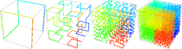
Space-filling curves are usually constructed via recursive algorithms, and, in the limit, they are true self-similar fractals. Of course, unlike in the mathematical literature where the nature of the limit is the central aspect, for our purposes we only need a high, but finite level of iteration. In this sense, for instance, the 3D Hilbert or Moore curves in Fig. 1 represent true fractal globules: they are unknotted and self-similar by construction.
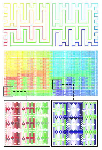
Classical curves, such as Peano, Hilbert, Moore and Sierpinski space-filling curves, are characterized by pretty smooth surfaces between neighboring folds, which corresponds to in 3D (or more generally in -dimensional space). As we will show below, this yields (or ). In the context of chromatin models, the natural question is this: is smoothness of surfaces, expressed by the value , an inherent property of regular space-filling curves? Recently, two of us (JS and AYG) answered this question by explicitly constructing the entire novel family of fractal unknotted space filling curves with very wiggly surfaces Smrek and Y.Grosberg (2013). A 2D example of such a curve is shown in Fig. 2. In fact, there are curves with index arbitrarily close to unity from below (and consequently arbitrarily close to unity from above). E. Lieberman-Aiden mentioned to one of us (AYG) a similar result of his own Lieberman-Aiden (2012).
Of course, the existence of one or a few fractal space-filling curves with various surface roughness (values of ) does not mean that there are enough conformations of this type to provide sufficient entropy and to make the state thermodynamically competitive. In this sense the crumpled globule as a thermodynamic (macro)state is still a hypothesis, and the entropically (or probabilistically) dominant values of and for a randomly chosen conformation are still not known.
IV.4 Simulation data for the melt of rings
The most detailed simulation results on the melt of unknotted nonconcatenated rings are presented in the works Vettorel et al. (2009a) using the Monte Carlo method for a lattice model and in back-to-back papers Halverson et al. (2011a, b) using off-lattice molecular dynamics. These data along with other available simulation results Müller et al. (1996); Brown and Szamel (1998); Suzuki et al. (2009); Hur et al. (2011) are summarized in Ref. J.D.Halverson et al. (2012, 2013). We do not attempt to repeat here all of the many results, but the main outline can be formulated as follows.
Much effort went into the proper equilibration of the samples, and convincing evidence was accumulated to claim that equilibration was achieved. For the lattice model, rings up to were considered ( were not fully equilibrated) whereas for the off-lattice systems it was done for rings up to . In the former case, the entanglement length (de Gennes (1979); Doi and F.Edwards (1986); Rubinstein and Colby (2005); Y.Grosberg and R.Khokhlov (1994, 2011)) was found to be versus in the latter case. Therefore, in terms of the number of entanglements per chain the achieved lengths were roughly comparable: up to about for lattice systems (and incompletely equilibrated) and up to about for the off-lattice case.
In both the lattice and off-lattice systems, the overall ring size, measured by either , the distance between beads apart along the ring, or , approaches with increasing an asymptotic behavior which seems consistent with . At the same time, the dependence exhibits an unexpectedly wide cross-over region between Gaussian behavior at the distance smaller than the entanglement length and asymptotic behavior at very large . In this cross-over region one expects the intermediate asymptotics close to the estimate of Eq. (5) or , cf. Eq. (7).
The comparison between different simulation models in terms of looking at their number of entanglements, , was justified for linear polymers in Everaers et al. (2004); Sukumaran et al. (2005); Uchida et al. (2008). This approach appears fruitful also for the rings. In a recent paper J.D.Halverson et al. (2012) it was shown how the data of many very different simulations collapse on a single master curve when plotted as against (the factor was introduced for historical reasons). This universal master dependence is observed over a very wide range: . It is this dependence that exhibits a very wide cross-over and eventually approaches the scaling.
When this paper was already written, a new simulation work appeared Rosa and Everaers (2013). In that article, the molecular dynamics simulation results for the melt of unconcatenated rings is generally in very good agreement with our simulations Halverson et al. (2011a). Additionally, the authors show that simulation results for the melt of rings are in quantitative agreement with more efficient simulations of lattice systems of annealed branched objects coarse grained above the scale. This gives more credence to the idea of the annealed lattice animal representation, discussed above (section IV.2.3). This is also highly promising in terms of simulating the larger systems.
Ostensibly an scaling indicates that different rings are segregated from one another. A closer look at the data indicates that despite the scaling of a compact object for very large , the rings do not look at all like smooth rounded globules. By contrast their shapes are very irregular, and their surfaces are very rough. In what follows we will pay major attention to the characteristics of this roughness and the attempts to relate it to the properties of chromatin.
IV.5 Simulation of other models with topological constraints
In the context of chromatin modeling, rings are only a tool. Their advantage is that the model is very cleanly formulated, allowing us to achieve very solid unambiguous results. The alternative approach is to simulate very long chains with open ends, following the logic that real chromatin fibers appear to have their ends available. Two groups followed this approach Rosa and Everaers (2008); Junier et al. (2010); Rosa et al. (2010), as we also discuss below. They prepared initial conformations of long linear worm-like chains in the form of a pretty densely packed and definitely un-knotted zig-zag conformation, confined to a thick cylinder. The overall shape was chosen to approximately match the shape of specific chromosomes (human chromosome 4 and drosophila chromosome 2L) in their condensed metaphase state. Then they placed the initial polymers into a box with periodic boundary conditions and followed the slow relaxation of the conformations. They intentionally examined only times shorter than the overall relaxation of the whole system, under the presumption that time scales were matched to biological ones and, therefore, one should not worry what happens on the time scale longer than the cell cycle. More recently they introduced defects or kinks into their worm-like chain model in order to better reproduce the observed contact probability behavior Rosa et al. (2010).
A rather different type of simulation was reported in the work Lieberman-Aiden et al. (2009) (particularly in its supplemental material) alongside experimental data. In that work, authors started from a coil conformation of a discrete worm-like chain and then forced it to collapse rapidly under the action of a steadily narrowing “potential well” acting on every monomer a distance from the mass center of the chain as . was adjusted at every Monte Carlo step to , where is the instantaneous maximal distance from the center of mass to any of the monomers. This strong confinement leads to a very fast collapse. Once the desired density was reached, was fixed and a standard MC simulation of a freely jointed chain with excluded volume was performed. The resulting conformation appeared to be a beautiful fractal globule with very well pronounced territorial segregation of different parts and virtually no knots despite open ends. The contact probability was found to decay as , which is very close to that observed experimentally. Notice that this algorithm forces the chain to collapse on a time scale significantly faster than the conformational relaxation. One of the truly interesting results of these kind of simulations, compared to those with rings where the conformations are fully equilibrated, is that the conformations are rather similar, although maybe not identical.
A more recent simulation study using the same model appears to indicate an unexpectedly speedy relaxation of this fractal structure. The interpretation of these findings and their association with chain length in relation to powers of and other relevant factors will have to wait until after the details of the work are made available.
Yet another model was simulated in the work Iyer and Arya (2012). It does not involve topological constraints explicitly, instead the authors simulated an annealed branched structure, similar to that described earlier for rings in Section IV.2.3 and in the works Rubinstein (2012); Grosberg (2013); Smrek and Grosberg (2014). As in Lieberman-Aiden et al. (2009), the model Iyer and Arya (2012) involves chain compression in real space, but an additional ad hoc assumption is a similarly strong compression of the “generation number” (number of branches).
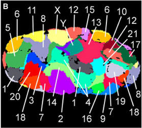
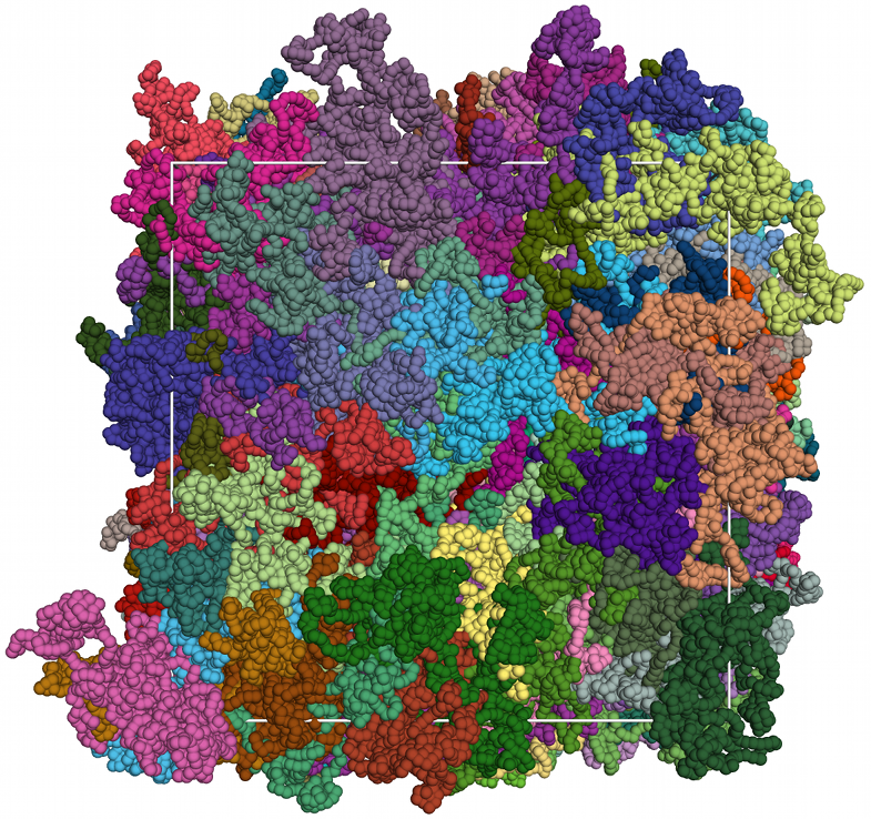
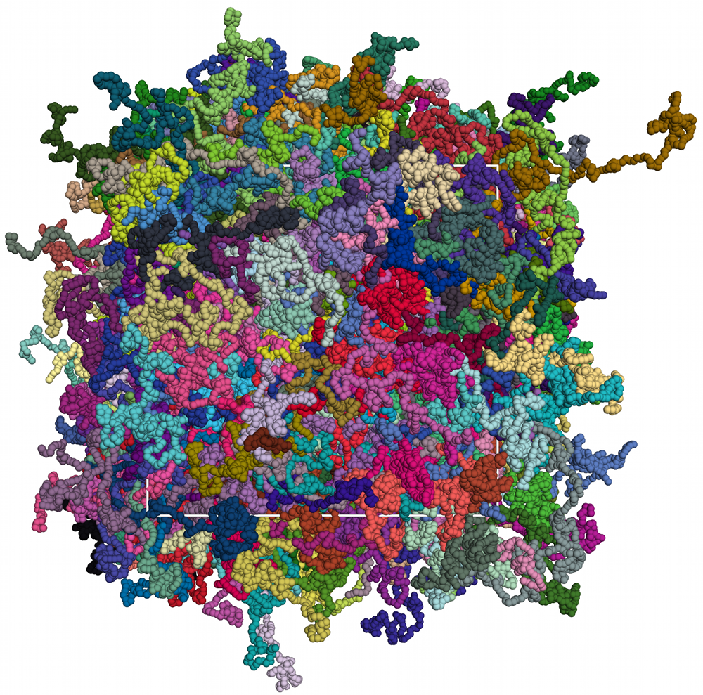
V “Territorial polymers” and chromosome territories: qualitative aspect
As we already mentioned, mutual segregation or the incomplete penetration of rings provides a simple generic natural explanation of chromosome territories Cremer and Cremer (2001); Cremer et al. (2006); Meaburn and Misteli (2007). As a matter of fact, territorial segregation of chromosomes was seen also in HiC data Lieberman-Aiden et al. (2009). But the most obvious view of territories was described in Refs. Cremer and Cremer (2001); Cremer et al. (2006); Meaburn and Misteli (2007). Recall that the microscopic image of an interphase nucleus, where each chromosome is stained a distinct color, looks like a political geographic map Cremer and Cremer (2001) or contiguous set of colorful patches with irregular shapes, covering space (see also reviews Cremer et al. (2006); Meaburn and Misteli (2007)). In this sense, the melt of rings exhibits a very similar behavior. If each ring is colored in its own distinct color then a something of a map emerges as published on the back cover of “Physics Today”Vettorel et al. (2009b). This is illustrated in Fig. 3. We want to emphasize that no segregation, no territories are observed in the system of linear polymers. This fact is known in polymer physics as the Flory theorem de Gennes (1979), and it is also illustrated by the lower panel in the Fig. 3. Thus, we see that topological constraints in a concentrated system of long polymers lead to territorial segregation.
This leads to the hypothesis that chromosomes do not intermix, do not tangle, and remain distributed over their respective territories mostly because of their topological constraints Rosa and Everaers (2008); Vettorel et al. (2009a). This idea was also suggested in the works T.Blackstone et al. (2011); Dorier and Stasiak (2009), where it was based directly on the topological repulsion of nonconcatenated loops in a dilute system, without any attention to the compact packing issue. The role of topological constraints was also emphasized in the experimental work Kawamura et al. (2010).
The initial formation of territory-like regions is also observed in computer simulations and to a certain extent by scattering experiments of polyelectrolyte gels and solutions Dobrynin et al. (1996); Micka et al. (1999); Spiteri et al. (2007); Mann et al. (2011). Upon change in solvent quality or counterion valency the systems start to shrink by chain contraction. This is facilitated by counterion condensation along the backbone of the chains leading to complexes, which still contain a (decreasing) net charge. At the early stages of this process the so-called pearl necklace structure is observed. Consequently these dense regions still repel each other, i.e., they stay segregated. For perfect gels without any dangling chain ends this segregation state seems to survive up to melt densities, while solution studies of (short) linear polymers suggest a relaxation into the Gaussian chain conformation once the density is sufficiently high that the counterions are no longer localized on the backbone and can diffuse freely throughout the melt or solution.
It is worth emphasizing the important differences in approach between Refs. Vettorel et al. (2009a); Halverson et al. (2011a), Rosa and Everaers (2008) and the simulation part of Ref. Lieberman-Aiden et al. (2009). In Vettorel et al. (2009a); Halverson et al. (2011a), a system of rings was considered to enforce topological constraints. This system was carefully equilibrated. That is, the authors made sure that the entropically dominant or statistically most probable set of conformations was observed and studied. By contrast, the authors of Rosa and Everaers (2008) simulated (mostly) long linear chains for a pre-reptation time, therefore, not even attempting to equilibrate their system. What they have done instead is to take advantage of having mapped the parameters of the simulated model onto those of a real chromatin fiber. Accordingly, they were able to argue that their molecular dynamics run was over a biologically reasonable amount of time over which the interphase stage of the cell cycle exists in nature. These authors showed also that their results for linear chains on a pre-reptation time were similar to those of the rings. It is interesting to realize that some sort of territorial segregation was observed in three rather different simulation schemes. In Rosa and Everaers (2008) the authors start with compact unknotted crumpled-like polymers and run the simulation much shorter than the reptation time. In the simulation part of Lieberman-Aiden et al. (2009) the authors start from a very open coil conformation, force it to collapse very fast, and then run it for a short time. While in Vettorel et al. (2009a); Halverson et al. (2011a) dense systems of rings were equilibrated, which is to say it could be run indefinitely long. The similarity in the results of these three very different systems is a powerful demonstration of the validity of the very idea that topological constraints are the key to segregation.
This idea is also supported by the study undertaken in a different context by Vettorel and one of us (KK) Vettorel and Kremer (2010). In that work, the authors simulated melts of long collapsed polymers and found that relaxation into the entangled state is significantly delayed to times larger than the reptation time once the chain lengths reach of the order of about . As shown above, typical chromatin fibers exceed this length significantly.
We should repeat here the arguments why we think topological constraints to be so important for the chromatin fiber. Of course, the chromatin fiber is not a ring. Nevertheless, un-crossability is likely to play a huge role. The simplest argument is that the cell life time is by far not sufficient to realize complete relaxation via reptation Rosa and Everaers (2008); Sikorav and Jannink (1994). Also, telomere regions at the ends of chromosomes are likely to form bulges which are not conducive for reptation. The additional factor which slows down the reptation is that the chromatin fiber is probably attached in some places to the nuclear envelope (its inner surface). If that is the case then regular reptation becomes problematic and the situation becomes reminiscent of star-branched macromolecules, where the relaxation time is controlled by the arm retraction mechanism and is exponentially long Rubinstein and Colby (2005).
We should also comment here about the role of topological enzymes (topoisomerase) which are capable of cutting both strands of dsDNA, passing another piece of DNA through the cut, and then gluing the cut back together. This seems to make dsDNA effectively phantom or self-crossable Sikorav and Jannink (1994). Simple estimates show, however, that the amount of enzymes in the nucleus and ATP (free energy) supply in the cell is by far insufficient to forget about topological enzymes. Nevertheless, this subject remains somewhat controversial in the literature, as emphasized recently, e.g., by H. Schiessel Schram et al. (2013). Sure enough, topoisomerase enzymes play their role, but mostly in the metaphase. In fact, apparently there is no indication of topoisomerase activity during the interphase Mirny (2012). In any case, the activity of topological enzymes does not cancel the fact that to the first approximation the chromatin fiber is not crossable Rosa and Everaers (2008). Thus, it is sensible to hypothesize that instead of using the sophisticated machinery of topological enzymes to make many random crossings, nature would make good use of polymer un-crossability to control territorial segregation.
Territorial segregation of chromatin fibers appears to be a common feature of higher eukaryotes, including humans. Lower eukaryotes, such as yeast seem to have much less pronounced territories, or no territories at all Meaburn and Misteli (2007); Duan et al. (2010); Therizols et al. (2010). Given the relatively short genome of yeast (see Table 1), this observation is at least qualitatively consistent with our above estimates of the number of entanglement lengths of the yeast genome. It is interesting to mention that yeast has about as many genes (and about as large genes) as humans. In other words, the amount of coding DNA is nearly the same for both yeast and human. However, yeast has almost no introns while we have plenty. This is why human DNA is two orders of magnitude longer than that of yeast.
Since the qualitative picture of political maps agrees rather well for the model system of a melt of rings and for real chromosomes in cell nuclei, it makes sense to look at more details for both cases.
VI “Territorial polymers” and chromosome territories: quantitative considerations
VI.1 Modern experimental techniques of chromatin investigation
In recent years, quantitative experimental information about chromatin organization in space was delivered mostly by two groups of methods, called “FISH” and “3C” with derivatives up to HiC. Recall that FISH basically measures , the subchain size, while HiC measures , the loop factor or contact probability. We will discuss these results in greater detail below in this section, but it is useful to remind briefly the basic ideas behind these two methods.
The essence of the FISH method Bridger and Volpi (2010) is to label a few loci on the genome with small fluorescent molecules. The spatial distance of the fluorescing spots is then measured under a microscope. Recent advances in microscopy increase the resolution significantly beyond the wavelength of the light used (see, e.g., Weiland et al. (2011); Markaki et al. (2012)). By doing this for many pairs of loci, one can – ideally – relate the genetic distance between the loci or, in polymer physics parlance, the contour distance between the monomers to their spatial Euclidean distance Neer et al. (1977); Höfers et al. (1993a, b); Yokota et al. (1995); Solovei et al. (2002); Gilbert et al. (2004); Jhunjhunwala et al. (2008); Weiland et al. (2011); Markaki et al. (2012).
The central idea of the “C” methods is to chemically cross-link at some particular time pieces of the chromatin fiber which happen to be close to each other in space at that very moment. Then the DNA is chopped into pieces by a restriction enzyme, and (omitting important technical details here!) the parts which happened to be cross-linked are sequenced. Cross-linking is usually realized by formaldehyde which connects histones, but not DNA. Thus the cross-linking acts on the level of the fiber, not on the level of DNA. The cross-linked parts are then separated, for instance, in the HiC version by using biotin labeled ends, followed by deep sequencing. In any case, since the entire genome sequence is known, and since the experiment is done on many cells in parallel, the result allows one to reconstruct the contact map or, in polymer physics language, the probability of contact between any two monomers of the same chain and , say . It is usually assumed that on average depends only on the distance , and so one can find the so-called loop factor, equivalently known as contact probability, which is the probability that two monomers a contour distance apart meet in space.
There is also one work Lebedev et al. (2005) which carried out small angle neutron scattering experiments on the entire cell nucleus (of a chicken erythrocyte). The global spatial arrangement of the chromatin strands were analyzed. We comment on this experiment later in Section VI.6.1.
Since the essential quantitative information at our disposal comes from measuring subchain sizes and contact probabilities , we start with a discussion of a simple approximate relation between these two quantities.
VI.2 Mean field relation between subchain size and loop factor
For any fractal conformation with the subchain size scaling as , the loop factor can be estimated by the following argument. Let us imagine that we hold one end of the subchain fixed in space. The second end is located at a distance of about , i.e., it is dispersed over the volume of the order of . Therefore, the probability, , to find it in a small volume around the first end is estimated as
| (9) |
This assumes that the second end is more or less uniformly distributed over the volume , i.e, we make a mean field argument. Given that , this argument yields for the critical exponent :
| (10) |
For the Gaussian coil (with ) this gives the well known correct answer in 3D (or in dimension ). For the crumpled fractal globule (8) and, therefore, . This simple theoretical estimate is in excellent agreement with HiC data for both human Lieberman-Aiden et al. (2009) and mouse chromosomes Y.Zhang et al. (2012).
However nice the agreement between theory and experiment is, the exact scaling for the loop factor cannot be correct, at least not over an infinite range of length scales . Take the total number of neighbors of any given monomer, namely the sum , which diverges for , i.e., a monomer would have a diverging number of close neighbors. For a chain of length , this would mean that the number of spatial neighbors for a given monomer becomes of order . This is surely impossible in the limit .
Nevertheless, from a theoretical point of view it is important to understand whether we are talking about a real fractal, which remains self-similar over an infinite range of scales and which approximates real chromatin over some finite range, or whether there is even theoretically no real fractal. Aesthetically the first alternative is more attractive, of course. But even logically, we can imagine arbitrarily long rings in the melt, much longer than any DNA in any real genome, and we want to understand the loop factor for such system and find out whether this system is fractal or not. Thus, to summarize, the mean field estimate (or ), which is based on the assumption of statistical independence between the subchain ends within the volume , cannot be accurate. There should be some correlation between subchain ends for any .
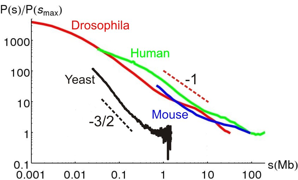
VI.3 Contact probability (loop factor) of chromatin: experiments
The contact probability of chromatin is the probability that two DNA loci on the same chromosome, separated by the genetic (contour) distance (measured, for instance, in the number of base pairs), will be neighbors in real 3D space. This probability and its dependence on was measured for human fibroblasts Lieberman-Aiden et al. (2009), mice Y.Zhang et al. (2012), fruit flies Sexton et al. (2012), and yeast Duan et al. (2010). All these data are collected in Fig. 4. We see that the contact probability behaves rather similarly for human, mouse, and drosophila cells. In particular, Lieberman-Aiden et al. (2009) reports that the loop factor scales as in the interval of roughly , with a “critical exponent” for the human genome. In a similar interval, the data for mouse Y.Zhang et al. (2012) indicate . However, the implicit error bar of these measurements is hard to estimate. This scaling appears to be consistent with the crumpled globule model, based on the previous mean field estimate of Eq. (10), as also emphasized in Rosa et al. (2010). Based on this the authors of the experimental work Lieberman-Aiden et al. (2009) claimed agreement with the theoretical predictions based on crumpled globules.
We leave aside the questions of error bars associated with the above mentioned values of as well as the question of the range of where the power law fitting is considered. We refer the interested reader to the original papers as well as review articles A.Mirny (2011); Fudenberg and Mirny (2012); Dekker and van Steensel (2013). Instead, we concentrate on the following observations. First, yeast definitely belongs to a separate class compared to all other organisms presented in Fig. 4. Second, the contact probability for yeast is roughly consistent with equilibrium globule behavior, both because the slope is close to and because of the leveling-off at larger , at least for the largest chromosome. Third, the contact probability for human, mice, and drosophila is definitely inconsistent with the equilibrium globule, both in terms of the slope being nowhere near and the definite lack of leveling off.
The situation with yeast was significantly clarified in the recent work Wong et al. (2012), where the authors computationally examined an elaborate, but straightforward polymer model of the yeast nucleus. The estimated values for the Kuhn segment and linear density of yeast chromatin fiber were and . They took into account the real lengths of all chromosome arms, the fact that each chromosome has a centromere attachment, and the fact that chromosome ends (telomere regions) are preferably located next to the nuclear envelope. Importantly, they simulated the equilibrium properties of the system, and obtained a very good account of the contact probability as well as a number of other structural data. The agreement thus obtained gives a convincing proof that folding of chromatin fiber in yeast can be reasonably considered as an equilibrium state of a confined polymer. In particular, this explains why the contact probability in yeast, as seen in Fig. 4, behaves very much like that of an equilibrium globule.
Fig. 4 demonstrates beyond a doubt that the chromatin of other species is organized differently compared to yeast. Our main hypothesis, already stated several times in different ways, suggests that the main difference is due to topological constraints. The relatively short genome of yeast does not need to reptate and, therefore, is equilibrated, as confirmed by the above discussed conclusions of the work Wong et al. (2012). We argue that genomes of higher eukaryotes do not have enough time to reptate. This of course goes along with our assertion that chromosome territories have topological origin. We now continue and hypothesize that the contact probabilities of the higher eukaryotes seen in Fig. 4 do not follow the equilibrium globule behavior also for the same topological reason. This topology-controlled deviation from standard equilibrium globule behavior is a manifestation of non-equilibrium of very long linear chains, or in a ring model it may be viewed as an equilibrium manifestation of topology.
VI.4 Subchain sizes in chromatin
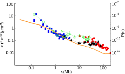
The FISH method, which measures the subchain sizes , was developed significantly earlier than the “C” methods. From the polymer physics prospective, the value of is also well known for regular equilibrium globules: it behaves as up to , i.e., up to the distance where the chain can cross the available volume by a random walk. Then levels off at and remains independent of at all larger .
The first FISH data were consistent with the Gaussian scaling . For values of up to about base pairs the FISH data for were satisfactorily approximated as a straight line in coordinates . This tempted the authors of Ref. van den Engh et al. (1992) to formulate the “random walk” model of chromatin organization. In fitting the data there was a significant element of uncertainty for any analysis beyond the bare power law. Namely, if we believe that chromatin is a Gaussian polymer on a certain length scale, with a Kuhn segment of length , if the number of base pairs per unit contour length is then because is the contour length. Although the Kuhn segment of bare DNA is known to be about , it is not easy to determine a priori the relevant Kuhn segment for chromatin fiber, so the practical approach is to view it as an adjustable parameter. FISH data, apart from the very scaling , allow only to determine the combination of parameters , not each of them separately. Nevertheless, more detailed FISH data for larger showed some sign of slowing down in the growth of which was interpreted as the signature of spatial confinement. Accordingly, the work Hahnfeldt et al. (1993) introduced the model of a random walk in a confined volume. From a polymer physics point of view, this means that Refs. van den Engh et al. (1992); Hahnfeldt et al. (1993) interpreted the FISH data as pointing to an equilibrium globule organization of chromatin. The difficulties of making this model consistent with excluded volume constraints, topological constraints, and the lack of reptation relaxation were not addressed in the early papers van den Engh et al. (1992); Hahnfeldt et al. (1993). Also, a fully developed saturation of was not observed. Therefore, an alternative interpretation of the slowing down of the growth of might be appropriate.
A more recent FISH study Mateos-Langerak et al. (2009) makes the point that , at least for some chromosomes, saturates at a value of significantly smaller than the nucleus size, as is clearly visible in Fig. 5. This suggests some mechanism of saturation other than volume confinement. The authors of Mateos-Langerak et al. (2009) argue that this is evidence of what they call loop formation. They refer then to the well known fact of entropic (topological in nature) repulsion between closed polymeric loops in dilute solution, as first reported in the work Vologodski et al. (1974). We note in passing that the territorial segregation of rings in a concentrated system, which is the central subject of our work, can be viewed as the result of the same repulsion in a very dense system. Importantly, this repulsion exists only for loops which maintain their closeness and remain nonconcatenated over the entire longevity of the experiment, i.e., in physics language, the loops have to be quenched. Annealed loops, which close and open in the course of thermal motion, exhibit no repulsion at all. Meanwhile, the fact that two monomers and gave a signal in a particular Hi-C experiment suggests that the piece of the chain between them formed a “loop” at the time of the experiment, but it does not mean this loop was quenched before the experiment, in vivo. In our opinion, the interpretation of each Hi-C signal as an indication of a real quenched loop in the system is a mistake.
Another interpretation would relate this effect to the territorial segregation. Namely, we can expect that a smaller chromosome gets topologically confined among bigger ones, just as one ring in the melt gets compressed by its neighbors. Such an explanation would of course make sense if and only if the size of even a small chromosome significantly exceeds the entanglement length . That is one of the reasons why we discussed the estimate of in detail before. According to the summary given in Table 2, even the sizes of small chromosomes, whose saturates at distances of about (cf. Fig. 5), are long enough for the topological mechanism to be functional. The overall dependence of being at least roughly consistent with the expected crumpled globule scaling (8) is visible in Fig. 5 in the region.
In recent years, much more detailed models have been developed to fit the FISH data. In most cases, the authors talk about delicately organized and balanced loops supported and controlled by special long lived cross-links (see, for instance, Ostashevsky (1998, 2002); Mateos-Langerak et al. (2009); Iyer and Arya (2012)). The idea is that these cross-links are created by special biological mechanisms. There is no way for us to negate such a hypothesis. At the same time, one should first exhaust all possibilities to explain observations based on simple robust physics before resorting to biological machinery and the special actions of such machinery. Indeed, it seems that nature is “opportunistic” in the sense that it employs simple physics whenever possible. Although this question can perhaps be argued in different ways, here we concentrate on the possibility to explain observations regarding the chromatin organization based on topological constraints and polymer physics.
VI.5 Theoretical issues: contacts between subchains
Let us return for a moment to the mean field relation (10), and to the discussion of the fact that this relation cannot be exact for a true fractal over the unrestricted range of scales. The latter conclusion means that the critical exponent is not determined by the value of , i.e., it is a different, independent exponent. The existence of critical indexes independent of is not so surprising given our experience in polymer physics des Cloizeaux and Jannink (2010); de Gennes (1979); Y.Grosberg and R.Khokhlov (1994); Rubinstein and Colby (2005).
Thus, for the rings in the melt we have to turn our attention to exponents independent of . That means, we should take a more detailed view of conformations. One useful perspective opens up if we look at surfaces between neighboring rings in the melt of rings, or surfaces between subchains, i.e., to consider the index which describes the roughness of the boundaries of territorial polymers. Furthermore, there are two closely related surface exponents and . They are defined in the following way:
-
•
Consider an -long part of the chain. How many contacts does this part have with all other monomers? This scales with index :
(11) This index characterizes the “surface area” of the blob.
-
•
Consider two blobs of length each, located next to one another in space. How many contacts do they have? This scales with index :
(12) This index describes the “contact area” between two touching blobs.
The relation between indexes and has to do with the number of blobs which can simultaneously be neighbors to one another:
| (13) |
To estimate we can resort to the physics of polymer solutions (see, e.g., book de Gennes (1979)). Each subchain of length is spread over volume (in dimensions). This volume can contain monomers, but since each subchain has monomers we arrive at
| (14) |
which yields
| (15) |
For the dense fractal system with we get , which is why we used simply in the work Halverson et al. (2011a), deriving the relation
| (16) |
Given that there are two indices and , there are two relations:
| (17) | |||||
| (18) |
Because of Eq. (15) these two relations are equivalent, and each of them gets reduced to the familiar result (16) in the case . Below we briefly outline arguments leading to the relations (17) and (18). The arguments in Sections VI.5.1 and VI.5.3 were developed during a discussion with R.Everaers and M.Rubinstein Everaers et al. (2011).
VI.5.1 Counting argument Everaers et al. (2011)
Consider a polymer chain which is a space-filling fractal (i.e., ), and suppose its contact probability is , with large asymptotics being , . For one monomer, the number of “foreign” contacts (i.e., with monomers a distance or greater away along the chain) is proportional to , and for the whole subchain this number is times larger. Thus
| (19) | |||||
which is the result (17).
VI.5.2 Self-similarity argument
Here we paraphrase the original argument given in Halverson et al. (2011a). Let us count how many pairs of monomers, a distance apart along the chain, are in contact. There are pairs a distance apart each, and with the probability of contact for each pair being , the number of pairs is .
Let us now re-count this same number in a different manner by using blobs of some monomers each. We have pairs of blobs, and the probability of contact between blobs is , yielding blob contacts. Each blob contact delivers monomer contacts. However, these monomer contacts are between monomers a chemical distance apart, and only the fraction of those are a distance apart. Thus, the number of monomer contacts is now expressed as . This must coincide with the original estimate because the number of monomer contacts cannot depend on how we count them. By equating the two
| (20) |
one arrives at formula (18).
VI.5.3 Blob contact argument Everaers et al. (2011)
The probability of contact between two monomers a distance apart, for instance, and , is . Consider now two subchains of length each, namely, one from to and another from to . They are centered at the two monomers of interest. These two subchains have contacts. What is the probability that one of these contacts is between the monomers of interest, and ? There are possible choices of pairs of contacting monomers, therefore, the probability that one of these pairs will be chosen among the actual pairs is proportional to . Thus this argument yields
| (21) |
which is again the result (18).
VI.5.4 Examples and comparisons
It is instructive to see how all the relations above work in various known cases. First, consider a melt of linear chains. In this case, , , (controls contacts between two overlapping blobs), and (controls contacts of a given chain with monomers of any other), so all relations above work perfectly.
It is worth emphasizing that the relations (17) and (18) represent general properties of self-similarly organized polymers. They do not rely on any specific properties of crumpled globules. In particular, they are not based on the mean field estimate of . Since we know already that the mean field estimate is not accurate, we need to make our considerations not assuming anything specific about the value of .
For the “naive” crumped globule, best exemplified by a Hilbert curve or its circular analog called Moore curve Sagan (1994) in 3D (Fig. 1), we have and also all surfaces are pretty smooth, so and then .
By contrast, if we accept the mean field estimate , Eq. (10), and then take for a dense fractal globule, we get and also . That means in this approximation, the surfaces of a fractal globule include some finite fraction of all monomers. In fact, these surfaces are so “rough” that they can hardly be called surfaces. Quite apart from the terminological question of whether we can call different rings with to be territorially segregated, we point out that such structures cannot be fractal with an unlimited spectrum of scales, which is the same conclusion we made based on the logarithmic divergence of the number of contacts.
In general, there are several obvious conditions on the indexes: in dimensions and , while , and . It follows then from the relations above that in general , while specifically for the compact case a stricter bound holds: .
The numerical results for the melt of rings J.D.Halverson et al. (2013) indicate quite complex finite size behavior of different quantities. The total number of surface monomers for either the full ring or its subchains demonstrates a very accurate power law behavior throughout the studied range of polymer length, and shows . This value is just below unity, so one is tempted to suspect that it is unity. However, the fraction of surface monomers does decrease with chain length, so it is indeed possible that . Even more convincing data suggesting are obtained from the scaling of the static structure factor, as we discuss below. At the same time, the contacts between blobs exhibit a very different finite size behavior, for the seeming power , i.e., the slope of a plot of monomer-monomer contacts between blobs against blob length increases monotonically from well below to somewhere close to unity at the largest accessed in simulation so far. At the same time, the contact probability exhibits complementary behavior with the seeming value of (in the same sense) increasing with , while the number of blob neighbors seems to saturate at large in accordance with (14). The nature of this finite size behavior remains to be understood.
VI.6 Static structure factor and scattering off chromatin
VI.6.1 Spatial nuclear density (in-)homogeneity: scattering results
The whole line of quantitative arguments above relies on the assumption that there are no strong density variations on scales of the order of the entanglement length and beyond. The classical color pictures of chromatin territories (cf. Fig. 3) suggest a homogeneous density throughout the cell nucleus. This however might be misleading as the wavelength of visible light is too large to easily allow one to resolve greater local density variation by eye. To be more accurate, one could resort to scattering experiments. For that we can start from the structure of the “semidilute chromatin solution”, which can in principle be determined by scattering experiments de Gennes (1979); Rubinstein and Colby (2005).
In a melt or dense solution there are no density fluctuations expected to exist beyond the scale of the blob size. On scales smaller than the blob diameter, one can resolve the single chain structure. This, however, requires sufficient scattering contrast between the chromatin fiber and its immediate surrounding, i.e., typically salty water and proteins. However, even within the scale of the blobs, this scattering contrast usually is very weak. For X-rays to be used efficiently the electron density is too small and for neutrons the overall density of H atoms does not display enough variation. To improve this, one can create an artificial contrast. For instance, in the case of neutron scattering of polymer systems, the usual procedure is deuteration, i.e., replacement of hydrogen atoms for deuterium in one of the components of the system. Here this would correspond to a (partial) deuteration of the water. Neutron scattering is a perfect tool to study subtle variations in H and D densities, since H and D have dramatically different scattering lengths Borsali and Pecora (2008); Richter (2000).
We are aware of only one piece of experimental work Lebedev et al. (2005) examining chromatin structure (of a chicken erythrocyte) by small angle neutron scattering (see also the work Ilatovskiy et al. (2011) by the same group attempting to simulate the data ). In this experiment, scattering indeed was based on the contrast between DNA and surrounding water, a mixture of and . On length scales within the blob size one expects a variation of the scattering intensity as a function of momentum transfer, providing information about the structure of the chromatin fiber. For larger scales both the density of the solvent and chromatin are averaged out, suppressing any change in the scattering signal. From this crossover one can directly read off the blob size, at least in principle. The scattering experiment Lebedev et al. (2005) indicates a homogeneous smeared out DNA density on length scales above to or, equivalently, for momentum transfers between and . While this confirms the concept of a homogeneous, concentrated solution, the quoted blob size certainly is too large to accommodate the contour length of the chromatin fiber with and about . Indeed, standard polymer physics arguments de Gennes (1979); Y.Grosberg and R.Khokhlov (1994); Rubinstein and Colby (2005) imply that the blob size is about (because implies that the fiber is nearly straight inside the blob). For the parameters of the human cell nucleus discussed above, this would be about . For the higher density chicken erythrocyte should be even smaller. This discrepancy between measured blob size and theoretical estimates can result from different sources. One possible reason is due to the overall small accuracy of the data because the system is much more complex than a regular semidilute polymer solution, where such methods have been employed very successfully. In addition, experiments have been performed on the nuclei of chicken erythrocytes, which are quite different from other eukaryotic cells.
The experiments of Ref. Lebedev et al. (2005) also provide the first information about the fractal property (on scales below the blob diameter) of the DNA fold itself, but cannot reveal any information of the global indices , , or . It also does not give information about the relevant contour length and the Kuhn segment of the chromatin fiber inside the blob, which would provide an independent estimate of the entanglement length.
VI.6.2 Scaling of the static structure factor
Although a few scattering experiments have been performed so far, this is a very promising technique, which motivates us to discuss what we can expect theoretically. In general, the static structure factor is the most informative characteristic of conformations. We denote it , with the scattering wave vector (, where is the scattering angle and is the wavelength of scattered waves). Usually, in the most interesting interval of , namely , the static structure factor scales as . Accordingly, one would expect to be able to distinguish a Gaussian coil () or a ball with smooth surface (Porod law, ) from a crumpled globule with expected scaling .
This conclusion, however appealing, holds only on scales below the blob size, and its application is tricky because of the fractal surfaces between territories, which smear out the scattering contrast. Corresponding analysis was presented in Halverson et al. (2011a) based on the ideas of the works B.Duplantier (1989); H.Meyer et al. (2010); Carmesin and Kremer (1990) for 2D melts. The argument was originally given without making the distinction between and by assuming that they were the same. The essence of the argument can be formulated in the following way.
Imagine that we labeled (for instance, deuterated) some monomers. The static structure factor is defined by the formula:
| (22) |
For the moment let us assume that the labeled part is either one entire ring () or some part of it (). In this case, the static structure factor (22) has the following properties. First, at we always obtain . Second, in the intermediate range of we expect some power law dependence , where the power is to be found. Moreover, since the overall size is the only relevant length scale (since we deal with one ring or its part), we can write . Third, and this is the most delicate part of the argument, the only place where scattering can take place is the surface of the labeled part. Therefore, the total scattered intensity, which is equal to , must depend on the number of labeled monomers as . Comparing, we conclude that . Therefore, the structure factor scales as
| (23) |
For instance, for the linear chain in a regular polymer melt all monomers belong to “surface”, as they contact with other chains, so and we return to the familiar result (where for the melt). But for the melt of rings and the use of a more sophisticated scaling (23) is necessary. Computational data for both lattice and off-lattice systems were fit using formula (23), and the result was found J.D.Halverson et al. (2013). The fitting procedure used in our works Halverson et al. (2011a); J.D.Halverson et al. (2013) was criticized and placed under doubt in a recent comment Wittmer et al. (2013) (see also our reply in Halverson et al. (2013)). However, it is worth emphasizing that the scaling formula (23) was not questioned, only the numerical fitting procedure. In light of this criticism, extracting reliable estimates of and from static structure factor measurements will have to wait for more accurate data. Nevertheless, our present understanding, which is based on the several different measurements discussed above, remains that is slightly smaller than unity while is slightly larger than one.
VII Dynamics
From a comprehensive polymer physics viewpoint, we should consider the dynamics of the ring melts and not only the statistics of equilibrium conformations. This is a difficult task, because reptation de Gennes (1979); Doi and F.Edwards (1986); Rubinstein and Colby (2005); Y.Grosberg and R.Khokhlov (1994, 2011), the workhorse of polymer dynamics theory, does not apply for the rings as they have no ends.
The simplest model, the dynamics of a single ring in a lattice of immobile obstacles, can be analyzed in great detail (see, e.g., the problems for Chapter 9 in the book Rubinstein and Colby (2005) as well as papers Rubinstein (1986); Obukhov et al. (1994)). However, since the lattice of obstacles concept is not proven even for statics, its usefulness for dynamics remains questionable. Recent works Rubinstein (2012); Grosberg (2013); Smrek and Grosberg (2014) may change this situation and give more credence to the generalized lattice animal model based on annealed branched polymer theory. It is perhaps too early to decide how successful this theory is.
So far we have to rely on simulations. We can summarize the main findings Halverson et al. (2011b) as follows:
If is the time dependent position vector of one ring (say, its mass center, with the convention that ), then at very long times , and we can address the -dependence of the diffusion coefficient. It was found Halverson et al. (2011b) that , which is not dramatically different from the result of similar simulations and experiments for linear chains, . At the same time the viscosity of the melt of rings increases with dramatically slower than for linear chains, as (instead of for linear chains).
At smaller times, before the cross-over to regular diffusion, was found to exhibit sub-diffusion, although somewhat faster sub-diffusion (roughly for the ring sizes considered in the simulations) than for linear chains ().
For linear chains the cross-over between the sub-diffusion and diffusion regimes for the displacement of the center of mass of the chains is observed at about the Rouse time , well below the time of stress relaxation, . Surprisingly, this is not the case for the ring systems, where molecules continue to sub-diffuse long after the stress has completely relaxed, as far as the accuracy of the present simulations can determine this. An explanation of this observation, beyond the fact that shape fluctuations do not necessarily contribute to the overall ring diffusion, remains to be found.
In terms of mapping polymer models to chromatin, an interesting estimate can be performed based on the fact that the diffusion coefficients behave nearly identically while viscosities behave so dramatically different for the melt of territorial globules versus a melt of entangled linear chains. This difference indicates that the relaxation mechanism, which controls viscosity, in the territorial system is decoupled from translational motion, which is manifested in the diffusion coefficient. Therefore, the ratio of viscosities for territorial globules and for entangled linear chains, which scales as according to our computational result, reflects the ratio of the relevant relaxation times. This ratio is about for the longest chains for which both linear and ring polymers were tested in Halverson et al. (2011b) (, ). Therefore, simple extrapolation suggests that even for short human chromosomes, for which , the relaxation time is shortened by about 5 orders of magnitude (a factor of about ) because of territorial segregation. Obviously, this is a very significant advantage.
Much more attention to dynamics was paid by Rosa and Everaers in their work Rosa and Everaers (2008).
Another interesting aspect in which the melt of rings model can be compared to chromatin data has to do with sub-diffusion at shorter time scales. We here only point out that most reported observations of sub-diffusion in the cell nucleus deal with rather small length scales, below about 200 nm Bancaud et al. (2012, 2009); Golding and Cox (2006); Banks and Fradin (2005); Bronstein et al. (2009), and it may be unrelated to the topic of our interest, which is the role of topological constraints. Or in polymer language, these phenomena are likely to happen below the concentration blob and tube diameter scales.
VIII Role of sequence
The fractal crumpled globule hypothesis is attractive because it is based on very generic properties of polymers and does not involve any assumptions of an evolutionary developed special machinery. But we know of course that DNA does have a sequence of base pairs, which cannot be ignored. It is a particularly relevant and intriguing question in regards to the non-coding DNA, the introns. Their role in general is unclear, and one cannot exclude the possibility that they are somehow involved in the control of genome folding. If there is any relation between large scale features of sequence and structure, its casual nature can be speculated to be in any of two directions. On the one hand, since chromatin conformations are to some extent fractal for the reasons of topology discussed above, it is possible to imagine DNA sequences which in the course of evolution develop some degree of self similarity. On the other hand, it is also possible that a particular conformation or group of conformations of chromatin can be additionally stabilized by selecting the proper DNA sequence, provided that protein-mediated interactions are sufficiently complex and diverse.
It is instructive to compare this situation with the relation between sequences and structures known for proteins Shakhnovich (2006). The use and applicability of these types of ideas for chromatin is in no way self-evident. We can speculate that the role of volume interactions in the chromatin case will be relegated to the complex relations between chromatin pieces realized via histone and non-histone proteins and nucleosomes. We point out in this regard that the relation between fractal properties of sequences and fractal arrangement of conformations is known even for models with very simple interactions Shakhnovich (2006); Govorun et al. (2001); Nazarov et al. (2013); it seems, therefore, very reasonable to expect that much more sophisticated interactions, involving architectural proteins and the like, are also likely to lead to some sort of connections between sequences and structures.
IX Concluding remarks
The genome of a eukaryotic cell is stored in the cell nucleus as a very long chromatin fiber. It is confined rather densely in the limited space of the nucleus, but at the same time all (or at least many) parts of it are accessible for cell machinery manipulations. This means, chromatin fiber is incomparably less tangled than a random thread of comparable length packed into a comparably small volume. The underlying principles governing this efficient genome organization are poorly understood.
We argue that at least a significant fraction of this puzzle belongs to the realm of polymer physics. This follows from a very generic argument: chromatin fiber has a one dimensional connectivity and it obeys the excluded volume constraint. Therefore, its conformation in space belongs to the class of so-called self-avoiding walks (SAW). However, there are a huge variety of SAWs, and this raises questions about the SAWs that chromatin follows and why they are found in the cell.
Two types of SAWs are most frequently considered in polymer physics, neither of which is a good candidate to model chromatin: isolated SAWs (dilute set of coils in a good solvent) or a dense tangle of SAWS (as in a melt). In both cases the SAWs are self-similar fractals, but different ones, characterized by different swelling exponents for their gyration radii ( with for an isolated SAW and for the melt). The third commonly considered type, the so-called equilibrium globule, is typically a single chain SAW confined in a volume comparable to the joint excluded volume of its monomers. This is also not a good candidate to model chromatin because it is very heavily tangled.
The selection of a specific type of SAW in real chromatin is controlled in two ways. First, chromatin fiber is not naked DNA but a complex of DNA with a large variety of proteins Dekker and van Steensel (2013). The pieces of such a complex “polymer” do obey the excluded volume, but they also interact in a complex way via their associated proteins. Second, the chromatin fiber is thermodynamically not an equilibrium system and its folding is under some sort of a dynamic control. Following the work Grosberg et al. (1993), we hypothesize that these mechanisms select SAW conformations which are weakly tangled, because it is necessary for biological functions, but otherwise random. In other words, the hypothesis is that conformations, although not equilibrated, correspond to conditional maximum of entropy, conditioned on weak tangling.
This leads then to the workhorse model of nonconcatenated rings, first suggested in Rosa and Everaers (2008). It cannot be overemphasized that we do not assume chromatin fibers to be rings. The model of rings is motivated only by the above hypothesis of partial, incomplete entropy maximization, restricted by weak tangling. Of course, this is consistent with the fact that the estimated relaxation time (“reptation time”) for the chromatin fiber is much longer than the life time of any cell.
Because of this logic, we reviewed in this article two interconnected but distinct lines of research. On the one hand, there is the pure polymer physics problem: a melt of nonconcatenated unknotted rings. It turned out to be a challenging problem in its own right. On the other hand, we attempted to gain insight into chromatin based on the newly acquired knowledge about rings. Our theoretical thinking in this direction was informed by the new generation of experiments, including those on chromosome territories, FISH, and “C” (from 3C to Hi-C) van den Engh et al. (1992); Yokota et al. (1995); Cremer and Cremer (2001); Dekker et al. (2002); Gilbert et al. (2004); Simonis et al. (2006); Jhunjhunwala et al. (2008); Lieberman-Aiden et al. (2009); Kawamura et al. (2010); Weiland et al. (2011); Y.Zhang et al. (2012); Sexton et al. (2012); Markaki et al. (2012) as well as computational work Rosa and Everaers (2008); Junier et al. (2010); Iyer and Arya (2012); Wong et al. (2012); Mateos-Langerak et al. (2009).
The results presented indicate that rings in the melt are segregated for topological reasons, i.e., they correspond to SAWs with . This gives a very natural explanation of the phenomenon of chromosome territories. Furthermore, conformations of rings in the melt appear to be self-similar fractals consistent with the crumpled globule idea Grosberg et al. (1988). The fractal properties of chromatin were noted and discussed in a number of contexts Bancaud et al. (2012); Burnecki et al. (2012). The melt of rings model sheds some light on these observations. More detailed experiments, such as Hi-C, address properties which from a theoretical viewpoint go beyond the index and have to do with an independent set of indices, denoted above as and . Our theoretical understanding of the corresponding properties is incomplete even on the level of rings. Simulations indicate that appears to be slightly above unity, meaning that the contact probability between DNA loci decays as , somewhat faster than which seems to be consistent with observations for higher eukaryotes such as humans and mice Lieberman-Aiden et al. (2009); Y.Zhang et al. (2012); Sexton et al. (2012). At the same time, Hi-C data for yeast are closer to which is characteristic of an equilibrated polymer globule and consistent with the fact that yeast chromatin is significantly shorter, owing to the lack of non-coding DNA.
These successes seem sufficiently encouraging to continue the study of the topological properties of weakly tangled dense polymer systems with the idea in mind to learn more about chromatin. This is why we would like to conclude by listing a set of open questions pertaining to both the melt of rings model system and its more general application to chromatin. We start with relatively simple, well-defined problems and then continue to increasingly tough questions.
It is established that one can construct space-filling curves with any desirable value of . However, what are the entropically dominant values of the critical exponents and for a polymer system without knots? In other words, what are and for the majority of all non-tangled space-filling curves? (We emphasize here that the powers and are not very accurately measured in experiments and characterize a relatively narrow window of scales; however, we view them as an important tool to understand the system, which is why we view them as relevant.) How can one understand the sub-diffusion of rings in dense, nonconcatenated systems and how can one understand the fact that sub-diffusion continues for a time longer than the apparent stress relaxation time? How could this relate to the properties of chromatin fibers in territories? How does it relate to the observations of sub-diffusion (of telomeres and of inserted particles) in chromatin? What is the role of DNA sequence in the formation and support of chromatin folding (via its effect on histones and other proteins in and around chromatin fiber and thus on the fiber-fiber interactions)? In particular, what is the connection between the fractal organization of chromatin in space and long-range correlations in non-coding DNA sequences Peng et al. (1992)? (Although arguments in this direction in the work Grosberg et al. (1993) were based on the naive assumption , the question remains valid and open.) How do crumpled globules or other polymer models react to chemical “signal” modifications, such as methylation, which formally means perturbing the parameters of volume interactions? How do particles diffuse through the chromatin matrix?
Another group of questions pertains to the bridge between polymer physics and chromatin folding. As we discussed, the physical parameters of chromatin fiber are poorly known at present, including the simple ones such as persistence length or even linear density (the number of base pairs per unit contour length). We also pointed out that the entanglement length depends on these parameters, but also very sensitively on the density, which in this case is controlled by the cell nucleus size. The volume of the nucleus can easily be different by a factor of two for different cells. For nonentangled territorial chromatin fibers this factor only weakly affects the friction, and thus the prefactor to the relevant relaxation times. For an entangled system the resulting change of would alter the relaxation times dramatically, asking for different relaxation mechanisms for different cells. We view this as a powerful argument supporting our main topological hypothesis.
This list of open questions is easy to continue. The important conclusion that we want to emphasize is that modern quantitative experimental methods allow one to place the serious physical analysis of chromatin folding on rather solid footing. The first impressive steps have been taken in the context of FISH and HiC experiments, to name the two most prominent approaches. As physicists, we expect scattering experiments to pick-up and to provide valuable information as well.
We also see grounds to expect a large role for simulations. They appear to be extremely promising for a thorough analysis of the static and dynamic properties of well-defined model systems. They allow a direct comparison to either experiment or theory or both. In the case of melts of nonconcatenated rings, the simulations so far provide the only way to generate data of perfectly controlled, equilibrated systems. They impressively demonstrate the crucial role of topological constraints. Already a simple visual inspection illustrates the similarity of ring polymer territories and chromosome territories. The quite close agreement of numerical estimates of the exponent from simulation data of long, concentrated ring polymers and those from “C” and “FISH” experiments, further supports our view that polymer physics concepts can provide very helpful arguments on our way to better understand the complex morphology of the chromatin fiber in the interphase cell nucleus.
Acknowledgements
The authors acknowledge useful discussions with Robijn Bruinsma, Job Dekker, Ralf Everaers, Yuval Garini, Gary Grest, Maxim Imakaev, Erez Lieberman-Aiden, Ron Milo, Leonid Mirny, Sergei Nechaev, Rob Phillips, Yitzhak Rabin, Michael Rubinstein, Jean-Louis Sikorav, Jean-Marc Victor, Edouard Yeramian, Alexandra Zidovska, Christophe Zimmer. This research was supported in part by the National Science Foundation under Grant No. NSF PHY11-25915. KK, JS, and AYG acknowledge the hospitality of KITP Santa Barbara where part of this work was completed. KK acknowledges the hospitality of the Center for Soft Matter Research at NYU and AYG acknowledges the support from the Alexander von Humboldt Foundation through a Senior Scientist Award to spend time at the MPI for Polymer Research. Research carried out in part at the Center for Functional Nanomaterials, Brookhaven National Laboratory, which is supported by the US Department of Energy, Office of Basic Energy Sciences, under contract no. DE-AC02-98CH10886.
References
- Vettorel et al. (2009a) T. Vettorel, A. Y. Grosberg, and K. Kremer, Physical Biology 6, 025013 (2009a).
- Halverson et al. (2011a) J. D. Halverson, W. B. Lee, G. S. Grest, A. Y. Grosberg, and K. Kremer, The Journal of Chemical Physics 134, 204904 (pages 13) (2011a).
- Halverson et al. (2011b) J. D. Halverson, W. B. Lee, G. S. Grest, A. Y. Grosberg, and K. Kremer, The Journal of Chemical Physics 134, 204905 (pages 10) (2011b).
- J.D.Halverson et al. (2012) J.D.Halverson, G.S.Grest, A.Y.Grosberg, and K.Kremer, Physical Review Letters 108, 038301 (2012).
- J.D.Halverson et al. (2013) J.D.Halverson, K.Kremer, and A.Y.Grosberg, Journal of Physics A: Mathematical & Theoretical 46, 065002 (2013).
- van den Engh et al. (1992) G. van den Engh, R. Sachs, and B. Trask, Science 257, 1410 (1992).
- Yokota et al. (1995) H. Yokota, G. van den Engh, J. Hearst, R. Sachs, and B. Trask, J. Cell Biol. 130, 1239 (1995).
- Cremer and Cremer (2001) T. Cremer and C. Cremer, Nature Reviews Genetics 2, 292 (2001).
- Dekker et al. (2002) J. Dekker, K. Rippe, M. Dekker, and N. Kleckner, Science 295, 1306 (2002).
- Gilbert et al. (2004) N. Gilbert, S. Boyle, H. Fiegler, K. Woodfine, N. Carter, and W. Bickmore, Cell 118, 555 (2004).
- Simonis et al. (2006) M. Simonis, P. Klous, E. Splinter, Y. Moshkin, R. Willemsen, E. de Wit, B. van Steensel, and W. de Laat, Nature Genetics 38, 1348 (2006).
- Jhunjhunwala et al. (2008) S. Jhunjhunwala, M. C. van Zelm, M. M. Peak, S. Cutchin, R. Riblet, J. J. van Dongen, F. G. Grosveld, T. A. Knoch, and C. Murre, Cell 133, 265 (2008).
- Lieberman-Aiden et al. (2009) E. Lieberman-Aiden, N. L. van Berkum, L. Williams, M. Imakaev, T. Ragoczy, A. Telling, I. Amit, B. R. Lajoie, P. J. Sabo, M. O. Dorschner, et al., Science 326, 289 (2009).
- Kawamura et al. (2010) R. Kawamura, L. H. Pope, M. O. Christensen, M. Sun, K. Terekhova, F. Boege, C. Mielke, A. H. Andersen, and J. F. Marko, Journal of Cell Biology 188, 653 (2010).
- Weiland et al. (2011) Y. Weiland, P. Lemmer, and C. Cremer, Chromosome Research 19, 5 (2011).
- Y.Zhang et al. (2012) Y.Zhang, R.McCord, Y.-J.Ho, B.Lajoie, D.Hildebrand, A.Simon, M.Becker, F.Alt, and J.Dekker, Cell 148, 908 (2012).
- Sexton et al. (2012) T. Sexton, E. Yaffe, E. Kenigsberg, F. Bantignies, B. Leblanc, M. Hoichman, H. Parrinello, A. Tanay, and G. Cavalli, Cell 148, 458 (2012).
- Markaki et al. (2012) Y. Markaki, D. Smeets, S. Fiedler, V. J. Schmid, L. Schermelleh, T. Cremer, and M. Cremer, Bioassays 34, 412 (2012).
- Rosa and Everaers (2008) A. Rosa and R. Everaers, PLoS Computational Biology 4, e1000153 (2008).
- Junier et al. (2010) I. Junier, O. Martin, and F. Képès, PLoS Computational Biology 6, e1000678 (2010).
- Iyer and Arya (2012) B. V. S. Iyer and G. Arya, Phys Rev E 86, 011911 (2012).
- Wong et al. (2012) H. Wong, H. Marie-Nelly, S. Herbert, P. Carrivain, H. Blanc, R. Koszul, E. Fabre, and C. Zimmer, Current Biology 22, 1881 (2012).
- Mateos-Langerak et al. (2009) J. Mateos-Langerak, M. Bohn, W. de Leeuw, O. Giromus, E. M. M. Manders, P. J. Verschure, M. H. G. Indemans, H. J. Gierman, D. W. Heermann, R. van Driel, et al., Proc. Nat. Ac. Sci. USA 106, 3812 (2009).
- Bohn and Heermann (2010) M. Bohn and D. W. Heermann, Macromolecules 43, 2564 (2010).
- Bohn and W.Heermann (2010) M. Bohn and D. W.Heermann, Journal of Chemical Physics 132, 044904 (2010).
- Bohn and W.Heermann (2011) M. Bohn and D. W.Heermann, PLoS One 6, e14428 (2011).
- Fritsche et al. (2012) M. Fritsche, L. G. Reinholdt, M. Lessard, M. A. Handel, J. Bewersdorf, and D. W. Heermann, PLoS One 7, e36282 (2012).
- Cremer et al. (2006) T. Cremer, M. Cremer, S. Dietzel, S. Müller, I. Solovei, and S. Fakan, Current Opinion in Cell Biology 18, 307 (2006).
- A.Mirny (2011) L. A.Mirny, Chromosome Research 19, 37 (2011).
- Fudenberg and Mirny (2012) G. Fudenberg and L. A. Mirny, Current Opinion in Genetics & Development 22, 115 (2012).
- Dekker and van Steensel (2013) J. Dekker and B. van Steensel, in Handbook of Systems Biology: Concepts and Insights, edited by M. Walhout, M. Vidal, and J. Dekker (Elsevier, 2013), chap. 7, pp. 137 – 151.
- van Steensel (2011) B. van Steensel, The EMBO Journal 30, 1885 (2011).
- Rosa and Zimmer (2014) A. Rosa and C. Zimmer, in International Review of Cell and Molecular Biology, edited by R. Hancock and K. W. Jeon (Elsevier, 2014), vol. 307, chap. 9, pp. 275–349.
- de Graaf and van Steensel (2013) C. A. de Graaf and B. van Steensel, Current Opinion in Genetics & Development 23, 185 (2013).
- Hahnfeldt et al. (1993) P. Hahnfeldt, J. E. Hearst, D. J. Brenner, R. K. Sachs, and L. R. Hlatky, Proc. Nat. Ac. Sci. USA 90, 7854 (1993).
- Sachs et al. (1995) R. Sachs, B. G. van den Engh, Trask, H. Yokota, and J. Hearst, Proc. Nat. Ac. Sci. USA 92, 2710 (1995).
- Marko and Siggia (1997) J. F. Marko and E. D. Siggia, Molecular Biology of the Cell 8, 2217 (1997).
- Ostashevsky (1998) J. Ostashevsky, Molecular Biology of the Cell 9, 3031 (1998).
- Schiessel et al. (2001) H. Schiessel, W. M. Gelbart, and R. Bruinsma, Biophysical Journal 80, 1940 (2001).
- Ostashevsky (2002) J. Ostashevsky, Molecular Biology of the Cell 13, 2157 (2002).
- Cook and Marenduzzo (2009) P. R. Cook and D. Marenduzzo, The Journal of Cell Biology 186, 825 (2009).
- Tark-Dame et al. (2011) M. Tark-Dame, R. van Driel, and D. W. Heermann, Journal of Cell Science 124, 839 (2011).
- Grosberg et al. (1993) A. Y. Grosberg, Y. Rabin, S. Havlin, and A. Neer, Europhysics Letters 23, 373 (1993).
- Sikorav and Jannink (1994) J.-L. Sikorav and G. Jannink, Biophysical Journal 66, 827 (1994).
- Vettorel et al. (2009b) T. Vettorel, A. Y. Grosberg, and K. Kremer, Physics Today 62, 72 (2009b).
- Rosa et al. (2010) A. Rosa, N. B. Becker, and R. Everaers, Biophysical Journal 98, 2410 (2010).
- T.Blackstone et al. (2011) T.Blackstone, R.Scharein, B.Borgo, R.Varela, Y.Diao, and J.Arsuaga, Journal of Mathematical Biology 62, 371 (2011).
- Dorier and Stasiak (2009) J. Dorier and A. Stasiak, Nucleic Acids Research 37, 6316 (2009).
- Bohn et al. (2010) M. Bohn, D. W.Heermann, O. Lourenco, and C. Cordeiro, Macromolecules 43, 2564 (2010).
- de Gennes (1979) P.-G. de Gennes, Scaling Concepts in Polymer Physics (1979).
- Doi and F.Edwards (1986) M. Doi and S. F.Edwards, The Theory of Polymer Dynamics (Oxford Universiy Press, 1986).
- Rubinstein and Colby (2005) M. Rubinstein and R. Colby, Polymer Physics (Oxford University Press, 2005).
- Y.Grosberg and R.Khokhlov (1994) A. Y.Grosberg and A. R.Khokhlov, Statistical Physics of Macromolecules (American Institute of Physics Press, 1994).
- Y.Grosberg and R.Khokhlov (2011) A. Y.Grosberg and A. R.Khokhlov, Giant Molecules (World Scientific, 2011).
- Takahashi (1989) M. Takahashi, Journal of Theoretical Biology 141, 117 136 (1989).
- Lebedev et al. (2005) D. Lebedev, M. Filatov, A. Kuklin, A. Islamov, E. Kentzinger, R. Pantina, B. Toperverg, and V. Isaev-Ivanov, FEBS Letters 579, 1465 (2005).
- Bancaud et al. (2012) A. Bancaud, C. Lavelle, S. Huet, and J. Ellenberg, Nucleic Acids Research 40, 8783 (2012).
- Bancaud et al. (2009) A. Bancaud, S. Huet, N. Daigle, J. Mozziconacci, J. Beaudouin, and J. Ellenberg, EMBO J. 28, 3785 (2009).
- Marshall et al. (1997) W. F. Marshall, A. Straight, J. Marko, J. Swedlow, A. Dernburg, A. Belmont, A. W. Murray, D. A. Agard, and J. W. Sedat, Current Biology 7, 930 (1997).
- Bornfleth et al. (1999) H. Bornfleth, P. Edelmann, D. Zink, T. Cremer, and C. Cremer, Biophysical Journal 77, 2871 (1999).
- Golding and Cox (2006) I. Golding and E. Cox, Phys. Rev. Lett. 96, 098102 (2006).
- Banks and Fradin (2005) D. Banks and C. Fradin, Biophysical Journal 89, 2960 (2005).
- Heun et al. (2001) P. Heun, T. Laroche, K. Shimada, P. Furrer, and S. M. Gasser, Science 294, 2181 (2001).
- Tseng et al. (2004) Y. Tseng, J. S. H. Lee, T. P. Kole, I. Jiang, and D. Wirtz, Journal of Cell Science 117, 2159 (2004).
- Dahl et al. (2005) K. N. Dahl, A. J. Engler, J. D. Pajerowski, and D. E. Discher, Biophysical Journal 89, 15619 (2005).
- Levi et al. (2005) V. Levi, Q. Ruan, M. Plutz, A. S. Belmont, and E. Gratton, Biophysical Journal 89, 4275 (2005).
- Meshorer et al. (2006) E. Meshorer, D. Yellajoshula, E. George, P. J. Scambler, D. T. Brown, and T. Misteli, Developmental Cell 10, 105 (2006).
- Chuang et al. (2006) C.-H. Chuang, A. E. Carpenter, B. Fuchsova, T. Johnson, P. de Lanerolle, and A. S. Belmont, Current Biology 16, 825 (2006).
- de Vries et al. (2007) A. H. B. de Vries, B. E. Krenn, R. van Driel, V. Subramaniam, and J. S. Kanger, Nano Letters 7, 1424 (2007).
- Pajerowski et al. (2007) J. D. Pajerowski, K. N. Dahl, F. L. Zhong, P. J. Sammak, and D. E. Discher, Proc. Natl. Acad. Sci. U.S.A. 104 (2007).
- Bronstein et al. (2009) I. Bronstein, Y. Israel, E. Kepten, S. Mai, Y. Shav-Tal, E. Barkai, and Y. Garini, Phys. Rev. Lett. 103, 018102 (2009).
- Celedon et al. (2011) A. Celedon, C. M. Hale, and D. Wirtz, Biophysical Journal 101, 1880 (2011).
- Hameed et al. (2012) F. M. Hameed, M. Rao, and G. V. Shivashankar, PLoS One 7, e45843 (2012).
- Hinde et al. (2012) E. Hinde, F. Cardarelli, A. Chen, M. Khine, and E. Gratton, Epigenetics & Chromatin 5, 20 (2012).
- Weber et al. (2012) S. Weber, A. Spakowitz, and J. Theriot, Proc. Natl. Acad. Sci. U.S.A. 109, 7338 (2012).
- Burnecki et al. (2012) K. Burnecki, E. Kepten, J. Janczura, I. Bronshtein, Y. Garini, and A. Weron, Biophysical Journal 103, 1839 1847 (2012).
- Kepten et al. (2013) E. Kepten, I. Bronshtein, and Y. Garini, Phys. Rev. E 87, 052713 (2013).
- Zhu et al. (2013) J. Zhu, M. Adli, J. Zou, G. Verstappen, M. Coyne, X. Zhang, T. Durham, M. Miri, V. Deshpande, P. De Jager, et al., Cell 152, 642 (2013).
- Meaburn and Misteli (2007) K. J. Meaburn and T. Misteli, Nature 445, 379 (2007).
- Höfers et al. (1993a) C. Höfers, T. M. Jovin, G. Hummer, and D. J. Arndt-Jovin, Bioimaging 1, 107 (1993a).
- Höfers et al. (1993b) C. Höfers, P. Baumann, G. Hummer, T. M. Jovin, and D. J. Arndt-Jovin, Bioimaging 1, 96 (1993b).
- Solovei et al. (2002) I. Solovei, A. Cavallo, L. Schermelleh, F. Jaunin, C. Scasselati, D. Cmarko, C. Cremer, S. Fakan, and T. Cremer, Experimental Cell Research 276, 10 (2002).
- Bridger and Volpi (2010) J. Bridger and E. Volpi, eds., Fluorescence in situ Hybridization (FISH): Protocols and Applications, vol. 659 (Humana Press Inc., Totowa, NJ, 2010).
- Duan et al. (2010) Z. Duan, M. Andronescu, K. Schutz, S. McIlwain, Y. J. Kim, C. Lee, J. Shendure, S. Fields, C. A. Blau, and W. S. Noble, Nature 465, 363 (2010).
- Kiermer (2006) V. Kiermer, Nature Methods 3, 872 (2006).
- Knobler and Gelbart (2009) C. M. Knobler and W. M. Gelbart, Annu Rev Phys Chem 60, 367 (2009).
- Micheletti et al. (2011) C. Micheletti, D. Marenduzzo, and E. Orlandini, Physics Reports 504, 1 (2011).
- Thanbichler et al. (2005) M. Thanbichler, S. Wang, and L. Shapiro, Journal of Cell Biochemistry 96, 506 521 (2005).
- Benza et al. (2012) V. G. Benza, B. Bassetti, K. D. Dorfman, V. F. Scolari, K. Bromek, P. Cicuta, and M. C. Lagomarsino, Reports on Progress in Physics 75, 076602 (2012).
- Gruber (2011) S. Gruber, Molecular Microbiology 81, 855 859 (2011).
- Dame et al. (2011) R. T. Dame, M. Tark-Dame, and H. Schiessel, Molecular Microbiology 82, 1311 (2011).
- Alberts et al. (2008) B. Alberts, A. Johnson, J. Lewis, M. Raff, K. Roberts, and P. Walter, Molecular Biology of the Cell (New York: Garland Science, 2008).
- L.Wolfe (1993) S. L.Wolfe, Molecular and Cellular Biology (Wadsworth Publishing Company, 1993).
- Chr (2012) Web Site Wikipedia, Chromatin Remodeling: http://en.wikipedia.org/wiki/Chromatin_remodeling (2012).
- Chakravarthy et al. (2005) S. Chakravarthy, Y. Park, J. Chodaparambil, R. Edayathumangalam, and K. Luger, FEBS Letters 579, 895 898 (2005).
- Meyer et al. (2011) S. Meyer, N. B. Becker, S. H. Syed, D. Goutte-Gattat, M. S. Shukla, J. J. Hayes, D. Angelov, J. Bednar, S. Dimitrov, and R. Everaers, Nucleic Acids Research 39, 9139 9154 (2011).
- Segal et al. (2006) E. Segal, Y. Fondufe-Mittendorf, L. Chen, A. C. Thastrom, Y. Field, I. K. Moore, J.-P. Z. Wang, and J. Widom, Nature 442, 772 (2006).
- Emanuel et al. (2009) M. Emanuel, N. H. Radja, A. Hendriksson, and H. Schiessel, Physical Biology 6, 025008 (2009).
- Hansen (2012) J. C. Hansen, The EMBO Journal 31, 1621 (2012).
- Bloom and Joglekar (2010) K. Bloom and A. Joglekar, Nature 463, 446 (2010).
- Phillips-Cremins et al. (2013) J. Phillips-Cremins, M. Sauria, A. Sanyal, T. Gerasimova, B. Lajoie, J. Bell, C.-T. Ong, T. Hookway, C. Guo, Y. Sun, et al., Cell 153, 1281 (2013).
- Bodnar and Spector (2013) M. Bodnar and D. Spector, Cell 153, 1187 (2013).
- Milo (2012) R. Milo, Web Site BioNumbers: http://bionumbers.hms.harvard.edu/KeyNumbers.aspx (2012).
- (104) R. Phillips and R. Milo, Cell Biology by Numbers (????).
- A.Y.Grosberg et al. (1993) A.Y.Grosberg, Y.Rabin, S.Havlin, and A.Neer, Biofizika (Moscow) 38, 75 (1993).
- Grosberg et al. (1988) A. Y. Grosberg, S. K. Nechaev, and E. I. Shakhnovich, Journal de Physique (France) 49, 2095 (1988).
- Dixon et al. (2012) J. R. Dixon, S. Selvaraj, F. Yue, A. Kim, Y. Li, Y. Shen, M. Hu, J. S. Liu, and B. Ren, Nature 485, 376 (2012).
- Vettorel and Kremer (2010) T. Vettorel and K. Kremer, Macromolecular Theory and Simulations 19, 44 56 (2010).
- T.C.B.McLeish (2007) T.C.B.McLeish, Soft Matter 3, 83 (2007).
- A.M.Olovnikov (1973) A.M.Olovnikov, J. Theor. Biol. 41, 181 (1973).
- Szostak and Blackburn (1982) J. Szostak and E. Blackburn, Cell 29, 245 (1982).
- Greider and Blackburn (1985) C. Greider and E. Blackburn, Cell 43, 405 (1985).
- Greider and Blackburn (1989) C. Greider and E. Blackburn, Nature 337, 331 (1989).
- de Gennes (1968) P.-G. de Gennes, Biopolymers 6, 715 (1968).
- Sulkowska et al. (2012) J. I. Sulkowska, E. J. Rawdon, K. C. Millett, J. N. Onuchic, and A. Stasiak, Proceedings of the National Academy of Sciences 109, E1715 (2012).
- Colby et al. (1992) R. Colby, M. Rubinstein, and J. Viovy, Macromolecules 25, 996 (1992).
- Fetters et al. (1999) L. Fetters, D. Lohse, and W. Graessley, J. Pol. Sci. Part B: Pol. Phys. 37, 1023 (1999).
- Everaers et al. (2004) R. Everaers, S. K. Sukumaran, G. S. Grest, C. Svaneborg, A. Sivasubramanian, and K. Kremer, Science 303, 823 (2004).
- Sukumaran et al. (2005) S. K. Sukumaran, G. S. Grest, K. Kremer, and R. Everaers, Journal of Polymer Science, Part B: Polymer Physics 43, 917 (2005).
- Uchida et al. (2008) N. Uchida, G. S. Grest, and R. Everaers, The Journal of Chemical Physics 128, 044902 (pages 6) (2008).
- J.Barham and M.Sadler (1991) P. J.Barham and D. M.Sadler, Polymer 32, 393 (1991).
- de Gennes (1995) P.-G. de Gennes, C. R. Acad. Sci. (Paris) 321(II b), 363 (1995).
- Rastogi et al. (2005) S. Rastogi, D. R. Lippits, G. W. M. Peters, R. Graf, Y. Yao, and H. W. Spiess, Nature Materials 4, 635 (2005).
- Kim et al. (2012) J. U. Kim, Y.-B. Yang, and W. B. Lee, Macromolecules 45, 3263 (2012).
- Cates and Deutsch (1986) M. Cates and J. Deutsch, Journal de Physique (France) 47, 2121 (1986).
- Moore and Grosberg (2005) N. T. Moore and A. Y. Grosberg, Phys. Rev. E 72, 061803 (2005).
- Sakaue (2011) T. Sakaue, Phys. Rev. Lett. 106, 167802 (2011).
- Sakaue (2012) T. Sakaue, Phys. Rev. E 85, 021806 (2012).
- Y.Grosberg (1993) A. Y.Grosberg, Macromolecules 26, 3200 (1993).
- Vologodski et al. (1974) A. V. Vologodski, A. V. Lukashin, and M. D. Frank-Kamenetski, Sov. Phys. JETP 67, 1875 (1974).
- Nechaev and Vasilyev (2005) S. Nechaev and O. Vasilyev, Journal of Knot Theory and Its Ramifications 14, 243 (2005).
- S.K.Nechaev et al. (1996) S.K.Nechaev, A.Y.Grosberg, and A.M.Vershik, Journal of Physics A: Math. & General 29, 2411 (1996).
- Lua et al. (2004) R. Lua, A. L. Borovinskiy, and A. Y. Grosberg, Polymer 45, 717 (2004).
- Schram et al. (2013) R. D. Schram, G. T. Barkema, and H. Schiessel, J. Chem. Phys. 138, 224901 (2013).
- Alipour and Marko (2012) E. Alipour and J. F. Marko, Nucleic Acids Research 40, 11202 (2012).
- S.F.Edwards (1977) S.F.Edwards, British Polymer Journal 9, 140 (1977).
- Khokhlov and Nechaev (1985) A. Khokhlov and S. Nechaev, Physics Letters A 112, 156 (1985).
- Rubinstein (1986) M. Rubinstein, Phys. Rev. Lett. 57, 3023 (1986).
- Nechaev et al. (1987) S. K. Nechaev, A. N. Semenov, and M. K. Koleva, Physica A: Statistical and Theoretical Physics 140, 506 (1987).
- Obukhov et al. (1994) S. P. Obukhov, M. Rubinstein, and T. Duke, Phys. Rev. Lett. 73, 1263 (1994).
- Grosberg (2013) A. Y. Grosberg, Soft Matter 10, 560 (2013).
- Sagan (1994) H. Sagan, Space-Filling Curves (Springer Verlag, Heidelberg, 1994).
- Platzman and Bartholdi III (1989) L. Platzman and J. Bartholdi III, Journal of the Association for Computing Machinery 36, 719 (1989).
- Smrek and Y.Grosberg (2013) J. Smrek and A. Y.Grosberg, Physica A 392, 6375 (2013).
- Lieberman-Aiden (2012) E. Lieberman-Aiden (2012), seminar talk at Courant Institute, New York.
- Müller et al. (1996) M. Müller, J. P. Wittmer, and M. E. Cates, Phys. Rev. E 53, 5063 (1996).
- Brown and Szamel (1998) S. Brown and G. Szamel, J. Chem. Phys. 109, 6184 (1998).
- Suzuki et al. (2009) J. Suzuki, A. Takano, T. Deguchi, and Y. Matsushita, J. Chem. Phys. 131, 144902 (2009).
- Hur et al. (2011) K. Hur, C. Jeong, R. G. Winkler, N. Lacevic, R. H. Gee, and D. Y. Yoon, Macromolecules 44, 2311 (2011).
- Rosa and Everaers (2013) A. Rosa and R. Everaers (2013), arXiv:1310.7641.
- Rubinstein (2012) M. Rubinstein (2012), talk in Leiden Conference on “Genome Mechanics at the Nuclear Scale”, December 11.
- Smrek and Grosberg (2014) J. Smrek and A. Y. Grosberg (2014), in preparation.
- Dobrynin et al. (1996) A. V. Dobrynin, M. Rubinstein, and S. P. Obukhov, Macromolecules 29, 2974 (1996).
- Micka et al. (1999) U. Micka, C. Holm, and K. Kremer, Langmuir 15, 4033 (1999).
- Spiteri et al. (2007) M. Spiteri, C. E. Williams, and F. Boue, Macromolecules 40, 6679 (2007).
- Mann et al. (2011) B. Mann, K. Kremer, O. Lenz, and C. Holm, Macromolecular Theory and Simulations 20, 721 (2011).
- Mirny (2012) L. Mirny (2012), private communication.
- Therizols et al. (2010) P. Therizols, T. Duong, B. Dujon, C. Zimmer, and E. Fabre, Proc. Nat. Ac. Sci. USA 107, 2025 (2010).
- T.A.Bolzer et al. (2005) T.A.Bolzer, G.Kreth, I.Solovei, D.Koehler, K.Saracoglu, C.Fauth, S.Muller, R.Eils, C.Cremer, M.R.Specher, et al., PLoS Biology 3, e157 (2005).
- Neer et al. (1977) A. Neer, N. Baran, and H. Manor, Cell 11, 65 (1977).
- des Cloizeaux and Jannink (2010) J. des Cloizeaux and G. Jannink, Polymers in Solution: Their Modelling and Structure (Oxford University Press, Oxford, 2010).
- Everaers et al. (2011) R. Everaers, A. Grosberg, and M. Rubinstein (2011), unpublished results of discussions at KITP, Santa Barbara.
- Borsali and Pecora (2008) R. Borsali and R. Pecora, eds., Soft Matter Characterization, vol. I-II (Springer-Verlag, Berlin Heidelberg, 2008).
- Richter (2000) D. Richter, Physica B 276, 22 (2000).
- Ilatovskiy et al. (2011) A. V. Ilatovskiy, D. V. Lebedev, M. V. Filatov, M. Grigoriev, M. G. Petukhov, and V. V. Isaev-Ivanov, Journal of Applied Physics 110, 102217 (2011).
- B.Duplantier (1989) B.Duplantier, Journal of Statistical Physics 54, 581 (1989).
- H.Meyer et al. (2010) H.Meyer, J.P.Wittmer, T.Kreer, A.Johner, and J.Baschnagel, Journal of Chemical Physics 132, 184904 (2010).
- Carmesin and Kremer (1990) I. Carmesin and K. Kremer, Journal de Physique 51, 915 (1990).
- Wittmer et al. (2013) J. P. Wittmer, H. Meyer, A. Johner, S. Obukhov, and J. Baschnagel, J. Chem. Phys. 139, 217101 (2013).
- Halverson et al. (2013) J. D. Halverson, W. B. Lee, G. S. Grest, A. Y. Grosberg, and K. Kremer, J. Chem. Phys. 139, 217102 (2013).
- Shakhnovich (2006) E. Shakhnovich, Chemical Reviews 106, 1559 (2006).
- Govorun et al. (2001) E. Govorun, V. Ivanov, A. Khokhlov, P. Khalatur, A. Borovinsky, and A. Grosberg, Phys. Rev. E 64, 040903 (2001).
- Nazarov et al. (2013) L. Nazarov, M. Tamm, S. Nechaev, and V. Avetisov (2013), arXiv:1311.7689.
- Peng et al. (1992) C.-K. Peng, S.V., Buldyrev, A. Goldberger, S. Havlin, F. Sciortino, M. Simons, and H. Stanley, Nature 356, 168 (1992).