Dilute Magnetism and Spin-Orbital Percolation Effects in Sr2Ir1-xRhxO4
Abstract
We have used a combination of resonant magnetic x-ray scattering (RMXS) and x-ray absorption spectroscopy (XAS) to investigate the properties of the doped spin-orbital Mott insulator Sr2Ir1-xRhxO4 (0.07 x 0.70). We show that Sr2Ir1-xRhxO4 represents a unique model system for the study of dilute magnetism in the presence of strong spin-orbit coupling, and provide evidence of a doping-induced change in magnetic structure and a suppression of magnetic order at xc 0.17. We demonstrate that Rh-doping introduces Rh3+/Ir5+ ions which effectively hole-dope this material. We propose that the magnetic phase diagram for this material can be understood in terms of a novel spin-orbital percolation picture.
pacs:
75.25.-j, 78.70.Ck, 64.60.ah, 71.70.EjI Introduction
The physics of iridium-based transition metal oxides has sparked significant interest due to the potential for exotic electronic and magnetic ground states driven by strong spin-orbit coupling (SOC). Due to the large atomic mass and broad electronic wavefunctions associated with 5d iridium, these materials tend to display strong relativistic SOC and crystal electric field (CEF) effects, but relatively weak electronic correlations (U). As a result, the properties of the 5d iridates are often dramatically different from those of their lighter 3d counterparts. The layered perovskite Sr2IrO4 has attracted particular attention as the first experimental realization of a = 1/2 spin-orbital Mott insulator Kim_PRL_2008 ; Kim_Science_2009 . The magnetism in this compound arises from Ir4+ ions with a 5d5 electronic configuration. However, unlike conventional S = 1/2 magnetic moments, the = 1/2 moments of Sr2IrO4 possess mixed spin and orbital character, with magnetic exchange interactions that are strongly bond and lattice-dependent. For the bond geometry of Sr2IrO4, these interactions can be described by an effectively isotropic Heisenberg Hamiltonian Jackeli_PRL_2009 ; Kim_PRL_2012 .
Sr2IrO4 has a tetragonal crystal structure (space group I41/acd, a = 5.499 Å, c = 25.79 Å) which consists of stacked layers of corner-sharing IrO6 octahedra Crawford_PRB_1994 ; Huang_JSSC_1994 . This structure is a variant of the K2NiF4 structure shared by La2-x(Ba,Sr)xCuO4 and Sr2RuO4, differing only by a staggered 11∘ rotation of IrO6 octahedra about the c-axis. The structural and magnetic similarities between these compounds have led to natural associations with superconductivity, and recent theoretical proposals Wang_PRL_2011 ; Watanabe_PRL_2013 have spurred renewed interest in the properties of doped Sr2IrO4. Although many forms of electron, hole, and isoelectronic doping have been experimentally tested to date Qi_PRB_2012 ; Lee_PRB_2012 ; Klein_JPCM_2008 ; Klein_JEM_2009 ; Cao_APS_2013 ; Calder_PRB_2012 ; Carter_PRB_1995 ; Cava_PRB_1994 ; Gatimu_JSSC_2012 ; Ge_PRB_2011 ; Korneta_PRB_2010 ; Shimura_PRB_1995 , there is still much to learn about the impact of doping on the spin-orbital Mott insulating ground state.
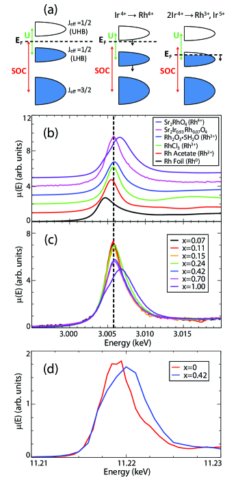
Sr2Ir1-xRhxO4 represents an ideal candidate for experimental doping studies. Rh is situated directly above Ir in the periodic table, and Sr2RhO4 is a paramagnetic metal which is isostructural to Sr2IrO4 (with slightly reduced lattice parameters and an octahedral rotation of 9.7∘) Perry_NJP_2006 ; Martins_PRL_2011 ; Itoh_JSSC_1995 . Bulk characterization measurements on Sr2Ir1-xRhxO4 have revealed a rich phase diagram with multiple electronic and magnetic transitions Qi_PRB_2012 . At low concentrations (x 0.16), Sr2Ir1-xRhxO4 is an antiferromagnetic insulator, while at higher dopings it becomes a paramagnetic metal/semiconductor (0.16 x 0.24), a frustrated magnetic insulator (0.24 x 0.85), and a paramagnetic correlated metal (x 0.85). In the simplest scenario, one expects Rh-doping to result in an isoelectronic substitution of Ir4+ (5d5) for Rh4+ (4d5). Such a substitution would tune the SOC of the system from the strong 5d regime to the moderate 4d regime, but leave the band filling unaffected Qi_PRB_2012 ; Lee_PRB_2012 . However, it has also been proposed that the dopant ions may adopt a Rh3+ (4d6) oxidation state, creating nearby Ir5+ (5d4) ions in order to preserve charge neutrality Klein_JPCM_2008 ; Klein_JEM_2009 ; Cao_APS_2013 . Such a substitution would not only tune SOC, but would also alter the band filling via hole-doping. A comparison of these two mechanisms is provided in Fig. 1(a). Rh4+ and Rh3+ substitution will also have very different effects on the magnetism of Sr2IrO4, with Rh4+ doping resulting in an exchange of effective S = 1/2 moments, and Rh3+ doping introducing pairs of non-magnetic vacancies (Rh3+ and Ir5+ are both non-magnetic due to fully filled t2g [Rh] and = 3/2 [Ir] electronic configurations).
In this article, we present complementary resonant magnetic x-ray scattering (RMXS) and x-ray absorption spectroscopy (XAS) measurements on single crystal samples of Sr2Ir1-xRhxO4 (0.07 x 0.70). Our results clearly demonstrate that Sr2Ir1-xRhxO4 must be considered as a hole-doped and magnetically diluted system. We show that Rh-doping results in a change of magnetic structure, a rapid decrease in magnetic transition temperatures, and a suppression of magnetic order at xc 0.17. In contrast to diluted La2CuO4, we show that the magnetic phase diagram of Sr2Ir1-xRhxO4 cannot be described by a conventional spin-only percolation picture. We propose that this discrepancy may reflect the importance of both spin and orbital percolation effects, which arise due to the strong SOC inherent to this system.
II Experimental Details
Single crystal samples of Sr2Ir1-xRhxO4 (0 x 1.0) were prepared using self-flux techniques, as described elsewhere Cao_PRB_1998 ; Qi_PRB_2012 . The samples used in this experiment had typical dimensions of 2.0 1.0 0.1 mm3. Detailed magnetization, resistivity, and specific heat measurements on these samples have previously been reported by Qi et al Qi_PRB_2012 . The Rh content of each sample was determined by energy dispersive x-ray (EDX) spectroscopy using a combined Hitachi/Oxford SwiftED 3000 unit. Crystal quality was assessed by x-ray rocking scans, which revealed sample mosaicities of 0.01∘ to 0.15∘ full-width at half-maximum (FWHM). In particular, the three samples which lie within the magnetically-ordered region of the phase diagram (x = 0.07, 0.11, and 0.15) all displayed FWHM of 0.02∘ or better.
X-ray absorption spectroscopy measurements were performed using the Soft X-ray Microcharacterization Beamline (SXRMB) at the Canadian Light Source (CLS) and Beamline 9-ID-B at the Advanced Photon Source (APS) at Argonne National Laboratory. Measurements on SXRMB were carried out at the Rh L3 (2p3/2 4d) and L2 (2p1/2 4d) absorption edges, which occur at energies of 3.004 keV and 3.146 keV respectively. Data was collected using Total Electron Yield (TEY) and Fluorescence Yield (FY) detection modes, and energy calibration was verified by a comparison of Ar K-edge features observed at E = 3.206 keV. Measurements on 9-ID-B were carried out at the Ir L3 absorption edge (2p3/2 5d, E = 11.215 keV), using Partial Fluorescence Yield (PFY) detection mode. PFY-XAS is a form of resonant x-ray emission spectroscopy, which involves tuning the incident energy to the Ir L3-edge, and monitoring the intensity of the Ir L emission line (3d3/2 2p3/2, E = 9.099 keV) as a function of energy. By suppressing the spectral broadening due to 2p core-hole lifetime effects, PFY-XAS can provide a significant improvement in experimental energy resolutionHamalainen_PRL_1991 ; deGroot_PRB_2002 . These measurements were carried out using a double-bounce Si-(1,1,1) primary monochromator, a channel-cut Si-(3,3,3) secondary monochromator, and a spherical (1m radius) diced Ge-(3,3,7) analyzer crystal to obtain an instrumental energy resolution of 225 meV (FWHM). Measurements were collected using horizontal scattering geometry, with a scattering angle close to 2 = 90∘.
Resonant magnetic x-ray scattering measurements were performed using Beamline 6-ID-B at the APS. Measurements were carried out at the Ir L3 (2p3/2 5d) and L2 (2p1/2 5d) absorption edges, which occur at energies of 11.215 keV and 12.824 keV respectively. Samples were mounted in a closed-cycle cryostat with a base temperature of T = 6 K. Measurements were performed in vertical scattering geometry, with the polarization of the incident beam perpendicular to the scattering plane defined by ki and kf (i.e. a -polarized beam). The polarization of the scattered beam was analyzed using the (0,0,8) and (0,0,10) reflections from a pyrolytic graphite (PG) analyzer crystal. These reflections correspond to analyzer scattering angles of = 82.33∘ at the Ir L3-edge and = 92.04∘ at the Ir L2-edge, respectively. In this configuration, the scattering term with a rotated polarization vector (i.e. -) is magnetic in origin, while the term with an unrotated polarization vector (i.e. -) is due to charge scattering. The intensity of the magnetic scattering contribution is proportional to (kf M)2 [Ref. 27].
High-resolution non-resonant x-ray diffraction measurements were performed using Beamline X21 at the National Synchrotron Light Source (NSLS) at Brookhaven National Laboratory. Measurements were carried out in vertical scattering geometry, using x-rays with an incident energy of 11.000 keV. A Ge-(1,1,1) analyzer was used to improve angular resolution and reduce experimental background.
III Experimental Results
III.1 Ionic Composition of Sr2Ir1-xRhxO4
To investigate the role of the Rh dopant ions in Sr2Ir1-xRhxO4 we performed x-ray absorption spectroscopy (XAS) measurements at the Rh L3-edge. The position of the sharp “white-line” peak at the absorption edge is very sensitive to oxidation state, and displays a chemical shift which is proportional to the ionic charge. Fig. 1(b) shows representative x-ray absorption spectra for a series of Rh-based reference samples, with oxidation states ranging from 0 to 4+. The white-line peak for Sr2Ir0.93Rh0.07O4 clearly coincides with the Rh3+ reference samples, and is shifted by -1.4 eV with respect to Rh4+. The doping dependence of the absorption spectra (Fig. 1(c)) indicates that the position of the white-line peak remains fixed for x = 0.07 to x = 0.70.
By performing similar XAS measurements at the Ir L3-edge (Fig. 1(d)) we can also characterize the doping dependence of the Ir oxidation state. These measurements reveal a broadening of the Ir L3-edge white-line peak and a positive shift in spectral weight with increasing Rh concentration. Both of these features are consistent with a mixed population of Ir4+ and Ir5+ ions introduced by doping. It should be noted that the doping dependence of the Ir L3-edge absorption spectra is difficult to observe with conventional XAS methods due to the effect of core-hole lifetime broadening, which is more than twice as large for Ir (5.3 eV) as it is for Rh (2.1 eV)Fuggle_and_Inglesfield . It is only by utilizing the PFY-XAS technique, which suppresses such core-hole lifetime effects, that we can resolve these features in the present study.
The combination of Rh and Ir XAS results allow us to draw four main conlusions: (1) The Rh dopant ions in Sr2Ir1-xRhxO4 adopt a 3+ rather than 4+ oxidation state. (2) This oxidation state persists across almost the entire Rh-doped phase diagram. (3) The electronic effect of Rh-doping is to tune band-filling via hole-doping. (4) The magnetic effect of Rh-doping is to introduce quenched non-magnetic vacancies (2 per dopant ion).
We must emphasize that while the Rh3+/Ir5+ picture will accurately describe Sr2Ir1-xRhxO4 at low dopings, and within the percolation regime that our RMXS measurements will focus on (i.e. for 0 x 0.24), at higher dopings this picture must be modified. The complication arises from the fact that once the concentration of Rh reaches x = 0.50 there will no longer be enough potential Ir5+ ions available to balance the electronic charge. As a result, while the lower dopings will be dominated by Rh3+ ions, the higher dopings must contain some mixture of 3+ and 4+ oxidation states. This scenario appears to be consistent with the XAS fit parameters provided in Table I. Although the position of the Rh L3 edge white-line does not change between x = 0.07 and x = 0.70, the width of the white-line peak becomes significantly broader for x = 0.42 and x = 0.70. This broadening is consistent with the development of a high-energy shoulder on the white-line peak, as one would expect for an increasing, but still minority, population of Rh4+ ions.
| Rh Concentration | Peak Position (eV) | Peak Width (eV) |
|---|---|---|
| x = 0.07 | 3005.8 0.1 | 3.2 0.1 |
| x = 0.11 | 3005.8 0.1 | 3.1 0.1 |
| x = 0.15 | 3005.8 0.1 | 3.3 0.1 |
| x = 0.24 | 3005.9 0.1 | 3.4 0.1 |
| x = 0.42 | 3005.9 0.1 | 4.1 0.1 |
| x = 0.70 | 3005.9 0.1 | 3.9 0.1 |
| x = 1.00 | 3007.2 0.1 | 3.9 0.2 |
III.2 Magnetic Structure of Sr2Ir1-xRhxO4
The impact of Rh-doping on magnetic structure was investigated using resonant magnetic x-ray scattering (RMXS). The magnetic ground state of pure Sr2IrO4 is known to be a canted ab-plane antiferromagnet in which magnetic moments follow the rotations of IrO6 octahedra Kim_Science_2009 ; Ye_PRB_2013 . This structure, AF-I, is illustrated in Fig. 2(a). The magnetic structure of Sr2Ir1-xRhxO4 was determined using three different elements from the RMXS data: (1) the magnetic selection rule, (2) the magnetic structure factor, and (3) the azimuthal dependence of the magnetic Bragg peaks.
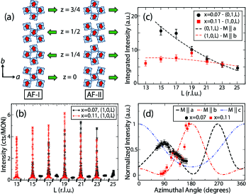
The simplest of these elements is the magnetic selection rule, which is illustrated in Fig. 2(b). This panel shows the characteristic magnetic Bragg peaks which develop in Sr2Ir1-xRhxO4 for dopings of x = 0.07 and 0.11. Note that on the basis of this selection rule alone, one can immediately identify that a doping-induced magnetic phase transition takes place between x = 0 and x = 0.07. The AF-I magnetic structure which develops in undoped Sr2IrO4 (x = 0) gives rise to magnetic Bragg peaks at (1,0,L)/(0,1,L) wave vectors for L = even, and (0,0,L) wave vectors for L = odd. A single AF-I domain produces magnetic peaks at (1,0,4n+2) and (0,1,4n) wave vectors for all integer n. However, given the tetragonal crystal structure of Sr2IrO4, it is natural for a two-domain magnetic structure to develop, with the second domain giving rise to peaks at (1,0,4n) and (0,1,4n+2). This selection rule is clearly inconsistent with the data in Fig. 2(b), allowing us to rule out the possibility of an AF-I spin configuration for x = 0.07 and x = 0.11.
The magnetic Bragg peaks observed in Sr2Ir1-xRhxO4 (0.07 x 0.15) appear at (1,0,L)/(0,1,L) wave vectors for L = odd. Scans along other high symmetry directions in reciprocal space, such as [0,0,L], [1,1,L], [1/2,1/2,L], and [1/2,0,L], reveal no evidence of additional magnetic peaks, either at commensurate or incommensurate wave vectors. These magnetic peaks are consistent with a k = (0,0,0) magnetic propagation vector. Following a similar approach to Calder et al Calder_PRB_2012 , we used representational analysis to identify potential magnetic structures for Sr2Ir1-xRhxO4. This analysis was performed using the SARAh Representational Analysis software package SARAh . For a crystal structure with I41/acd symmetry and magnetic moments located on the Ir 8a site, there are only six irreducible representations consistent with a propagation vector of k = (0,0,0): , , , , , and . Two of these representations can immediately be discarded as they fail to reproduce the observed magnetic Bragg peaks - (which describes a magnetic structure with ferromagnetic in-plane coupling, ferromagnetic out-of-plane coupling, and moments oriented along the c-axis), and (which describes a magnetic structure with ferromagnetic in-plane coupling, antiferromagnetic out-of-plane coupling, and moments oriented along the c-axis). The four remaining irreducible representations (, , , and ) are all characterized by antiferromagnetic in-plane coupling, and antiferromagnetic out-of-plane coupling. The chief distinction between these representations is the choice of magnetic easy axis. and describe magnetic structures with moments oriented along the c-axis (as in the doping-induced state observed in Sr2Ir0.9Mn0.1O4 [Ref. 14], while and describe structures with moments in the ab-plane (as in the field-induced state of Sr2IrO4 [Ref. 2]). The magnetic structure corresponding to and , which we will label AF-II, is illustrated in Fig. 2(a).
In order to distinguish between these possible structures, we can model both the magnetic structure factor and the azimuthal dependence of the magnetic Bragg peaks. These quantities are both sensitive to the orientation of the magnetic moments, and can be calculated using the FDMNES software package FDMNES . For simplicity, we have employed a single-domain model for these calculations, which assumes one dominant magnetic domain. The integrated intensity of the magnetic Bragg peaks (obtained from or rocking scans) is plotted as a function of L in Fig. 2(c). These measurements were performed with the sample aligned such that the [1,1,0] and [0,0,1] directions are coincident with the vertical scattering plane. In this orientation, there will be non-zero magnetic scattering contributions from moments aligned along the a, b, or c-axes. However, a satisfactory fit to the magnetic structure factor can only be obtained for moments oriented within the ab-plane - either along the a-axis (x = 0.07) or along the b-axis (x = 0.11). Note that because the crystal structure of Sr2Ir1-xRhxO4 has tetragonal symmetry, there is no physical distinction between these two axes. Hence the apparent 90∘ rotation of moment direction between x = 0.07 and x = 0.11 is simply due to a spontaneous choice of [1,0,0]/[0,1,0] orientation adopted by the dominant grain upon cooling through TN1.
The azimuthal dependence of the magnetic Bragg peaks in Sr2Ir1-xRhxO4 (Fig. 2(d)) is also consistent with magnetic moments oriented in the ab-plane. Here = 0∘ has been defined as the sample orientation for which [0,1,0] is coincident with the vertical scattering plane defined by ki and kf. The modeling of the azimuthal dependence indicates that the magnetic easy axis is along the a-axis for x = 0.07, and along the b-axis for x = 0.11, in full agreement with the results of the structure factor calculation. In particular, two qualitative features of the azimuthal dependence - the 180∘ oscillation period and the vanishing of magnetic intensity at specific angles - cannot be reproduced by a magnetic structure which has a c-axis spin configuration.
The results of our magnetic structure analysis indicate that: (1) Sr2IrxRh1-xO4 undergoes a doping-induced magnetic phase transition at x 0.07, (2) the magnetic ground state of Sr2IrO4 is very sensitive to a variety of external perturbations, and (3) the effects of quenched magnetic (Mn) and non-magnetic (Rh) impurities are significantly different. It should be noted that a full magnetic structure factor and azimuthal dependence measurement was not completed for the x = 0.15 sample. We have attributed the same AF-II magnetic structure to this compound based purely on the magnetic selection rule. As in the case of the x = 0.07 and x = 0.11 compounds, this sample displays magnetic Bragg peaks at (1,0,L) and (0,1,L) wave vectors for L = odd, but not for L = even. Additional follow-up measurements would be required for an unambiguous determination of the magnetic structure and moment direction for this doping.
III.3 Magnetic Order Parameter and Correlation Lengths in Sr2Ir1-xRhxO4
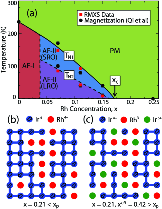
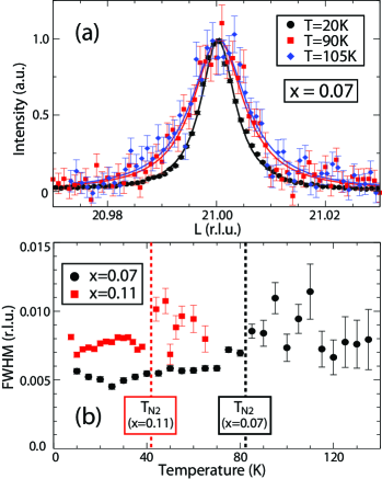
The temperature dependence of the magnetic peak intensity (Fig. 3) provides a direct measure of the antiferromagnetic order parameter (I M2). The magnetic peak intensity closely tracks the bulk magnetization Qi_PRB_2012 , with TN1 marking the appearance of magnetic Bragg peaks and a net ferromagnetic moment, and TN2 marking a dramatic increase in peak intensity and a magnetization kink. Although the magnetic peaks persist between TN1 and TN2, they display a broadened lineshape which is indicative of finite magnetic correlation lengths. By combining our RMXS measurements with previously reported magnetization dataQi_PRB_2012 , we can construct the magnetic phase diagram provided in Fig. 4(a).
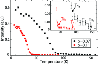
These results suggest that the magnetic phase diagram of Sr2Ir1-xRhxO4 is characterized by two distinct regions of AF-II magnetic order; a long-range-ordered (LRO) phase below TN2, and a short-range-ordered (SRO) phase between TN1 and TN2. The change in magnetic correlation lengths at TN2 is reflected in the width of the magnetic Bragg peaks, as shown in Fig. 5. The magnetic peaks within the SRO phase are significantly weaker than those observed in the LRO phase, and appear to be broader along both the in-plane ([H,0,0] and [0,K,0]) and out-of-plane ([0,0,L]) directions. The experimentally measured peak width (expressed as the FWHM, ) represents a convolution of the intrinsic peak width () and the instrumental resolution function (). In this case, an experimental resolution function was determined by measuring the lineshape of a nearby structural Bragg peak. For the L-scans provided in Fig.5, the width of the experimental resolution function was 0.0066 r.l.u. The magnetic correlation length () is inversely proportional to the intrinsic peak width through the relation: = [(2/d)(/2)]-1. This allows us to determine the average magnetic correlation lengths within the SRO phase, which are found to be 1500 Å (x = 0.07) and 1400 Å (x = 0.11) in-plane, and 1000 Å (x = 0.07) and 800 Å (x = 0.11) out-of-plane. Note that in both of these samples the average magnetic correlation length is substantially longer than the average distance between Rh dopant ions ( 95 Å and 60 Å, respectively).
To summarize, the magnetic phase diagram of Sr2Ir1-xRhxO4 is distinguished by three major features: (1) the disappearance of magnetic order at a critical doping of xc 0.17, (2) a doping-induced change in magnetic structure between x = 0 and x = 0.07, and (3) a thermally-driven transition between long-range (LRO) and short-range (SRO) magnetic order at TN2.
III.4 Robustness of the = 1/2 Ground State
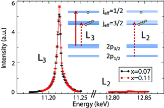
The RMXS data also allows us to address the question of how Rh-doping affects the = 1/2 character of Sr2IrO4. In previous work Kim_Science_2009 ; Boseggia_PRB_2012 ; JWKim_PRL_2012 ; Calder_PRB_2012 ; Boseggia_PRL_2013 ; Ohgushi_PRL_2013 , the = 1/2 ground state has been identified on the basis of an anomalously large L3/L2 magnetic intensity ratio, which arises due to the selection rules and transition matrix elements associated with the L2 (2p1/2 5d3/2) and L3 (2p3/2 5d3/2, 5d5/2) RMXS processes. The energy dependence of the (0,1,21) magnetic Bragg peak in Sr2Ir1-xRhxO4 is provided in Fig. 6. Note that extremely large L3/L2 intensity ratios are observed for both the x = 0.07 and x = 0.11 samples. In fact, no magnetic Bragg peaks could be detected at the L2 edge for either sample, indicating that I(L3)/I(L2) 200. A similar result has also been reported for Sr2Ir0.9Mn0.1O4 [Ref. 14], suggesting that the = 1/2 character of Sr2IrO4 is very robust against doping in general. This persistence of strong character implies that the electronic transition at x 0.16 is not driven by the tuning of SOC effects, but rather by a combination of hole-doping and/or doping-induced structural changes Qi_PRB_2012 .
It should be noted that the interpretation of the L3/L2 magnetic intensity ratio has recently been questioned by Chapon and Lovesey Chapon_JPCM_2011 and Moretti Sala et al Moretti_arXiv_2013 . In particular, it has been suggested that the magnetic intensity at the L2-edge may vanish if Ir4+ magnetic moments are aligned within the ab-plane, regardless of the splitting of the t2g levels. This point is relevant to both Sr2IrO4 and Sr2Ir1-xRhxO4, as both systems adopt canted ab-plane antiferromagnetic ground states below TN. However, in the case of Sr2Ir0.90Mn0.10O4, which displays a collinear c-axis antiferromagnetic structure Calder_PRB_2012 , this objection does not apply. In addition, Mn-doping represents an even stronger pertubation to magnetism ( = 1/2 S = 3/2) and SOC (5d 3d) than Rh-doping. Although the signatures of the = 1/2 state in Sr2Ir1-xRhxO4 may still require further investigation, the analogy with Sr2Ir1-xMnxO4 suggests that, at least on a qualitative level, these conclusions will still hold true.
III.5 Octahedral Rotations and Structural Disorder in Sr2Ir1-xRhxO4
The rotations of IrO6 octahedra are known to play an important role in shaping the physics of Sr2IrO4. These rotations break the inversion symmetry between nearest-neighbor Ir ions, giving rise to an antisymmetric Dzyaloshinskii-Moriya interaction Jackeli_PRL_2009 . In addition, the orientation of the ordered moments in Sr2IrO4 appears to be strongly coupled to the octahedral rotations, with the canted antiferromagnetic ground state displaying a spin-canting angle of 8∘ [Refs. 2, 24]. Given the difference in rotation angles between Sr2IrO4 ( 11∘) and Sr2RhO4 ( 9.7∘), the disorder of IrO6/RhO6 octahedral rotations has been proposed as one possible explanation for the suppression of magnetic order in Sr2Ir1-xRhxO4 [Ref. 9].
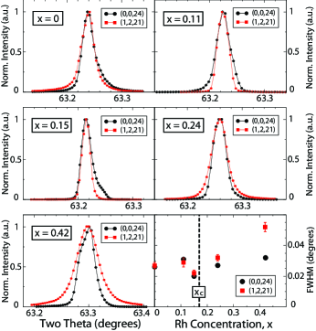
In order to investigate how the IrO6/RhO6 octahedral rotations in Sr2Ir1-xRhxO4 evolve as a function of doping, we performed a series of high-resolution non-resonant x-ray diffraction measurements. Fig. 7 provides a comparison of longitudinal (-2) scans taken through the (0,0,24) structural Bragg peak and the (1,2,21) superlattice Bragg peak for samples with x = 0, 0.11, 0.15, 0.24, and 0.42. The (1,2,21) superlattice peak arises due to the correlated rotations of the IrO6 octahedra, and it is one of the distinguishing characteristics of the I41/acd spacegroup Crawford_PRB_1994 ; Huang_JSSC_1994 . In the absence of correlated octahedral rotations, the superlattice peaks at (1,2,L)/(2,1,L), L = odd, disappear and Sr2IrO4 can be described by an I4/mmm spacegroup, with a unit cell reduced by in the ab-plane and halved along the c-axis. As a result, the width of the (1,2,21) superlattice peak provides a window into the correlation lengths associated with these octahedral rotations. For dopings of x = 0 to x = 0.24 the width of the superlattice peak is essentially the same as that of the structural Bragg peak, implying that the correlation lengths are long-ranged and the octahedral rotations are well-ordered. At higher dopings (x = 0.42), the superlattice peak becomes significantly broader than the Bragg peak, indicating reduced octahedral correlation lengths ( 500 Å) and increased rotational disorder. This suggests that while rotational disorder may play an important role in Sr2Ir1-xRhxO4 at higher dopings (x 0.42), it is not a significant effect at lower dopings (x 0.24), and is unlikely to drive the suppression of magnetic order at xc.
IV Discussion and Conclusions
It is very interesting to consider the mechanism responsible for the disappearance of magnetic order at xc 0.17. We have already touched upon two potential mechanisms for this transition in the preceding sections. From the lack of doping-dependence associated with the L3/L2 magnetic intensity ratio (Section III.D), we infer that this magnetic transition is not the result of spin-orbit tuning. Similarly, although it is possible for magnetic order to be disrupted by the disorder of IrO6/RhO6 octahedral rotations Qi_PRB_2012 , our measurements of the superlattice Bragg peaks associated with these rotations (Section III.E) reveal no significant change in correlation lengths at xc. Other doping-induced structural changes, such as a sudden jump in Ir-O-Ir bond angle, have previously been reported in the vicinity of xc [Ref. 9]. However, these structural changes appear to be discontinuous, while the observed decrease in TN1 and TN2 is clearly continuous.
An alternative explanation is provided by percolation theory (Figs. 4(b,c)), which has proven extremely successful at describing magnetism in doped cuprates such as La2Cu1-x(Zn,Mg)xO4 [Ref. 38]. This argument assumes that Sr2Ir1-xRhxO4 can be adequately described by a local moment picture for x 0.24, a claim which appears reasonably well-justified based on previous resistivity data Qi_PRB_2012 . As Sr2IrO4 is effectively a two-dimensional S = 1/2 Heisenberg antiferromagnet, we expect the conventional (i.e. spin-only) percolation threshold for this system to be xp = 0.407 [Ref. 39]. Since each Rh3+ dopant ion introduces two non-magnetic vacancies, the effective site dilution, xeff, will be equal to twice the nominal Rh concentration. We suggest that the apparent discrepancy between x = 2xc = 0.34 and xp = 0.407 may reflect novel percolation behavior arising from strong SOC effects. Recent theoretical work Tanaka_PRL_2007 ; Tanaka_PRB_2009 has shown that quantum orbital systems are much more sensitive to site dilution than pure spin systems, and can display considerably lower percolation thresholds. In a system where the spin and orbital degrees of freedom are strongly entangled, as in Sr2IrO4, it is therefore unsurprising that a spin-only percolation calculation overestimates the value of xp. This result suggests a full theoretical description of dilute magnetism in Sr2Ir1-xRhxO4 must account for both spin and orbital percolation effects, raising the possibility of exciting new percolation physics in the strong SOC regime.
In conclusion, we have used a combination of resonant x-ray techniques to investigate the chemical, electronic, and magnetic properties of the doped spin-orbital Mott insulator Sr2Ir1-xRhxO4. XAS measurements clearly demonstrate that Rh-doping introduces Rh3+ and Ir5+ ions into this material, leading to (1) hole-doping and (2) magnetic dilution of the system. RMXS measurements reveal a doping-induced change in magnetic structure at x 0.07, which leads to the development of a canted ab-plane antiferromagnetic state (AF-II) for x = 0.07, 0.11, and 0.15. Magnetic order is fully suppressed above xc 0.17 (or x 0.34), a result which suggests novel percolation effects and intriguing differences from diluted cuprates. We hope these results will help to motivate further theoretical and experimental work on Sr2Ir1-xRhxO4 and other doped 5d systems in the future.
Acknowledgements.
The authors would like to acknowledge valuable discussions with Y. Cao, D. Dessau, D. Haskel, and J.W. Kim. Work at the University of Toronto was supported by NSERC of Canada, the Banting Postdoctoral Fellowship program, and the Canada Research Chair program. Work at the University of Kentucky was supported by NSF through grants DMR-0856234, DMR-1265162, and EPS-0814194. Use of SXRMB at the Canadian Light Source is supported by NSERC of Canada, NRC of Canada, CIHR, and the University of Saskatchewan. Use of the Advanced Photon Source at Argonne National Laboratory and the National Synchrotron Light Source at Brookhaven National Laboratory is supported by the U.S. Department of Energy, Office of Science, Office of Basic Energy Sciences, under Contract Nos. DE-AC02-06CH11357 and DE-AC02-98CH10886.References
- (1) B.J. Kim, H. Jin, S.J. Moon, J.-Y. Kim, B.-G. Park, C.S. Leem, J. Yu, T.W. Noh, C. Kim, S.-J. Oh, J.-H. Park, V. Durairaj, G. Cao, and E. Rotenberg, Phys. Rev. Lett. 101, 076402 (2008).
- (2) B.J. Kim, H. Ohsumi, T. Komesu, S. Sakai, T. Morita, H. Takagi, and T. Arima, Science 323, 1329 (2009).
- (3) G. Jackeli and G. Khaliullin, Phys. Rev. Lett. 102, 017205 (2009).
- (4) J.H. Kim, D. Casa, M.H. Upton, T. Gog, Y.-J. Kim, J.F. Mitchell, M. van Veenendaal, M. Daghofer, J. van den Brink, G. Khaliullin, and B.J. Kim, Phys. Rev. Lett. 108, 177003 (2012).
- (5) M.K. Crawford, M.A. Subramanian, R.L. Harlow, J.A. Fernandez-Baca, Z.R. Wang, and D.C. Johnston, Phys. Rev. B 49, 9198 (1994).
- (6) Q. Huang, J.L. Soubeyroux, O. Chmaissen, I. Natali Sora, A. Santoro, R.J. Cava, J.J. Krajewski, and W.F. Peck, Jr., J. Solid State Chem. 112, 355 (1994).
- (7) F. Wang and T. Senthil, Phys. Rev. Lett. 106, 136402 (2011).
- (8) H. Watanabe, T. Shirakawa, and S. Yunoki, Phys. Rev. Lett. 110, 027002 (2013).
- (9) T.F. Qi, O.B. Korneta, L. Li, K. Butrouna, V.S. Cao, X. Wan, P. Schlottmann, R.K. Kaul, and G. Cao, Phys. Rev. B 86, 125105 (2012).
- (10) J.S. Lee, Y. Krockenberger, K.S. Takahashi, M. Kawasaki, and Y. Tokura, Phys. Rev. B 85, 035101 (2012).
- (11) Y. Klein and I. Terasaki, J. Phys.: Condens. Matter 20, 295201 (2008).
- (12) Y. Klein and I. Terasaki, J. Electron. Mater. 38, 1331 (2009).
- (13) Y. Cao, Bull. Amer. Phys. Soc. 58, C1.4 (2013); http://meetings.aps.org/link/BAPS.2013.MAR.C1.4.
- (14) S. Calder, G.-X. Cao, M.D. Lumsden, J.W. Kim, Z. Gai, B.C. Sales, D. Mandrus, and A.D. Christianson, Phys. Rev. B 86, 220403 (2012).
- (15) S.A. Carter, B. Batlogg, R.J. Cava, J.J. Krajewski, W.F. Peck, Jr., and L.W. Rupp, Jr., Phys. Rev. B 51, 17184 (1995).
- (16) R.J. Cava, B. Batlogg, K. Kiyono, H. Takagi, J.J. Krajewski, W.F. Peck, Jr., L.W. Rupp, Jr., and C.H. Chen, Phys. Rev. B 49, 11890 (1994).
- (17) A.J. Gatimu, R. Berthelot, S. Muir, A.W. Sleight, and M.A. Subramanian, J. Solid State Chem. 190, 257 (2012).
- (18) M. Ge, T.F. Qi, O.B. Korneta, D.E. De Long, P. Schlottmann, W.P. Crummett, and G. Cao, Phys. Rev. B 84, 100402 (2011).
- (19) O.B. Korneta, T.F. Qi, S. Chikara, S. Parkin, L.E. De Long, P. Schlottmann, and G. Cao, Phys. Rev. B 82, 115117 (2010).
- (20) T. Shimura, Y. Inaguma, T. Nakamura, M. Itoh, and Y. Morii, Phys. Rev. B 52, 9143 (1995).
- (21) M. Itoh, T. Shimura, Y. Inaguma, and Y. Morii, J. Solid State Chem. 118, 206 (1995).
- (22) R.S. Perry, F. Baumberger, L. Balicas, N. Kikugawa, N.J.C. Ingle, A. Rost, J.F. Mercure, Y. Maeno, Z.X. Shen, and A.P. Mackenzie, New J. Phys. 8, 175 (2006).
- (23) C. Martins, M. Aichhorn, L. Vaugier, and S. Biermann, Phys. Rev. Lett. 107, 266404 (2011).
- (24) G. Cao, J. Bolivar, S. McCall, J.E. Crow, and R.P. Guertin, Phys. Rev. B 57, R11039 (1998).
- (25) K. Hamalainen, D.P. Siddons, J.B. Hastings, and L.E. Berman, Phys. Rev. Lett. 67, 2850 (1991).
- (26) F.M.F. de Groot, M.H. Krisch and J. Vogel, Phys. Rev. B 66, 195112 (2002).
- (27) J.P. Hill and D.F. McMorrow, Acta Cryst. A52, 236 (1996).
- (28) J.C. Fuggle and J.E. Inglesfeld, Unoccupied Electronic States: Fundamentals for XANES, EELS, IPS, and BIS (Springer-Verlag, New York, 1992).
- (29) F. Ye, S. Chi, B.C. Chakoumakos, J.A. Fernandez-Baca, T.F. Qi, and G. Cao, Phys. Rev. B 87, 140406(R) (2013).
- (30) A.S. Wills, Physica B 276, 680 (2000).
- (31) O. Bunau and Y. Joly, J. Phys.: Condens. Matter 21, 345501 (2009).
- (32) S. Boseggia, R. Springell, H.C. Walker, A.T. Boothroyd, D. Prabhakaran, D. Wermeille, L. Bouchenoire, S.P. Collins, and D.F. McMorrow, Phys. Rev. B 85, 184432 (2012).
- (33) J.W. Kim, Y. Choi, J. Kim, J.F. Mitchell, G. Jackeli, M. Daghofer, J. van den Brink, G. Khaliullin, and B.J. Kim, Phys. Rev. Lett. 109, 037204 (2012).
- (34) S. Boseggia, R. Springell, H.C. Walker, H.M. Ronnow, Ch. Ruegg, H. Okabe, M. Isobe, R.S. Perry, S.P. Collins, and D.F. McMorrow, Phys. Rev. Lett. 110, 117207 (2013).
- (35) K. Ohgushi, J. Yamaura, H. Ohsumi, K. Sugimoto, S. Takeshita, A. Tokuda, H. Takagi, M. Takata, and T. Arima, Phys. Rev. Lett. 110, 217212 (2013).
- (36) L.C. Chapon and S.W. Lovesey, J. Phys.: Condens. Matter 23, 252201 (2011).
- (37) M. Moretti Sala, S. Boseggia, D.F. McMorrow, and G. Monaco, arXiv:1308.0128v1 (2013).
- (38) O.P. Vajk, P.K. Mang, M. Greven, P.M. Gehring, and J.W. Lynn, Science 295, 1691 (2002).
- (39) D. Stauffer and A. Aharony, Introduction to Percolation Theory, Revised 2nd Ed. (Taylor and Francis, Bristol, PA, 1994).
- (40) T. Tanaka and S. Ishihara, Phys. Rev. Lett. 98, 256402 (2007).
- (41) T. Tanaka and S. Ishihara, Phys. Rev. B 79, 035109 (2009).