Angle-resolved photoemission spectroscopy observation of anomalous electronic states in EuFe2As2-xPx
Abstract
We used angle-resolved photoemission spectroscopy to investigate the electronic structure of EuFe2As2, EuFe2As1.4P0.6 and EuFe2P2. We observed doubled core level peaks associated to the pnictide atoms, which are related to a surface state. Nevertheless, strong electronic dispersion along the axis, especially pronounced in EuFe2P2, is observed for at less one band, thus indicated that the Fe states, albeit probably affected at the surface, do not form pure two-dimensional surface states. However, this latter material shows reduced spectral weight near the Fermi level as compared to EuFe2As2 and EuFe2As1.4P0.6. An anomalous jump is also found in the electronic states associated with the Eu2+ states in EuFe2P2.
pacs:
74.70.Xa, 74.25.Jb, 79.60.-iAlthough not as extensively studied as the Ba1-xKxFe2As2 and BaFe2-xCoxAs2 archetype systems of 122-ferropnictides, the EuFe2As2-xPx compounds show very unique and exotic properties, which vary significantly upon AsP isovalent substitution. In addition to a magnetic Fe network, a large moment is observed on the Eu2+ ions H. Raffius, E. Mörsen, B. D. Mosel, W. Müeller-Warmuth, W. Jeitsschko, L. Terbüchte and T. Vomhof (1993); Z. Ren, Z. Zhu, S. Jiang, X. Xu, Q. Tao, C. Wang, C. Feng, G. Cao and Z. Xu (2008); Y. Xiao, Y. Su, M. Meven, R. Mittal, C. M. N. Kumar, T. Chatterji, S. Price, J. Persson, N. Kumar, S. K. Dhar, A. Thamizhavel, and Th. Brueckel (2009); C. Feng, Z. Ren, S. Xu, S. Jiang, Z. Xu, G. Cao, I. Nowik and I. Felner (2010); D. H. Ryan, J. M. Cadogan, Shenggao Xu, Zhu an Xu, and Guanghan Cao (2011). While the Eu sublattice exhibits A-type antiferromagnetism below K in EuFe2As2 Y. Xiao, Y. Su, M. Meven, R. Mittal, C. M. N. Kumar, T. Chatterji, S. Price, J. Persson, N. Kumar, S. K. Dhar, A. Thamizhavel, and Th. Brueckel (2009); J. Herrero-Martín, V. Scagnoli, C. Mazzoli, Y. Su, R. Mittal, Y. Xiao, T. Brueckel, N. Kumar, S. K. Dhar, A. Thamizhavel, and L. Paolasini (2009), a ferromagnetic structure with slightly canted Eu moments aligned along the -axis is observed in EuFe2P2 below K C. Feng, Z. Ren, S. Xu, S. Jiang, Z. Xu, G. Cao, I. Nowik and I. Felner (2010); D. H. Ryan, J. M. Cadogan, Shenggao Xu, Zhu an Xu, and Guanghan Cao (2011). The Eu atoms also seem to play an essential role in the anomalous compressibility effects observed in EuFe2As2 W. Uhoya, G. Tsoi, Y. K. Vohra, M. A. McGuire, A. S. Sefat, B. C. Sales, D. Mandrus and S. T. Weir (2010), and a Eu valence change under pressure has even been reported in superconducting EuFe2As1.4P0.6 L. Sun, J. Guo, G. Chen, X. Chen, X. Dong, W. Lu, C. Zhang, Z. Jiang, Y. Zou, S. Zhang, Y. Huang, Q. Wu, X. Dai, Y. Li, J. Liu and Z. Zhao (2010).
Especially following the discovery of reentrant superconductivity in EuFe2As1.3P0.7 coinciding with the ordering of the Eu2+ moments Z. Ren, Z. Zhu, S. Jiang, X. Xu, Q. Tao, C. Wang, C. Feng, G. Cao and Z. Xu (2008), the detail of the interplay between the Eu2+ and Fe2+ layers, as well as the precise role of the AsP isovalent substitution for the emergence of superconductivity, became important issues that are still debated and need proper experimental characterizations. With its capacity to resolve the one-particle electronic spectra of materials directly in the momentum space, angle-resolved photoemission spectroscopy (ARPES) is a powerful tool that may be used for such purposes. Indeed, the electronic structures of the EuFe2As2 parent compound B. Zhou, Y. Zhang, L.-X. Yang, M. Xu, C. He, F. Chen, J.-F. Zhao, H.-W. Ou, J. Wei, B.-P. Xie, T. Wu, G. Wu, M. Arita, K. Shimada, H. Namatame, M. Taniguchi, X. H. Chen and D. L. Feng (2010); G. Adhikary, N. Sahadev, D. Biswas, R. Bindu, N. Kumar, A. Thamizhavel, S. K. Dhar and K. Maiti (2013); S. Thirupathaiah, E. D. L. Rienks, H. S. Jeevan, R. Ovsyannikov, E. Slooten, J. Kaas, E. van Heumen, S. de Jong, H. A. Dürr, K. Siemensmeyer, R. Follath, P. Gegenwart, M. S. Golden and J. Fink (2011) and of EuFe2As1.56P0.44 S. Thirupathaiah, E. D. L. Rienks, H. S. Jeevan, R. Ovsyannikov, E. Slooten, J. Kaas, E. van Heumen, S. de Jong, H. A. Dürr, K. Siemensmeyer, R. Follath, P. Gegenwart, M. S. Golden and J. Fink (2011) have been studied by ARPES recently. Although it has been first synthesized R. Marchand and W. Jeitschko (1978) three decades before the discovery of Fe-based superconductivity in 2008 Y. Kamihara, T. Watanabe, M. Hirano and H. Hosono (2008), there is unfortunately no ARPES report in literature on the electronic structure of EuFe2P2.
In this paper, we present an ARPES study of the electronic structure of EuFe2As2-xPx, from EuFe2As2 to EuFe2P2. The photoemission spectra indicate that all these materials have at least 2 inequivalent pnictide sites, which is linked to a surface state possibly resulting from the pnictide-pnictide interactions occurring in short -axis 122 compounds. Nevertheless, we record strong modulations of the electronic structure along the perpendicular momentum () direction, which become more prominent upon AsP substitution. We also observe an unexplained jump in the energy position of the Eu2+ electrons in EuFe2P2.
Single-crystals of EuFe2As2, EuFe2As1.4P0.6 and EuFe2P2 were grown using conventional methods described in Refs. A. S. Sefat, R. Jin, M. A. McGuire, B. C. Sales, D. J. Singh and D. Mandrus (2008); L. Sun, J. Guo, G. Chen, X. Chen, X. Dong, W. Lu, C. Zhang, Z. Jiang, Y. Zou, S. Zhang, Y. Huang, Q. Wu, X. Dai, Y. Li, J. Liu and Z. Zhao (2010); P. C. Canfield and Z. Fisk (1992). While the typical size of the samples of the first two compounds exceeds mm2, much smaller samples (around m2) of EuFe2P2 were measured. Most of the ARPES measurements were performed at the PGM and APPLE-PGM beamlines of the Synchrotron Radiation Center (Wisconsin) equipped with a VG-Scienta R4000 analyzer and a SES 200 analyzer, respectively. The energy and angular resolutions for the angle-resolved data were set at 10 - 30 meV and 0.2, respectively. The samples were cleaved in situ and measured at 20 K in a vacuum better than Torr. Additional core level measurements of the EuFe2As2 surface under potassium evaporation were performed at the Merlin beamline of the Advanced Light Source (California). We label the momentum values with respect to the 1 Fe/unit-cell Brillouin zone (BZ), and use as the distance between two Fe planes.
In Fig. 1(a), we compare the core level spectra of EuFe2As2, EuFe2As1.4P0.6 and EuFe2P2, which show signatures of the elemental composition of these materials. We first describe the features observed above 20 eV of binding energy (). All spectra contain a very small peak detected around eV that we assign to the Fe 3 electronic state, as well as a well-defined and more intense peak associated with the Fe 3 electrons, which is observed at a of 52.4 eV, 52.6 eV and 52.9 eV in these three compounds, respectively. As emphasized in the middle inset, a broad bump absent in EuFe2As2 but increasing with P substitution is also observed at a slightly higher (60.7 eV). This bump is detected at the same kinetic energy of 115.3 eV, independently of the incident photon energy, and we thus attribute it to Auger electrons from P.
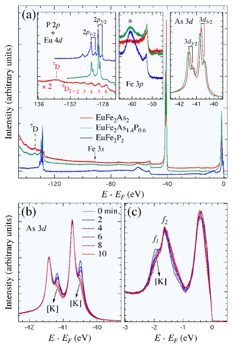
The most intense peaks observed in EuFe2As2 and EuFe2As1.4P0.6 correspond to the As and states. A zoom, displayed in the right inset of Fig. 1(a), shows an average shift of 75 meV towards the high in EuFe2As1.4P0.6 as compared to EuFe2As2. More importantly, we observe that these peaks are doubled, indicating the presence of two inequivalent As sites, suggesting a surface reconstruction affecting directly the As electronic states. When increasing the P content from to , the splittings between the peaks associated with the two sites increase slightly, from 237 meV to 248 meV, and from 225 meV to 240 meV for the As and As states, respectively. As expected from their equivalent role in the structure of EuFe2As2-xPx, double-peak features are also observed for the P and P electronic states in the P-substituted materials. As shown in the left inset of Fig. 1(a), the splitting between the two sites increases significantly as the P concentration is raised from to . Indeed, we record a splitting that increases from 206 meV to 522 meV for the P states, and a splitting that increases from 210 meV to 519 meV for the P states. It is important to note that while such effect on the pnictide atoms could potentially have a sizable impact on the Fe electronic states in EuFe2As2-xPx, our previous measurements M. Neupane, P. Richard, Y.-M. Xu, K. Nakayama, T. Sato, T. Takahashi, A. V. Federov, G. Xu, X. Dai, Z. Fang, Z. Wang, G.-F. Chen, N.-L. Wang, H.-H. Wen and H. Ding (2011) on a wide doping range of hole-doped Ba1-xKxFe2As2 and electron-doped BaFe2-xCoxAs2 did not evidence any of these doubled features.
To determine whether the doubled features observed in these materials are related to a surface state or to an impurity phase, we evaporated successively small amounts of K atoms in situ on the cleaved surface of a EuFe2As2 sample and investigated the As core levels. As illustrated in Fig. 1(b), the high- components of the As and As core levels are barely affected by this process, suggesting that they are related to bulk states. In contrast, the low- components are rapidly suppressed upon K evaporation, which is consistent with the destruction of a surface state. We also observe a slight shift towards high- attributed to a shift of the chemical potential at the surface due to the electron doping induced by the K dopant atoms. As discussed below, the states within 3 eV below the Fermi level () are also affected [see Fig. 1(c)], though in a more complicated way.
Although they cannot be assigned unambiguously, additional peaks in the spectra of the P-substituted samples are observed in the eV range. While some of them may also come from the P states, we should expect that others may be related to the Eu energy levels. Indeed, as shown in the left inset of Fig. 1(a), P-free EuFe2As2 exhibits a rather rich spectrum in this region: a series of peaks spaced by an average interval of 920 meV are observed at , 128.48, 129.40, 130.32 and 131.22 eV, in the proximity of a broader bump centered at eV. Interestingly, similar features have been already reported in Eu metal S. P. Kowalczyk, N. Edelstein, F. R. McFeely, L.Ley and D. A. Shirley (1974) as well as in EuTe D. A. Shirley (1978). Starting from an Eu2+ initial state, the spectrum of Eu metal was interpreted in terms of the and spectroscopic terms of the Eu3+ final state S. P. Kowalczyk, N. Edelstein, F. R. McFeely, L.Ley and D. A. Shirley (1974). As in Eu metal, while the multiplets of the term can be identified in EuFe2As2, the term remains unresolved.
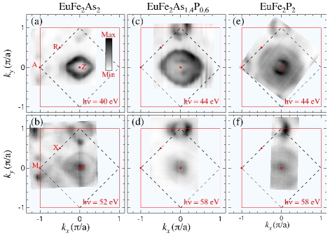
We now switch our attention to the electronic states forming the Fermi surface (FS) of EuFe2As2-xPx. In Fig. 2, we compare the FSs of EuFe2As2, EuFe2As1.4P0.6 and EuFe2P2, around and . As with BaFe2As2, the FS of EuFe2As2 exhibits stronger spots of intensity, mainly visible around the M/A [] point, which are attributed to the presence of Dirac cones induced by the antiferromagnetic ordering P. Richard, K. Nakayama, T. Sato, M. Neupane, Y.-M. Xu, J. H. Bowen, G. F. Chen, J. L. Luo, N. L. Wang, X. Dai, Z. Fang, H. Ding and T. Takahashi (2010). As a result of a surface reconstruction similar to the one reported in BaFe2-xCoxAs2 E. van Heumen, J. Vuorinen, K. Koepernik, F. Massee, Y. Huang, M. Shi, J. Klei, J. Goedkoop, M. Lindroos, J. van den Brink and M. S. Golden (2011) and Ca0.83La0.17Fe2As2 Y.-B. Huang, P. Richard, J.-H. Wang, X.-P. Wang, X. Shi, N. Xu, Z. Wu, A. Li, J.-X. Yin, T. Qian, B. Lv, C. W. Chu, S. H. Pan, M. Shi and H. Ding (2013), an extra pattern of intensity is observed at the X point.
As reported in a previous ARPES study of EuFe2As1.56P0.44 S. Thirupathaiah, E. D. L. Rienks, H. S. Jeevan, R. Ovsyannikov, E. Slooten, J. Kaas, E. van Heumen, S. de Jong, H. A. Dürr, K. Siemensmeyer, R. Follath, P. Gegenwart, M. S. Golden and J. Fink (2011) and commonly expected for a Fe-based superconductor with the 122 crystal structure and a non-magnetically ordered Fe network P. Richard, T. Sato, K. Nakayama, T. Takahashi and H. Ding (2011), the FS of EuFe2As1.4P0.6 and EuFe2P2 are composed by -centered hole FS pockets and M-centered electron FS pockets. Their size evolves with the P content, but in a non-symmetrical way. While the FS pattern at the M point becomes only a little smaller with increasing from 0 to 2, the size of the -centered pockets increases significantly, suggesting a hole-doping that cannot be explained by a simple chemical potential shift, as also pointed out in a previous ARPES study S. Thirupathaiah, E. D. L. Rienks, H. S. Jeevan, R. Ovsyannikov, E. Slooten, J. Kaas, E. van Heumen, S. de Jong, H. A. Dürr, K. Siemensmeyer, R. Follath, P. Gegenwart, M. S. Golden and J. Fink (2011). According to our core level data reported above, we cannot exclude the possibility that this non-trivial doping dependence might be related to a surface doping effect. However, the present case is quite different from the situations encountered for YBa2Cu3O7-x K. Nakayama, T. Sato, K. Terashima, H. Matsui, T. Takahashi, M. Kubota, K. Ono, T. Nishizaki, Y. Takahashi and N. Kobayashi (2007); V. B. Zabolotnyy, S. V. Borisenko, A. A. Kordyuk, D. S. Inosov, A. Koitzsch, J. Geck, J. Fink, M. Knupfer, B. Büchner, S.-L. Drechsler, V. Hinkov, B. Keimer and L. Patthey (2007) and the 1111-ferropnictides C. Liu, Y. Lee, A. D. Palczewski, J.-Q. Yan, T. Kondo, B. N. Harmon, R. W. McCallum, T. A. Lograsso and A. Kaminski (2010); I. Nishi, M. Ishikado, S. Ideta, W. Malaeb, T. Yoshida, A. Fujimori, Y. Kotani, M. Kubota, K. Ono, M. Yi, D. H. Lu, R. Moore, Z.-X. Shen, A. Iyo, K. Kihou, H. Kito, H. Eisaki, S. Shamoto and R. Arita (2011). In particular, the photoemission intensity mappings displayed in Fig. 2 for different photon energies indicate non-negligible electronic dispersion along , thus suggesting that the low-energy states probed by ARPES cannot be pure two-dimensional surface states.
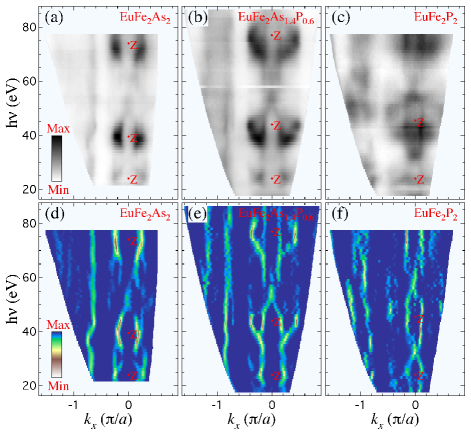
A better visualization of the electronic dispersion along is provided by the photon energy dependence of the photoemission intensity along the -M high-symmetry line of the three compounds measured in our study, which are shown in the top row of Fig. 3, as well as the curvature intensity plots P. Zhang, P. Richard, T. Qian, Y.-M. Xu, X. Dai and H. Ding (2011) given in the bottom row of Fig. 3. Within the free-electron approximation, there is indeed a monotonic relationship between the incident photon energy and the perpendicular momentum of the photoemitted electrons A. Damascelli (2004) that allows us to interpret these plots as FS mappings in the plane. One of the bands, centered at , exhibits strong dispersion along . In fact, the ARPES intensity plots displayed in Fig. 4 indicate that while that band has a very large Fermi wave vector () around (top panels of Fig. 4), it does not even cross around (bottom panels of Fig. 4) for any of the EuFe2As2-xPx materials studied here. In other words, this band forms a 3D hole pocket centered at Z, which becomes larger with the P content increasing, observation consistent with a large 3D FS suggested from de Hass van Alphen measurements in CaFe2P2 A. I. Coldea, C. M. J. Andrew, J. G. Analytis, R. D. McDonald, A. F. Bangura, J.-H. Chu, I. R. Fisher, and A. Carrington (2009), although that FS is even larger in the latter case. Interestingly, this band varies similarly to the band in Ba(Fe1-xRux)2As2 N. Xu, T. Qian, P. Richard, Y.-B. Shi, X.-P. Wang, P. Zhang, Y.-B. Huang, Y.-M. Xu, H. Miao, G. Xu, G.-F. Xuan, W.-H. Jiao, Z.-A. Xu, G.-H. Cao and H. Ding (2012); V. Brouet, F. Rullier-Albenque, M. Marsi, B. Mansart, M. Aichhorn, S. Biermann, J. Faure, L. Perfetti, A. Taleb-Ibrahimi, P. Le Fèvre, F. Bertran, A. Forget and D. Colson (2010); R. S. Dhaka, C. Liu, R. M. Fernandes, R. Jiang, C. P. Strehlow, T. Kondo, A. Thaler, J. Schmalian, S. L. Bud’ko, P. C. Canfield, and A. Kaminski (2011), which is another nominally non-doped 122-ferropnictide. We thus assign to this band the same main orbital character, being the even combination of the and orbitals. Our photon energy dependence also indicates that the distance in photon energy between two successive Z points increases, which is consistent with the decrease in the ’-axis parameter when As atoms are substituted by smaller P atoms.
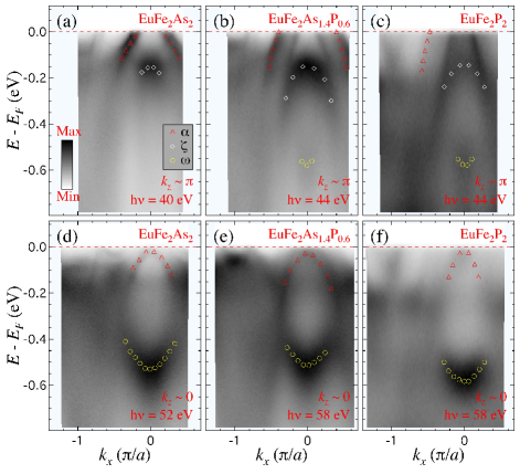
The spectral features shown in Figs. 3 are not as well defined in EuFe2P2 as in the samples with lower P content, an observation further evidenced by the ARPES intensity plots displayed in Fig. 4. In particular, the near- spectral intensity around the point of EuFe2P2 is quite weak [see Fig. 4(f)]. This contrasts with the intensity of the band found at higher binding energy, which remains high for all concentrations. In Fig. 5, we compare the photon energy dependence of the BZ center EDCs of EuFe2As2, EuFe2As1.4P0.6 and EuFe2P2. The lower energy part of the EDCs is dominated by two peaks coming from the and bands, which both have a dominant character Y. Zhang, F. Chen, C. He, B. Zhou, B. P. Xie, C. Fang, W. F. Tsai, X. H. Chen, H. Hayashi, J. Jiang, H. Iwasawa, K. Shimada, H. Namatame, M. Taniguchi, J. P. Hu and D. L. Feng (2011). Neglecting small and sample composition variations, these peaks are located around 170 and 530 meV below , respectively. Interestingly, their intensity oscillates with photon energy (or ) in anti-phase, as also illustrated in Fig. 4. While the intensity of the peak is the strongest around and the weakest around , the opposite behavior is found for the band. Although our study does not allow us to identify unambiguously the origin of the partial suppression of spectral intensity for the electronic states at low energy, it is possibly related to the surface state identified from the core level data presented in Fig. 1, which show significantly larger core level splittings in EuFe2P2. Distortion at the surface could eventually introduce additional scattering that would suppressed the coherence of the low-energy states. We caution though that an old study of structural characterization revealed the presence of FeP or Fe2P as secondary phase in the growth of some LnFe2P2 (Ln = lanthanide) compounds M. Reehuis and W. Jeitschko (1990), which could also alter the coherence of the low-energy states. However, we did not detect evidence for these impurities in our samples.
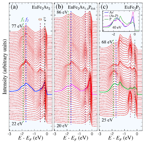
In addition to the and peaks, additional spectral intensity is found between 1 and 2.5 eV below . In particular, a peak labeled and a shoulder labeled are detected in EuFe2As2 at -1.7 eV and -1.9 eV, respectively. These dispersionless features are not observed in the more commonly studied Ba(Fe1-xCox)2-xAs2 and Ba1-xKxFe2As2 compounds M. Neupane, P. Richard, Y.-M. Xu, K. Nakayama, T. Sato, T. Takahashi, A. V. Federov, G. Xu, X. Dai, Z. Fang, Z. Wang, G.-F. Chen, N.-L. Wang, H.-H. Wen and H. Ding (2011). In agreement with a previous ARPES study G. Adhikary, N. Sahadev, D. Biswas, R. Bindu, N. Kumar, A. Thamizhavel, S. K. Dhar and K. Maiti (2013), we ascribe them to Eu electronic states. As indicated by the inset of Fig. 5(c), which compares the EDCs of the 3 measured compounds recorded with 40 eV photons, only small changes occur when the P content varies from to 0.6. While the peak position in EuFe2As1.4P0.6 is barely changed, the peak moves by only about 50 meV towards . As illustrated in Fig. 5(c), the peak shift is much larger in EuFe2P2. As compared to EuFe2As2, the and peaks in EuFe2P2 are located 80 meV and 320 meV closer to , respectively.
Such large splitting between the and peaks allows us to distinguish their spectral lineshapes, which are quite different. In contrast to the peak, which is quite broad and rounded, the peak is rather sharp. The latter is also asymmetric, with a tail on the low side possibly due to the presence of additional peaks, as mainly suggested from the spectra recorded at the highest photon energies. Interestingly, as shown in Fig. 1(c), our investigation of the electronic states in a EuFe2As2 sample upon K evaporation on the surface indicates that while the shape and intensity of the peak are barely modified by the evaporation of K atoms, the intensity of the peak is reduced, suggesting a surface state. However, this particular observation does not explain the large shift in the position of the peak in EuFe2P2. It is important to note that we did not observe any significant modification of the spectral lineshape across the Eu2+ magnetic ordering transition, thus suggesting that it is not relevant for this particular lineshape nor for the large - splitting in EuFe2P2. However, the inset of Fig. 5(c) seems to show that the shift of the peak in EuFe2P2 is accompanied by a spectral weight transfer from the near- states to the energy range. Whether this could be caused by enhanced Fe-Eu interactions is not excluded but would require confirmation from further theoretical investigations.
From the core levels to the electronic states in the vicinity of , our results indicate the presence of a surface state effect in EuFe2As2-xPx that does exist, or at least that does not manifest itself significantly, in the commonly studied Ba(Fe1-xCox)2-xAs2 and Ba1-xKxFe2As2 122-ferropnictides. We note that the size of the Eu2+ ion is significantly smaller than that of Ba2+, and the substitution of P by As contributes to reduce the -axis length even further. Previous studies showed that when the -axis becomes small and the As-As separation in the 122-ferropnictides becomes smaller than about 3 Å, either due to rare earth substitution S. R. Saha, N. P. Butch, T. Drye, J. Magill, S. Ziemak, K. Kirshenbaum, P. Y. Zavalij, J. W. Lynn and J. Paglione (2012) or AsP substitution in CaFe2As2-xPx H. L. Shi, H. X. Yang, H. F. Tian, J. B. Lu, Z. W. Wang, Y. B. Qin, Y. J. Song and J. Q. Li (2010); S. Kasahara, T. Shibauchi, K. Hashimoto, Y. Nakai, H. Ikeda, T. Terashima, and Y. Matsuda (2011), interactions between successive As layers induce a further decrease in the -axis length of these materials that thus encounter a tetragonal-collapsed tetragonal transition S. R. Saha, N. P. Butch, T. Drye, J. Magill, S. Ziemak, K. Kirshenbaum, P. Y. Zavalij, J. W. Lynn and J. Paglione (2012), as also supported by theoretical predictions T. Yildirim (2009). We point out that any material in the collapsed tetragonal phase or at the proximity of this transition may show a discontinuity in the pnictide-pnictide interactions at the surface, possibly altering the surface electronic properties. Indeed, a previous ARPES study revealed anomalies in conventional ARPES data recorded on LaRu2P2 as compared to bulk-sensitive soft-x-ray ARPES data E. Razzoli, M. Kobayashi, V. N. Strocov, B. Delley, Z. Bukowski, J. Karpinski, N. C. Plumb, M. Radovic, J. Chang, T. Schmitt, L. Patthey, J. Mesot, and M. Shi (2012). We predict that other 122 materials with similarly small -axis as EuFe2As2-xPx and LaRu2P2 may exhibit the same kind of anomalies reported here and in ref. E. Razzoli, M. Kobayashi, V. N. Strocov, B. Delley, Z. Bukowski, J. Karpinski, N. C. Plumb, M. Radovic, J. Chang, T. Schmitt, L. Patthey, J. Mesot, and M. Shi (2012). Yet, further theoretical and experimental investigations are necessary to confirm or infirm this conjecture.
In summary, we performed an ARPES study of EuFe2As2-xPx that shows the evolution of the electronic structure upon increasing the P content. Anomalies in the core level and near- state data suggest the existence of a surface state. Nevertheless, strong modulations enhanced with P substitution are observed in all studied samples for at least one band. Finally, a sudden and unexplained jump in the energy position of one peak associated to the Eu2+ states is observed in EuFe2P2.
We acknowledge M. Shi, E. Razolli and J.-X. Yin for useful discussions. This work was supported by grants from CAS (2010Y1JB6), MOST (2010CB923000, 2011CBA001000, 2011CBA00102, 2012CB821403 and 2013CB921703) and NSFC (10974175, 11004232, 11034011/A0402 and 11274362) from China. This work is based in part on research conducted at the Synchrotron Radiation Center, which is primarily funded by the University of Wisconsin-Madison with supplemental support from facility Users and the University of Wisconsin-Milwaukee. The Advanced Light Source is supported by the Director, Office of Science, Office of Basic Energy Sciences, of the U.S. Department of Energy under Contract No. DE-AC02-05CH112. The work at ORNL was supported by the Department of Energy, Basic Energy Sciences, Materials Sciences and Engineering Division.
References
- H. Raffius, E. Mörsen, B. D. Mosel, W. Müeller-Warmuth, W. Jeitsschko, L. Terbüchte and T. Vomhof (1993) H. Raffius, E. Mörsen, B. D. Mosel, W. Müeller-Warmuth, W. Jeitsschko, L. Terbüchte and T. Vomhof, J. Phys. Chem. Solids 54, 135 (1993).
- Z. Ren, Z. Zhu, S. Jiang, X. Xu, Q. Tao, C. Wang, C. Feng, G. Cao and Z. Xu (2008) Z. Ren, Z. Zhu, S. Jiang, X. Xu, Q. Tao, C. Wang, C. Feng, G. Cao and Z. Xu, Phys. Rev. B 78, 052501 (2008).
- Y. Xiao, Y. Su, M. Meven, R. Mittal, C. M. N. Kumar, T. Chatterji, S. Price, J. Persson, N. Kumar, S. K. Dhar, A. Thamizhavel, and Th. Brueckel (2009) Y. Xiao, Y. Su, M. Meven, R. Mittal, C. M. N. Kumar, T. Chatterji, S. Price, J. Persson, N. Kumar, S. K. Dhar, A. Thamizhavel, and Th. Brueckel, Phys. Rev. B 80, 174424 (2009).
- C. Feng, Z. Ren, S. Xu, S. Jiang, Z. Xu, G. Cao, I. Nowik and I. Felner (2010) C. Feng, Z. Ren, S. Xu, S. Jiang, Z. Xu, G. Cao, I. Nowik and I. Felner, Phys. Rev. B 82, 094426 (2010).
- D. H. Ryan, J. M. Cadogan, Shenggao Xu, Zhu an Xu, and Guanghan Cao (2011) D. H. Ryan, J. M. Cadogan, Shenggao Xu, Zhu an Xu, and Guanghan Cao, Phys. Rev. B 83, 123403 (2011).
- J. Herrero-Martín, V. Scagnoli, C. Mazzoli, Y. Su, R. Mittal, Y. Xiao, T. Brueckel, N. Kumar, S. K. Dhar, A. Thamizhavel, and L. Paolasini (2009) J. Herrero-Martín, V. Scagnoli, C. Mazzoli, Y. Su, R. Mittal, Y. Xiao, T. Brueckel, N. Kumar, S. K. Dhar, A. Thamizhavel, and L. Paolasini, Phys. Rev. B 80, 134411 (2009).
- W. Uhoya, G. Tsoi, Y. K. Vohra, M. A. McGuire, A. S. Sefat, B. C. Sales, D. Mandrus and S. T. Weir (2010) W. Uhoya, G. Tsoi, Y. K. Vohra, M. A. McGuire, A. S. Sefat, B. C. Sales, D. Mandrus and S. T. Weir, J. Phys.: Condens. Matter 22, 292202 (2010).
- L. Sun, J. Guo, G. Chen, X. Chen, X. Dong, W. Lu, C. Zhang, Z. Jiang, Y. Zou, S. Zhang, Y. Huang, Q. Wu, X. Dai, Y. Li, J. Liu and Z. Zhao (2010) L. Sun, J. Guo, G. Chen, X. Chen, X. Dong, W. Lu, C. Zhang, Z. Jiang, Y. Zou, S. Zhang, Y. Huang, Q. Wu, X. Dai, Y. Li, J. Liu and Z. Zhao, Phys. Rev. B 82, 134509 (2010).
- B. Zhou, Y. Zhang, L.-X. Yang, M. Xu, C. He, F. Chen, J.-F. Zhao, H.-W. Ou, J. Wei, B.-P. Xie, T. Wu, G. Wu, M. Arita, K. Shimada, H. Namatame, M. Taniguchi, X. H. Chen and D. L. Feng (2010) B. Zhou, Y. Zhang, L.-X. Yang, M. Xu, C. He, F. Chen, J.-F. Zhao, H.-W. Ou, J. Wei, B.-P. Xie, T. Wu, G. Wu, M. Arita, K. Shimada, H. Namatame, M. Taniguchi, X. H. Chen and D. L. Feng, Phys. Rev. B 81, 155124 (2010).
- G. Adhikary, N. Sahadev, D. Biswas, R. Bindu, N. Kumar, A. Thamizhavel, S. K. Dhar and K. Maiti (2013) G. Adhikary, N. Sahadev, D. Biswas, R. Bindu, N. Kumar, A. Thamizhavel, S. K. Dhar and K. Maiti, J. Phys.: Condens. Matter 25, 225701 (2013).
- S. Thirupathaiah, E. D. L. Rienks, H. S. Jeevan, R. Ovsyannikov, E. Slooten, J. Kaas, E. van Heumen, S. de Jong, H. A. Dürr, K. Siemensmeyer, R. Follath, P. Gegenwart, M. S. Golden and J. Fink (2011) S. Thirupathaiah, E. D. L. Rienks, H. S. Jeevan, R. Ovsyannikov, E. Slooten, J. Kaas, E. van Heumen, S. de Jong, H. A. Dürr, K. Siemensmeyer, R. Follath, P. Gegenwart, M. S. Golden and J. Fink, Phys. Rev. B 84, 014531 (2011).
- R. Marchand and W. Jeitschko (1978) R. Marchand and W. Jeitschko, J. Solid St. Chem. 24, 351 (1978).
- Y. Kamihara, T. Watanabe, M. Hirano and H. Hosono (2008) Y. Kamihara, T. Watanabe, M. Hirano and H. Hosono, J. Am. Chem. Soc. 130, 3296 (2008).
- A. S. Sefat, R. Jin, M. A. McGuire, B. C. Sales, D. J. Singh and D. Mandrus (2008) A. S. Sefat, R. Jin, M. A. McGuire, B. C. Sales, D. J. Singh and D. Mandrus, Phys. Rev. Lett. 101, 117004 (2008).
- P. C. Canfield and Z. Fisk (1992) P. C. Canfield and Z. Fisk, Philos. Mag. B 65, 1117 (1992).
- M. Neupane, P. Richard, Y.-M. Xu, K. Nakayama, T. Sato, T. Takahashi, A. V. Federov, G. Xu, X. Dai, Z. Fang, Z. Wang, G.-F. Chen, N.-L. Wang, H.-H. Wen and H. Ding (2011) M. Neupane, P. Richard, Y.-M. Xu, K. Nakayama, T. Sato, T. Takahashi, A. V. Federov, G. Xu, X. Dai, Z. Fang, Z. Wang, G.-F. Chen, N.-L. Wang, H.-H. Wen and H. Ding, Phys. Rev. B 83, 094522 (2011).
- S. P. Kowalczyk, N. Edelstein, F. R. McFeely, L.Ley and D. A. Shirley (1974) S. P. Kowalczyk, N. Edelstein, F. R. McFeely, L.Ley and D. A. Shirley, Chem. Phys. Lett. 29, 491 (1974).
- D. A. Shirley (1978) D. A. Shirley, In Photoemission in Solids, ed. by M. Cardona and L. Ley, Topics Appl. Phys., Vol. 26 (Springer, Berlin, Heidelberg), Chap. 4 (1978).
- P. Richard, K. Nakayama, T. Sato, M. Neupane, Y.-M. Xu, J. H. Bowen, G. F. Chen, J. L. Luo, N. L. Wang, X. Dai, Z. Fang, H. Ding and T. Takahashi (2010) P. Richard, K. Nakayama, T. Sato, M. Neupane, Y.-M. Xu, J. H. Bowen, G. F. Chen, J. L. Luo, N. L. Wang, X. Dai, Z. Fang, H. Ding and T. Takahashi, Phys. Rev. Lett. 104, 137001 (2010).
- E. van Heumen, J. Vuorinen, K. Koepernik, F. Massee, Y. Huang, M. Shi, J. Klei, J. Goedkoop, M. Lindroos, J. van den Brink and M. S. Golden (2011) E. van Heumen, J. Vuorinen, K. Koepernik, F. Massee, Y. Huang, M. Shi, J. Klei, J. Goedkoop, M. Lindroos, J. van den Brink and M. S. Golden, Phys. Rev. Lett. 106, 027002 (2011).
- Y.-B. Huang, P. Richard, J.-H. Wang, X.-P. Wang, X. Shi, N. Xu, Z. Wu, A. Li, J.-X. Yin, T. Qian, B. Lv, C. W. Chu, S. H. Pan, M. Shi and H. Ding (2013) Y.-B. Huang, P. Richard, J.-H. Wang, X.-P. Wang, X. Shi, N. Xu, Z. Wu, A. Li, J.-X. Yin, T. Qian, B. Lv, C. W. Chu, S. H. Pan, M. Shi and H. Ding, Chin. Phys. Lett. 30, 017402 (2013).
- P. Richard, T. Sato, K. Nakayama, T. Takahashi and H. Ding (2011) P. Richard, T. Sato, K. Nakayama, T. Takahashi and H. Ding, Rep. Prog. Phys. 74, 124512 (2011).
- K. Nakayama, T. Sato, K. Terashima, H. Matsui, T. Takahashi, M. Kubota, K. Ono, T. Nishizaki, Y. Takahashi and N. Kobayashi (2007) K. Nakayama, T. Sato, K. Terashima, H. Matsui, T. Takahashi, M. Kubota, K. Ono, T. Nishizaki, Y. Takahashi and N. Kobayashi, Phys. Rev. B 75, 014513 (2007).
- V. B. Zabolotnyy, S. V. Borisenko, A. A. Kordyuk, D. S. Inosov, A. Koitzsch, J. Geck, J. Fink, M. Knupfer, B. Büchner, S.-L. Drechsler, V. Hinkov, B. Keimer and L. Patthey (2007) V. B. Zabolotnyy, S. V. Borisenko, A. A. Kordyuk, D. S. Inosov, A. Koitzsch, J. Geck, J. Fink, M. Knupfer, B. Büchner, S.-L. Drechsler, V. Hinkov, B. Keimer and L. Patthey, Phys. Rev. Lett. 76, 024502 (2007).
- C. Liu, Y. Lee, A. D. Palczewski, J.-Q. Yan, T. Kondo, B. N. Harmon, R. W. McCallum, T. A. Lograsso and A. Kaminski (2010) C. Liu, Y. Lee, A. D. Palczewski, J.-Q. Yan, T. Kondo, B. N. Harmon, R. W. McCallum, T. A. Lograsso and A. Kaminski, Phys. Rev. B 82, 075135 (2010).
- I. Nishi, M. Ishikado, S. Ideta, W. Malaeb, T. Yoshida, A. Fujimori, Y. Kotani, M. Kubota, K. Ono, M. Yi, D. H. Lu, R. Moore, Z.-X. Shen, A. Iyo, K. Kihou, H. Kito, H. Eisaki, S. Shamoto and R. Arita (2011) I. Nishi, M. Ishikado, S. Ideta, W. Malaeb, T. Yoshida, A. Fujimori, Y. Kotani, M. Kubota, K. Ono, M. Yi, D. H. Lu, R. Moore, Z.-X. Shen, A. Iyo, K. Kihou, H. Kito, H. Eisaki, S. Shamoto and R. Arita, Phys. Rev. B 84, 014504 (2011).
- P. Zhang, P. Richard, T. Qian, Y.-M. Xu, X. Dai and H. Ding (2011) P. Zhang, P. Richard, T. Qian, Y.-M. Xu, X. Dai and H. Ding, Rev. Sci. Instrum. 82, 043712 (2011).
- A. Damascelli (2004) A. Damascelli, Phys. Scrypta T109, 61 (2004).
- A. I. Coldea, C. M. J. Andrew, J. G. Analytis, R. D. McDonald, A. F. Bangura, J.-H. Chu, I. R. Fisher, and A. Carrington (2009) A. I. Coldea, C. M. J. Andrew, J. G. Analytis, R. D. McDonald, A. F. Bangura, J.-H. Chu, I. R. Fisher, and A. Carrington, Phys. Rev. Lett. 103, 026404 (2009).
- N. Xu, T. Qian, P. Richard, Y.-B. Shi, X.-P. Wang, P. Zhang, Y.-B. Huang, Y.-M. Xu, H. Miao, G. Xu, G.-F. Xuan, W.-H. Jiao, Z.-A. Xu, G.-H. Cao and H. Ding (2012) N. Xu, T. Qian, P. Richard, Y.-B. Shi, X.-P. Wang, P. Zhang, Y.-B. Huang, Y.-M. Xu, H. Miao, G. Xu, G.-F. Xuan, W.-H. Jiao, Z.-A. Xu, G.-H. Cao and H. Ding, Phys. Rev. B 86, 064505 (2012).
- V. Brouet, F. Rullier-Albenque, M. Marsi, B. Mansart, M. Aichhorn, S. Biermann, J. Faure, L. Perfetti, A. Taleb-Ibrahimi, P. Le Fèvre, F. Bertran, A. Forget and D. Colson (2010) V. Brouet, F. Rullier-Albenque, M. Marsi, B. Mansart, M. Aichhorn, S. Biermann, J. Faure, L. Perfetti, A. Taleb-Ibrahimi, P. Le Fèvre, F. Bertran, A. Forget and D. Colson, Phys. Rev. Lett. 105, 087001 (2010).
- R. S. Dhaka, C. Liu, R. M. Fernandes, R. Jiang, C. P. Strehlow, T. Kondo, A. Thaler, J. Schmalian, S. L. Bud’ko, P. C. Canfield, and A. Kaminski (2011) R. S. Dhaka, C. Liu, R. M. Fernandes, R. Jiang, C. P. Strehlow, T. Kondo, A. Thaler, J. Schmalian, S. L. Bud’ko, P. C. Canfield, and A. Kaminski, Phys. Rev. Lett. 107, 267002 (2011).
- Y. Zhang, F. Chen, C. He, B. Zhou, B. P. Xie, C. Fang, W. F. Tsai, X. H. Chen, H. Hayashi, J. Jiang, H. Iwasawa, K. Shimada, H. Namatame, M. Taniguchi, J. P. Hu and D. L. Feng (2011) Y. Zhang, F. Chen, C. He, B. Zhou, B. P. Xie, C. Fang, W. F. Tsai, X. H. Chen, H. Hayashi, J. Jiang, H. Iwasawa, K. Shimada, H. Namatame, M. Taniguchi, J. P. Hu and D. L. Feng, Phys. Rev. B 83, 054510 (2011).
- M. Reehuis and W. Jeitschko (1990) M. Reehuis and W. Jeitschko, J. Phys. Chem. Solids 51, 961 (1990).
- S. R. Saha, N. P. Butch, T. Drye, J. Magill, S. Ziemak, K. Kirshenbaum, P. Y. Zavalij, J. W. Lynn and J. Paglione (2012) S. R. Saha, N. P. Butch, T. Drye, J. Magill, S. Ziemak, K. Kirshenbaum, P. Y. Zavalij, J. W. Lynn and J. Paglione, Phys. Rev. B 85, 024525 (2012).
- H. L. Shi, H. X. Yang, H. F. Tian, J. B. Lu, Z. W. Wang, Y. B. Qin, Y. J. Song and J. Q. Li (2010) H. L. Shi, H. X. Yang, H. F. Tian, J. B. Lu, Z. W. Wang, Y. B. Qin, Y. J. Song and J. Q. Li, J. Phys.: Condens. Matter 22, 125702 (2010).
- S. Kasahara, T. Shibauchi, K. Hashimoto, Y. Nakai, H. Ikeda, T. Terashima, and Y. Matsuda (2011) S. Kasahara, T. Shibauchi, K. Hashimoto, Y. Nakai, H. Ikeda, T. Terashima, and Y. Matsuda, Phys. Rev. B 83, 060505(R) (2011).
- T. Yildirim (2009) T. Yildirim, Phys. Rev. Lett. 102, 037003 (2009).
- E. Razzoli, M. Kobayashi, V. N. Strocov, B. Delley, Z. Bukowski, J. Karpinski, N. C. Plumb, M. Radovic, J. Chang, T. Schmitt, L. Patthey, J. Mesot, and M. Shi (2012) E. Razzoli, M. Kobayashi, V. N. Strocov, B. Delley, Z. Bukowski, J. Karpinski, N. C. Plumb, M. Radovic, J. Chang, T. Schmitt, L. Patthey, J. Mesot, and M. Shi, Phys. Rev. Lett. 108, 257005 (2012).