Surface Plasmon Resonance of Dumb-bell Nano-structure
Abstract
We present an intuitive theoretical description of the optical properties of complex metal nano-structure, consisting of two nano-shells connected by a nano-rod giving a dumb-bell like appearance. The simulations were done using Finite Element Method. The effect of the nano-rod length and radius as also the dimensions of the nano-shells were analyzed. The absorption spectra as in peak positions and intensities have been found to have a strong dependence on the geometrical parameters of the dumb-bell. This study provides evidence that the localized surface plasmon modes play a key role in the broadband light harvesting capabilities of these nanostructures and is promising for a wide range of practical applications, for example in surface-enhanced spectroscopies.
Central University of Kerala] Department of Physics, Central University of Kerala, Kasaragod, Kerala - 671 314, INDIA S.G.T.B. Khalsa College, University of Delhi] Mater. Sci. Res. Lab., S.G.T.B. Khalsa College, University of Delhi, Delhi 110007, INDIA
1 Introduction
Surface plasmons are electromagnetic surface waves originating due to the collective oscillation of the conduction electrons near the metal surface 1, 2. Light coupling with surface plasmons results in enhancement of the local electromagnetic field and strong resonance in the extinction profile of conductive nano-particles leading to the optical phenomenon known as localized surface plasmon resonance (LSPR) 3, 4. This interaction produces coherent localized plasmon oscillations with a resonant frequency that strongly depends on the shape and size, as also the dielectric media surrounding it 5. While the effect of size and influence of dielectric media have been studied quite extensively, both experimentally 6 and theoretically 7, this has not been the case when it comes to experimentally studying the effect of shape. Most of the nano particles usually obtained are spherical in nature or are rod like with circular cross-section. Theoretically, both shapes are interesting due to their symmetry. While the position of SPR peaks obtained from spherical grain is controllable by controlling the radius of sphere, the nano-rods present two variables, rod-length and radius of cross-section. Thus, one expects higher degree of control with more complex shaped metal clusters. One rarely comes across complex shaped metal clusters experimentally, thus making inroads along these lines difficult. However, with maturing of the methods used to theoretically simulate the optical properties of metal nano-clusters 8 exotic shapes have been reported, for example, nanorods with triangular cross-section 9, 10, pyramid shaped 11 grains etc.
While, nano-particles embedded in dielectric media with fairly constant dielectric constant over frequency ranges of interest are well studied, the case where it varies with frequency has not attracted much attention. However, situations in which a nanoparticle having another nanoparticle in its vicinity whose dielectric constant varies with frequency can lead to interesting LSPR features 12. For example the two nanoparticles would interact electromagnetically with each other to show high field enhancement in the gap. Recently, a bow tie nanoparticle, formed from equilateral triangle shaped nano particles, excited at its LSPR wavelength has been reported to generate extremely large fields within the gap 13, 4. This has been exploited to study Raman spectra for samples which would usually give weak signals in Surface-enhanced Raman Spectroscopy (SERS). This clearly shows studying exotic structures can also lead to applications of importance.
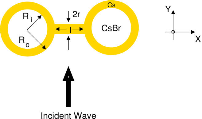
Hence, in this manuscript we undertake to further our understanding of dumb-bell shaped nano-particles that were experimentally reported recently. Wang et al 14 and Kuldeep et al 15 have reported experimental evidence of SPR in dumb-bell shaped clusters formed from spherical nano particles. While Wang et al’s clusters were of gold, Kuldeep et al reported two core-shell (core of Cesium Bromide and the shell of metal Cesium) connected by nano-rod bridges of Cesium. Though Kuldeep et al did not have control over the size of the dumb-bells (as compared to Wang et al), their results were interesting since the exotic shape allowed for four possible control parameters, namely, the length (l) of the nano-rod bridge separating the two spherical core-shell structure, the cross-sectional radius (r) of the nano-rod, the dielectric spherical core’s radius () and the core-shell’s radius (), see fig 1. The shell thickness would be given by ().
In this paper, the localized surface plasmon resonance of a dumb-bell structure is investigated using Finite Element Method. The dependence of plasmon resonance on various geometrical parameters are discussed. For field polarization orthogonal and parallel to the interparticle symmetry axis, the spectral properties of the structure are usually different. In this paper, the behavior of the dumbbell structure in presence of an unpolarized incident electromagnetic radiation is studied analogous to the usual experimental conditions. Nevertheless, studies with polarized light are included whenever required. The next section explains the simulation model used in the calculations. The section “Results and Discussion” provides the detailed analysis of the behavior of surface plasmon resonance on various geometrical parameters.
2 Simulation
The simulations were done using the commercially available Finite Element Method (FEM) package COMSOL Multiphysics (with the RF module) along with Matlab . The three dimensional simulation domain was composed of four spherical volumes: a core, a shell, an embedding medium, and a perfectly matched layer (PML) in addition to a cylinder connecting the shells as shown in fig 1. The nanoparticle core was taken to be , enclosed in a shell modeled using the empirically determined bulk dielectric constants provided by Palik 16 with linear interpolation. The medium surrounding the nanosystem was considered to be air. A plane wave was used for excitation of nanostructure and was inserted on the inside of PMLs surrounding the embedding medium. The dimensions of the embedding volume and the PML were chosen such that increase in the dimension of the nanoparticle further would not affect the simulation results. Discretization of the simulation domain was performed using the built-in meshing algorithm in COMSOL, which partitioned the simulation space into a collection of tetrahedral finite elements. Large field enhancements possible due to plasmon resonances, required the application of mesh parameters to ensure convergence of the simulation in the required frequency regime.
3 Results and Discussion
In the following passages we discuss the variation in the absorption spectra of a single dumb-bell shaped CsBr-Cs cluster. Since the absorption spectra of the structure depends on four variables, namely l, r, and , we have stimulated the spectra keeping three parameters fixed and one variable at a time.
3.1 Effect of Nanorod on Absorption
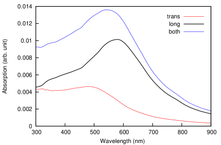
Before proceeding to the dumbbell structure, it is worthwhile to investigate the surface plasmon resonance peak due to the nano-rod. Under usual circumstances the nano-rod’s length is far greater than its radius, , then due to the anisotropy in its shape, we expect it to have two modes of oscillations, namely the transverse mode along the short-axis and the longitudinal mode along the long-axis. More explicitly, when light is incident along the ‘Y’-axis, as in our case (fig 1), the linear polarised light with electric field along ‘X’-axis () gives the longitudinal mode excitation while that along ‘Z’-axis () would result in transverse mode excitation. Such polarization dependency has been reported.17, 18 However, the simulation for a Cesium nano-rod having r and l as 10 nm and 40 nm respectively shows just one broad peak around 570 nm with no evidence of peaks in UV or IR region. To investigate why we get a broad peak, we have simulated SPR due to the transverse mode and longitudinal mode separately and find peaks at 500 nm and 600 nm due to the two modes respectively. The two peaks are close to each other because of the small dimensions of the nano-rod 19. Also, the longitudinal LSPR is also much stronger compared to the transverse mode due to the larger polarizability of the nanorod along the longitudinal direction. Hence, the aggregate of the two peaks give a single broad peak around 570 nm, i.e. tending towards the longitudinal mode’s peak position.
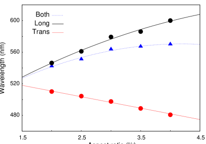
To further our understanding on the contribution from the nano-rod, we have investigated the influence of its size. An interesting observation made was that a redshift occurs in the SPR peak for increasing aspect ratio for the longitudinal mode while for the transverse mode excitation the peak blueshifts. For the unpolarized wave, the peak followed the trend shown by the longitudinal mode (fig 2b). As the Transmission Electron Microscope (TEM) images of Kuldeep et al15 suggest, their nano-rods were parallel to the substrate (‘XZ’ plane) and the absorption spectra were obtained in tranmission mode, implying for unpolarized light both transverse and longitudinal modes exist. The main results of their work was that as the as aspect ratio increases the SPR peaks shifted to lower wavelengths. Which would imply that the transverse mode played a dominant role there.
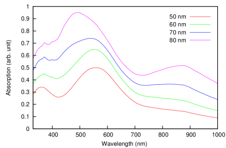
We now proceed to investigate the role of nano-rod bridges in the dumb-bell structure. For this, we have maintained the outer radius of the shells at nm, inner radius at nm while varying the length and radius of the nano-rod. The curves of fig 3 are distinctly different from that shown for a nano-rod without shells on its two sides (fig 2a). Not only has an additional peak appeared in the UV-region, the contributions from the two structures (shells and nano-rod) seem to interact constructively giving resonant absorption peaks whose intensities have increased (comparison shows increase). Such intensity/ field enhancements have been reported 20 with Nie et al showing enhancements due to constructive coupling of SPRs.22
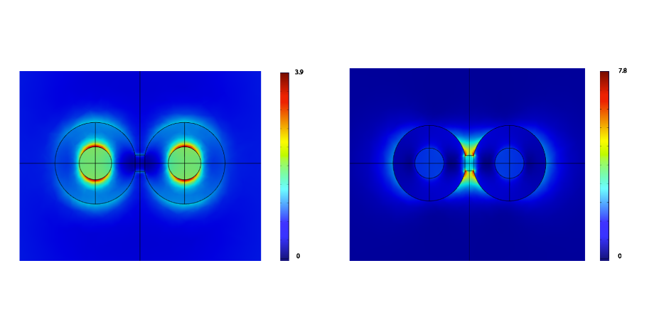
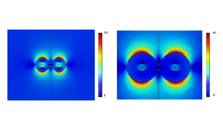
Figure 4 shows the field pattern corresponding to the peaks at UV (fig 4a) and visible (fig 4b) range of the spectra for the transverse modes. As explained in above passages, the visible peak is due to the plasmon resonance along the nanorod, which is enhanced by the two spherical shells, however, notice that the fields are not concentrated only on the cylinder. Infact a coupling of the localized plasmons of the shells along the cylinder is also observed possibly leading to the enhancement in absorption (fig 4b). Fig 4a show that the UV peak is essentially concentrated on the interface. The role of the shell’s thickness and details would be discussed in the next section.
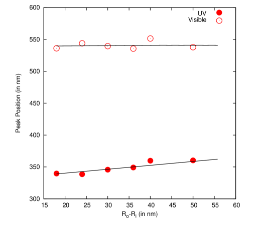
Compiling the results, we have shown the variation of absorption peak intensity and position for both the UV and visible peak with the nano-rod’s aspect ratio. As expected, the peak position () of the SPR’s visible peak shows a linear relation with aspect ratio (Fig 5). Such size dependent shifts (red-shift with increasing grain size) have been well documented.21 However, since the shell dimensions were kept fixed in these simulations, we do not observe any variation in the UV peak’s maxima position.
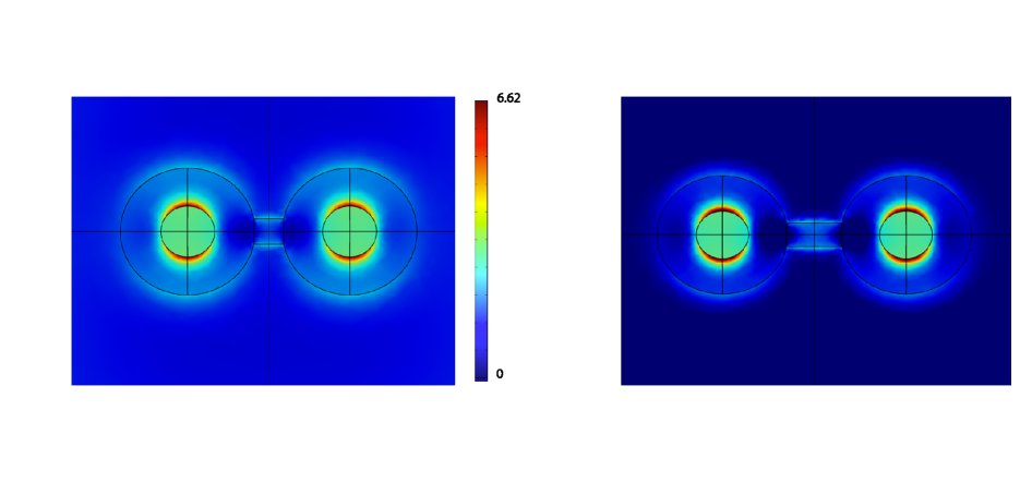
Fig 6 shows that there is a linear relationship between the absorption intensity of two peaks with the aspect ratio. The intensity is due to the amount of oscillating electrons present and hence related to the metal present in the cluster. As the aspect ratio of the nano-rod increases, metal content increases and hence an increase in the absorption intensity. Interestingly, these simulations were done for fixed shell dimensions and yet the contribution of UV absorption by these spheres increases with increasing aspect ratio of the nano-rods. However, one has to remember that the metallic nano-rod provides a conductive path. The existence of conductive path allows for charge transfer plasmons (CTP)23 to the charges distributed on the nano-shells. These charges appear due to charge accumulation from the longitudinal excitation mode. The large accumulation of charges of opposite signs on either sides of the nano-rod gives an enhancement of local fields (fig 4 b).24, 25 Also, the CTP is sensitive to the number of opposite signed charges present on either side of the nano-rod and hence on the nano-rod’s conductivity. Increasing nano-rod length decreases conductive path, leading to charge accumulation and hence enhanced absorption intensity.
3.2 Effect of Core-Shell
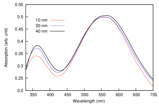
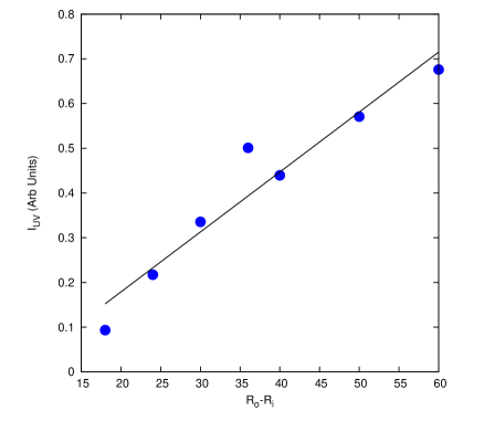
The previous section clearly showed that the shells act as amplifiers, increasing the plasmon oscillations and hence resonant absorption in the visible region caused by the nanorods. The shell also contributes a peak in the UV region whose absorption intensity is influenced by the aspect ratio of nano rod. This section will look into the effect of varying shell size on the absorption patterns. The simulations in this section were done keeping the nano-rod dimensions fixed at nm and varying and , the core-shell dimensions. Fig (7) shows some of the simulated absorption spectra, where we have varied the outer radius of shells (), keeping the radius of core () constant (nm). Alongside this, the compilation of our simulations shows the variation of peak position with the shell thickness. It can be seen that of the visible peak exhibits no shift with change in shell thickness. This shows that the visible SPR’s peak position depends solely on the variation of the nano-rod’s dimension. However, the UV peaks shows a linear relation with with the slope reflecting a red shift with increasing .
Fig (8) shows that there is a linear relation between the absorption intensity of the peak at UV range and the thickness of the metallic Cesium shell, (). As explained above, an accumulation of opposite charges takes place on either side of the conductive nano-rod due to the longitudinal mode of excitation. Thicker shells for a given nano-rod length mean larger accumulation of charges and hence more absorption as is reflected by increased absorption peak intensities. Interestingly, as the shell size increases (beyond nm), a SPR peak appears in the IR region. Oldenburg et al 26 have done extensive work on metal shells and have shown that each surface gives its own plasmon mode. The two modes couple across the shell thickness with the coupling strength based on the dipole model follow trend. Hence, with increase in shell thickness the coupling between the two surfaces, CsBr-Cs and Cs-air, would diminish. We believe this diminished coupling gives rise to the IR peak. We have investigated which surface gives the IR peak using the field pattern. The field pattern for the peak at the IR region is shown in fig 8 for different shell thickness. It can be seen that the IR peak corresponding to the plasmon resonance occur at the outer surface of the shell. It has been observed previously that the plasmon resonance peak of the sphere occurs in the far IR range.27 The above analysis shows that dumb-bell metal nano-structures are highly tunable and suitable for experimentalist who might require controllable geometrical parameters in developing light harvesting sensors etc.
4 Conclusion
We have investigated the localized surface plasmon resonance of a dumb-bell structure consisting of two core-shells connected with a nano-rod. As a primary step, the plasmon resonance of a nano-rod is studied and obtained that the plasmon absorption peak resides in the visible region. The study of the dumb-bell structure revealed that the two nano-shells enhances the absorption in the visible region in addition to contributing a peak in the UV range. As the shell thickness of the dumb-bell structure increased, another peak appeared in the IR region. Tunability of these resonance conditions with various geometrical parameters were appreciated.
Acknowledgement
We would like to acknowledge our gratitude to the University Grants Commission (UGC, Delhi) for its finanical assistance (F.No. 39-531/2010 SR) for carrying out this work. Also, the finanical travel grant given by University of Delhi (Innovation Projects, SGTB-101) is gratefully acknowledged.
References
- 1 Maier, S.A., “Plasmonics: Fundamentals and Applications", Springer (2007).
- 2 Raether, H., “Surface plasmons on smooth and rough surfaces and on gratings", Springer (1988).
- 3 Brongersma, M.L. and Kik, P.G., “Surface Plasmon Nanophotonics", Springer (2007).
- 4 Sarid, D. and Challener, W., “Modern Introduction to Surface Plasmons: Theory, Mathematica Modeling and Applications", Cambridge University Press (2010).
- 5 Maier S.A. and Atwater H.A., J Appl. Phys., 98 (2005) 011101.
- 6 Takele H., Greve H., Pochstein C., Zaporojtchenko V. and Faupel F., Nanotechnology 17 (2006) 3499.
- 7 Rodriguez-Gonzalez, Benito, Attouchi, Farah, Cardinal, Fernanda M., Myroshnychenko, Viktor, Stéphan, Odile, Garcia de Abajo, F. Javier, Liz-Marzán, Luis M., Kociak and Mathieu, Langmuir 28 (2012) 9063.
- 8 Pitarke J.M., Silkin V.M., Chulkov E.V. and Echenique P.M., Reports on Progress in Physics 70 (2007) 1.
- 9 Cao, Linyou, Fan, Pengyu, Vasudev, Alok P., White, Justin S., Yu, Zongfu, Cai, Wenshan, Schuller, Jon A., Fan, Shanhui, Brongersma and Mark L., Nano Letters 10 (2010) 439.
- 10 Wu, Yiying, Fan, Rong, Yang and Peidong, Nano Letters 2 (2002) 2.
- 11 Lee, Jeunghoon, Hasan, Warefta, Stender, Christopher L., Odom and Teri W., Accounts of Chemical Research 41 (2008) 1762.
- 12 Jain P.K. and Deeb C., “Handbook of Molecular Plasmonics", Ed Stefania D’ Agostino, Pan Stanford Publishing (2013).
- 13 Li G., Chen X., Huang L., Wang J., Hu W. and Lu W., Physica B 407 (2012) 2223.
- 14 Wang P, Liu M., Gao G., Zhang S., Shi H., Li Z., Zhang L. anf Fang Y., J. Mater. Chem. 22 (2012) 24006.
- 15 Kuldeep Kumar, P.Arun, Chhaya Ravi Kant and Bala Krishna Juluri, Appl. Phys. Lett. 100 (2012) 243106.
- 16 Palik E.D., “Handbook of Optical Constants of Solids, Volumes I, II, and III: Subject Index and Contributor Index", Academic Press Handbook Series (1985).
- 17 Jain, P.K., Huang W. and El-Sayed M.A., Nano Lett 7 (2007) 2080.
- 18 Gunnarsson L., Rindzevicius T., Prikulis J., Kasemo B., Kall M., Zou S. and Schtz G.C., J. Phys. Chem. B. 109 (2005) 1079.
- 19 Link, Stephan, El-Sayed and Mostafa A., J Phys Chem B 103 (1999) 8410.
- 20 Nie S. and Emory S. R., Science 275 (1997) 1102.
- 21 Cecilia Noguez, J. Phys. Chem. C, 111 (2007) 3806.
- 22 Micheals A.M., Jiang J and Brus L., J. Phys. Chem. B. 104 (2000) 11965.
- 23 Perez-Gonzale, O., Zabala N., Borisov A.G., Halsa N.J., Nordlander P., Aizpurua J., Nano Lett., 10 (2010) 3090.
- 24 Ward D.R., Halas N.J., Ciszek J.W., Tour J.M., Wu Y.P., Nordlander P., Natelson D., Nano Lett., 8 (2008) 919.
- 25 Zhang Z., Weber-Bargioni A., Wu S.W., Dhubey S., Cabrini S., Schuck P.J., Nano Lett., 9 (2009) 4505.
- 26 Oldenburg S.J., Averitt R.D., Westeott S.L. and Halas N.J., Chem. Phys. Lett. 28 (1998) 243.
- 27 Burchianti A, Bogi A., Marinelli C., Maibohm C., Mariotti E., Sanguinetti S. and Moi L., Eur. Phys. J. D., 49 (2008) 201.
Figure Captions
-
1.
Geometry of the dumb-bell structure. Light when incident along the ‘Y’-axis show excitation of the transverse mode due to the electric field component, , while contributes to excitation of the longitudinal mode.
-
2.
a) Variation of absorption with wavelength for a nanorod, and b) the variation in LSPR’s with aspect ratio of the nanorod.
-
3.
As an example we show simulated spectra of absorption with wavelength for different values of length of the rod connecting the shells.
-
4.
Field distribution in the plane for the structure for a) UV and b) visible peak.
-
5.
Figures shows the variation of the UV and visible peak’s position with metal Cesium nano-rod’s aspect ratio (l/r).
-
6.
Figure shows the variation of the UV and visible absorption peak’s intensity with nano-rod’s aspect ratio. Alongside the field distribution in the XZ plane is shown for two different nano-rod lengths.
-
7.
The simulated absorption spectra for increasing Cesium shell thickness keeping aspect ratio/ length of the nano-rod constant. Compiling the results of UV and visible peak positions () of the simulated spectra show no variation in visible peak position while UV’s peak position shows a steady red-shift.
-
8.
Figure shows the variation of the UV absorption peak’s intensity and the field distribution in the plane for the structure for outer radius of nm and nm.