Physarum wires:
Self-growing self-repairing smart wires
made from slime mould
Abstract.
We report experimental laboratory studies on developing conductive pathways, or wires, using protoplasmic tubes of plasmodium of acellular slime mould Physarum polycephalum. Given two pins to be connected by a wire, we place a piece of slime mould on one pin and an attractant on another pin. Physarum propagates towards the attract and thus connects the pins with a protoplasmic tube. A protoplasmic tube is conductive, can survive substantial over-voltage and can be used to transfer electrical current to lightning and actuating devices. In experiments we show how to route Physarum wires with chemoattractants and electrical fields. We demonstrate that Physarum wire can be grown on almost bare breadboards and on top of electronic circuits. The Physarum wires can be insulated with a silicon oil without loss of functionality. We show that a Physarum wire self-heals: end of a cut wire merge together and restore the conductive pathway in several hours after being cut. Results presented will be used in future designs of self-growing wetware circuits and devices, and integration of slime mould electronics into unconventional bio-hybrid systems.
Keywords: bio-wires, routing, slime mould, bio-electronics, unconventional computing
1. Introduction
The plasmodium of Physarum polycephalum (Order Physarales, class Myxomecetes, subclass Myxogastromycetidae) is a single cell, visible with the naked eye, with many diploid nuclei. The plasmodium feeds on bacteria and microscopic food particles by endocytosis. When placed in an environment with distributed sources of nutrients the plasmodium forms a network of protoplasmic tubes connecting the food sources. In 2000 Nakagaki et al [42] showed that the topology of the plasmodium’s protoplasmic network optimizes the plasmodium’s harvesting of nutrient resource from the scattered sources of nutrients and makes more efficient the transport of intra- cellular components. In [7] we have shown how to construct specialised and general purpose massively-parallel amorphous computers from the plasmodium (slime mould) of P. polycephalum that are capable of solving problems of computational geometry, graph-theory and logic.
Plasmodium’s foraging behaviour can be interpreted as a computation [42, 39, 40]: data are represented by spatial of attractants and repellents, and results are represented by structure of protoplasmic network [7]. Plasmodium can solve computational problems with natural parallelism, e.g. related to shortest path [39] and hierarchies of planar proximity graphs [3], computation of plane tessellations [47], execution of logical computing schemes [52, 5], and natural implementation of spatial logic and process algebra [49].
In the framework of our “Physarum Chip” EU project [10] we aim to experimentally implement a working prototype of a Physarum based general purpose computer. This computer will combine self-growing computing circuits made of a living slime mould with conventional electronic components. Data and control inputs to the Physarum Chip will be implemented via chemical, mechanical and optical means.
Aiming to develop a component base of future Physarum computers we designed Physarum tactile sensor [9] and undertook foundational studies towards fabrication of slime mould chemical sensors (Physarum nose) [19, 55]. We have uncovered memristive properties of the slime mould [21]: Physarum memristors could be a basic (logically universal) components of future slime mould computers.
To transfer data, elements of a program code and control signals in Physarum computers we must develop a reliable self-growing and self-healing conductive pathways made of the slime mould — Physarum wires.
In present paper we outline our scoping experimental results on evaluating properties of protoplasmic tubes as electrical wires. Our finding discussed in the paper complement previous studies on fabrication of organic wires and using living substrates to grow metallised conductive pathways: self-assembling molecular wires [54, 44], DNA wires [16], electron transfer pathways in biological systems [32], live bacteria templates for conductive pathways [17], bio-wires with cardiac tissues [18], golden wires with templates of fungi [45].
The paper is structured as follows. We outline experimental techniques in Sect. 2. Section 3 presents experimental results on resistivity of Physarum wires, behaviour of the wires under electrical load and potential division. In Sect. 4 we show how to route Physarum wires with chemo-attractants, chemo-repellents and electrical fields. An ability of Physarum wires to restore their integrity after damage is experimentally proved in Sect. 5. Section 6 exemplifies growth of Physarum wires on electronic boards. Insulation of Physarum wires is studied in Sect. 7. Section 8 discusses pros and cons of conductive pathways made from Physarum’s protoplasmic tubes.
2. Methods


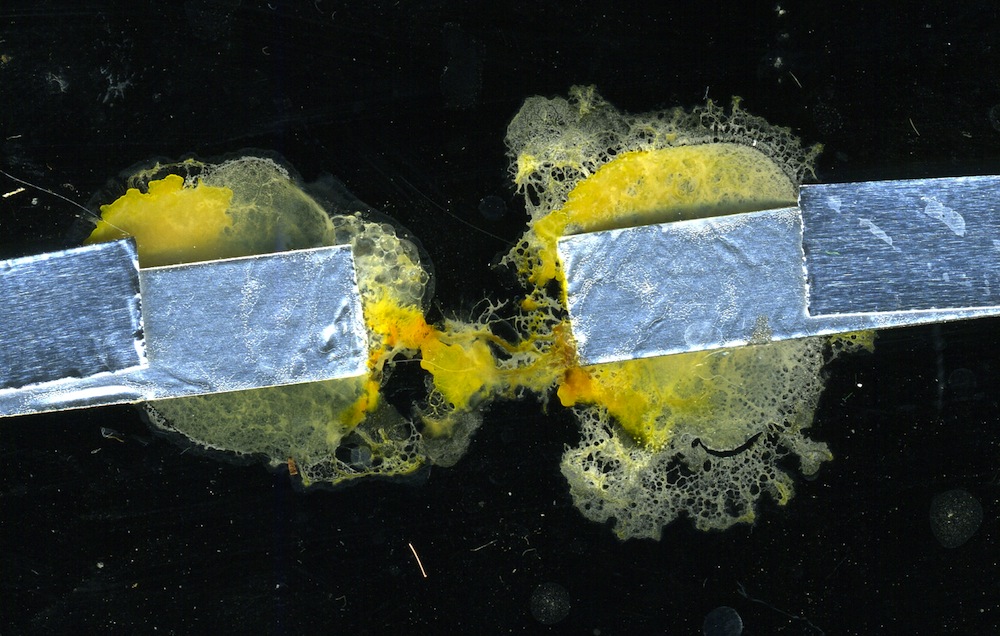
Plasmodium of Physarum polycephalum was cultivated in plastic lunch boxes (with few holes punched in their lids for ventilation) on wet kitchen towels and fed with oat flakes. Culture was periodically replanted to a fresh substrate. Electrical activity of plasmodium was recorded with ADC-24 High Resolution Data Logger (Pico Technology, UK). A scheme of experimental setup is shown in Fig. 1. Two blobs of agar 2 ml each (Fig. 1b) were placed on electrodes (Fig. 1c) stuck to a bottom of a plastic Petri dish (9 cm). Distance between proximal sites of electrodes is always 10 mm. Physarum was inoculated on one agar blob. We waited till Physarum colonised the first blob, where it was inoculated, and propagated towards and colonised the second blob. When second blob is colonised, two blobs of agar, both colonised by Physarum (Fig. 1a), became connected by a single protoplasmic tube (Fig. 1d). Resistivity and voltage over 2.5 V were measured on TTi 1604 Digital Multimeter; Physarum extracellular potential was measured using PicoTech ADC-20/24 logger. Voltage and current supplied to hybrid circuits incorporating Physarum wires was supplied using Iso-Tech IPS 4303 laboratory DC power supply unit. When growing Physarum on breadboard we made a layer of agar on one side of the board, to keep humidity higher, and inoculated Physarum on other side of the board. After Physarum reached its destination we removed the agar layer, thus no conductive substance apart of slime mould present on the breadboard. Images of Physarum were made using Fuji FinePix S6500 camera, EpsonPerfection 4490 scanner and DinoLite Computer Microscope.
3. Resistivity, overload and transfer function
In 25 experiments we measured resistance and calculated resistivity of protoplasmic tubes on agar blobs. In calculations we assumed length of a tube is 1 cm, and diameter is 0.03 cm. We found that minimum resistance recorded is 80 cm, maximum resistance is 2560 cm, median 625 cm, and average 825 cm. Resistivity of Physarum substantially varies from one experiment to another: standard deviation calculated is 776, which is just slightly below average of Physarum wire resistivity resistivity. Average resistivity of Physarum protoplasmic tubes is of the same rank as resistivity of a cardiac muscle of a dog, and skeletal muscles of a dog and a human [22].

An undisturbed Physarum exhibits more or less regular patterns of oscillations of its surface electrical potential. The electrical potential oscillations are more likely controlling a peristaltic activity of protoplasmic tubes, necessary for distribution of nutrients in the spatially extended body of Physarum [46, 24]. A calcium ion flux through membrane triggers oscillators responsible for dynamic of contractile activity [37, 20]. It is commonly acceptable that the potential oscillates with amplitude of 1 to 10 mV and period 50-200 sec, associated with shuttle streaming of cytoplasm [25, 26, 27, 37]. In our experiments we observed sometimes lower amplitudes because there are agar blobs between Physarum and electrodes and, also, electrodes were connected with a protoplasmic tube only. Exact characteristics of electric potential oscillations vary depending on state of Physarum culture and experimental setups [1].
In addition, low amplitude oscillations, in our experimental setup (two electrodes with agar blobs and Physarum on top) a potential difference between electrodes observed was usually 20-25 mV, and never exceeding 200 mV. The Physarum potential even in extreme situations, is low. Thus by applying over 1 V to a protoplasmic tube we cause over-voltage. How does Physarum reacts to over-voltage, will its protoplasmic tube disintegrate? We have conducted 12 experiments on incorporating Physarum living wire into a circuit which includes an array of 6 LEDs (15 V white 10,000 Mcd). In each experiment we applied c. 19 V to the circuit till the LED array was lighting bright. Potential on LED registered was 4.4-4.8 V and current 11-13 A. In all experiments Physarum wire was functioning for 24 h without loss of integrity (Fig. 2a).
Input voltage stayed unchanged during 24 h yet after 24 h functioning as wires under load protoplasmic tubes decreased their resistivity: voltage registered on the LED array was 10.2 V and current running 3.9 A. The decrease of resistivity is possibly due to increase of the overall mass of the Physarum wires, and, in some cases, growth of additional branches of tubes between the agar blobs. Typically, after one day of functioning under electrical load agar blobs start to dry out and Physarum gradually goes in the stage of sclerotisation (Fig. 2a).


Potential recorded on a Physarum wire is a fraction of a potential applied to the Physarum wire. That is a Physarum acts as a potential divider: see scheme of the circuit in Fig. 3a. The transfer function is linear (Fig. 3b), subject to usual fluctuations of Physarum impedances in laboratory experiments (Fig. 3c).
4. Routing Physarum wires
Growing Physarum circuits can be controlled by white [23] and blue [7] light, chemical gradients [33, 7], temperature gradients [56] and electrical fields [53]. In present section we evaluate routing in conditions far from an ‘idea experiment on slime mould taxis’. We grow and control Physarum grows on an almost bare breadboards.
4.1. Routing with chemical fields
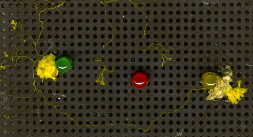
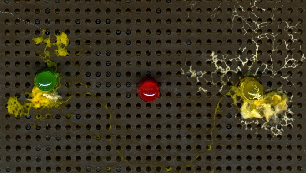

Results of two experiments are presented in Fig. 4. A task in both experiments was to route a Physarum wire from a position of yellow LED to a position of green LED but avoiding a position of red LED . In both experiments Physarum was inoculated nearby . An oat flake was placed east of . Chemoattractants released either by the oat flake or by bacteria colonising it diffused in the air and attracted Physarum. To prevent the slime mould going nearby we placed a grain of salt near . The salt absorbed water from humid environment of the experimental setup and diffused in the agar layer underlying the breadboard. Physarum is repelled by high concentration of salt in its substrate. Thus the Physarum moved towards attracting and, at the same time, avoided repelling (Fig. 4c).
After the Physarum wire was established and got into direct contact with pins of and we measured an electrical potential between the pins. Two experiments are illustrated in Fig. 4. In experiment shown in Fig. 4a) a protoplasmic tube connecting the and exhibited potential 37-40 mV, and in experiment shown in Fig. 4b the tube’s potential was 40-45 mV. These conform well with natural variance of potential in protoplasmic tubes of P. polycephalum. Resistivity measured between the pins was 1300-1500 cm, in a range of electrical resistivity of biological tissue [22].
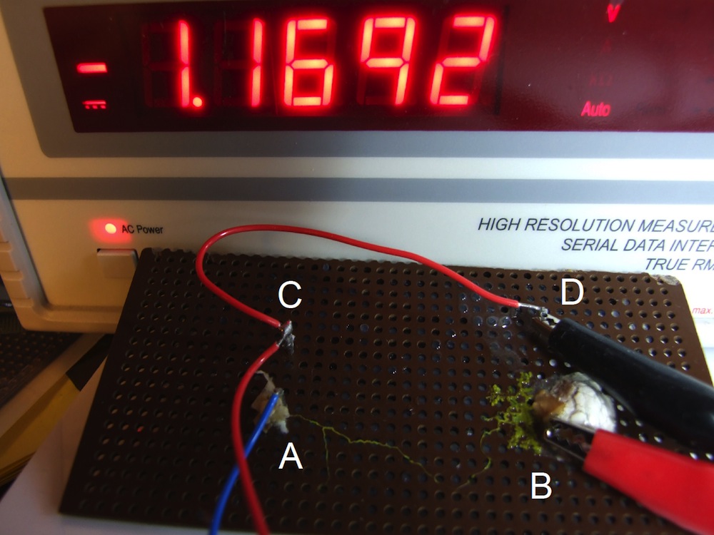
An example of routing with non-food chemo-attractants is shown in Fig. 5. To complete a circuit we needed to grow a Physarum wire between points ’A’ and ’B’. We inoculated Physarum so it was in a direct contact with pin ’A’ and placed half a pill of a valerian-containing herbal remedy Kalms, see details in [8], nearby pin ’B’. Being attracted to valerian Physarum propagated from pin ’A’ to pin ’B” and forms a conductive pathways between the pins. A difference of a membrane potential of the protoplasmic tube between ’A’ and ’B’ sites was in a range of 50 mV. When 8.5 V DC applied to inputs ’A’ an ’C’ of the hybrid circuit (Fig. 5) a potential slightly below 1.2 V was recorded between pins ’D’ and ’B’.
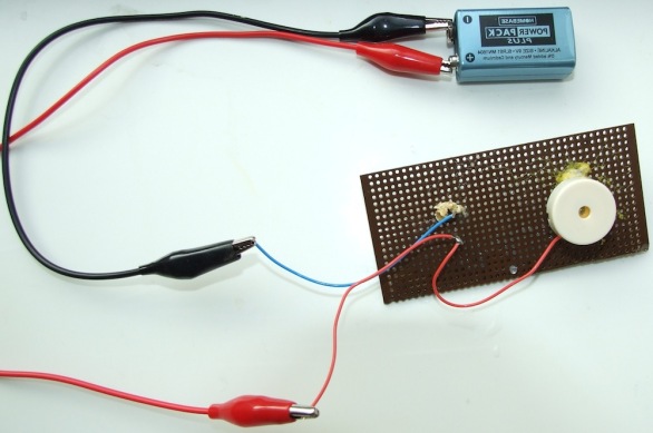
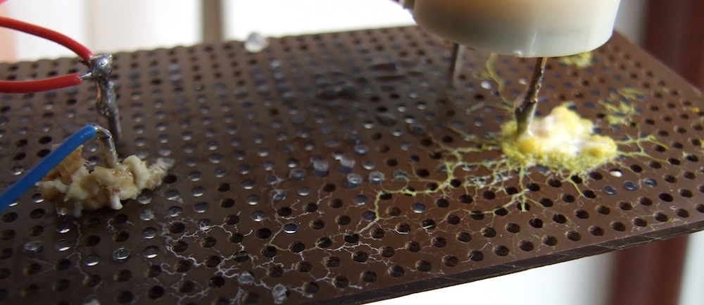
In an experiment illustrated in Fig. 6 Physarum developed a conductive pathway in a circuit including a DC voltage supply and a piezo audio transducer (Kingstate KPEG158-P5, operational for 1-20 V, current consumption max 7 A). On applying 8 V DC to inputs of the circuit we registered 2.2 V potential on the piezo transducer’s pins. The transducer produced sound near 30 dB for a minimum duration of 10 min. Physarum wire remained alive and did not change its morphology during the circuit’s operational mode.
4.2. Routing with electric fields
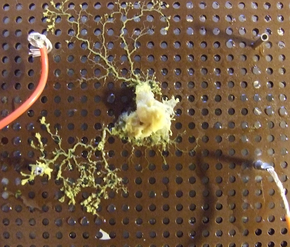
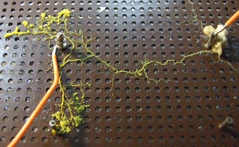
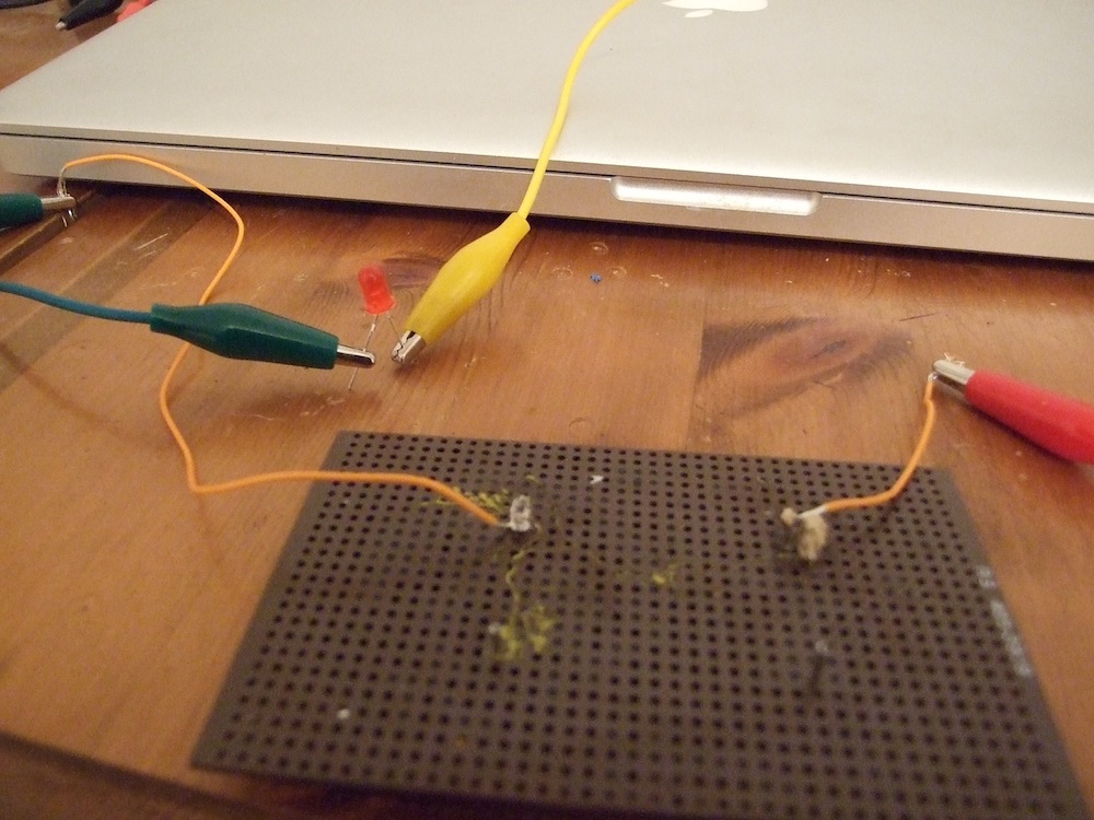
Controlling growth of Physarum with chemo-attractants and repellents is proved to be a reliable method of routing living wires. However, direct contact of attractants/repellents with growth and Physarum itself is undesirable: the substrate might become contaminated with the chemicals preventing further re-routing of the wires. Also, chemicals diffusing in a substrate might affect functioning of existing Physarum wires. Non-invasive control techniques, as e.g. with light [4] are appropriate yet unreliable and do not always give a predictable control of the growing slime mould. The Physarum plasmodium is known to shown negative galvanotaxis: the slime mould migrates towards cathode under an electric field [12, 13]. A fine pattern of Physarum growth can be achieved by culturing the slime mould on an array of probes, where direction, migration and obstacle avoidance behaviour of Physarum could be guided by the spatial distribution of electric currents formed by the probes [53].
To evaluate galvanotaxis based routing of Physarum wires in a conditions more close to realities of circuit design we undertook 15 experiments with Physarum wires growing on a breadboard with a limited agar substrate: agar plates were fixed underneath the breadboards, thus Physarum was only able to contact agar gel through the holes in the breadboards. In each experiment we placed 2-3 oat flakes colonised by Physarum onto a breadboard with several pins. Two of the pins were connected to a 1.6 V battery.
We found that in 10 of 15 experiments the slime mould propagated to cathode. Two experiments are illustrated in Fig. 7. In experiment Fig. 7a Physarum was inoculated between the pins. In experiment Fig. 7b the slime mould was placed nearby anode. In both experiments controllable propagation to cathode is recorded. Conductivity of Physarum wire was sufficient to light up a LED by applying 9 V DC to the circuit (Fig. 7c).
5. Self-repair of Physarum wires
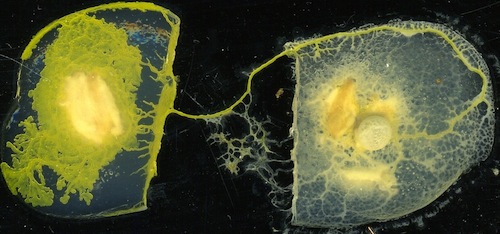
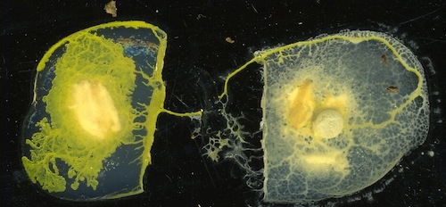
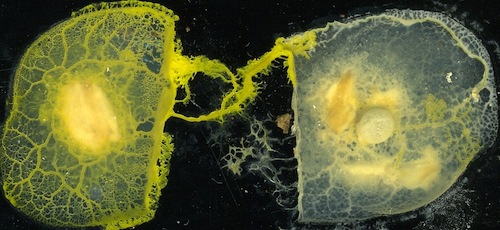
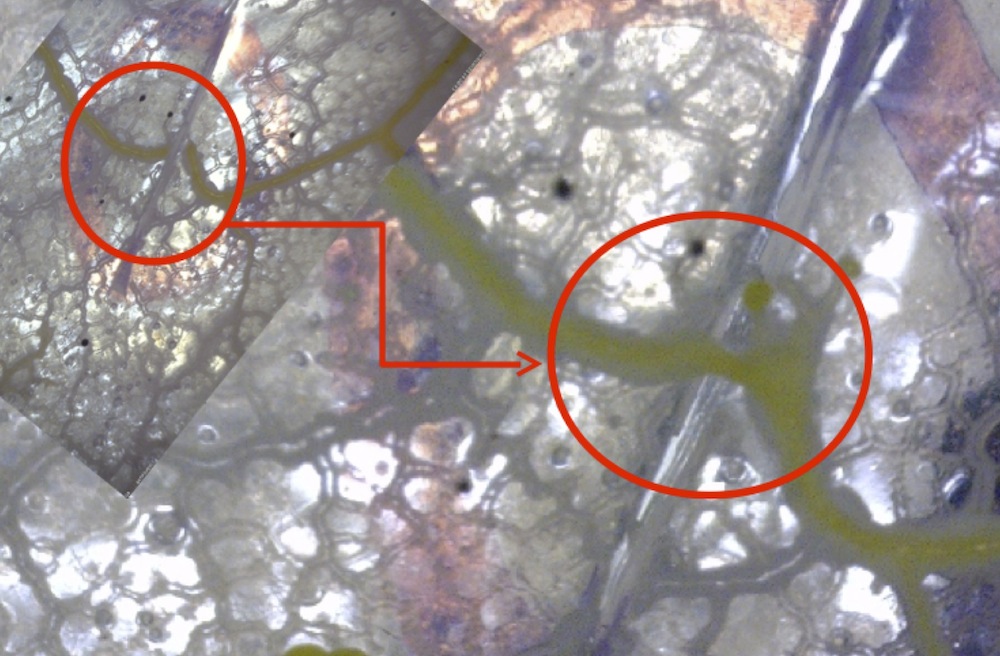
Physarum wires can self-repair after a substantial damage. In 12 experiments we found that after part of a protoplasmic tube is removed (1-2 mm segment) the tube restores its integrity in 6-9 hr. Typically, a cytoplasm from cut-open ends spills out on a substrate. Each spilling of cytoplasm becomes covered by a cell wall and starts growing. In few hours growing parts of the tube meet with each other and merge.
An example of tube’s self-repair is shown in Fig. 8a–c. In Fig. 8a we see an undamaged tube connecting two agar blobs. The tube propagates on a bare plastic substrate. A segment of the tube is removed, see Fig. 8b. In approximately 8 hr after the damage the tube restores its integrity, see Fig. 8c.
Growing ends of a damaged tube not necessarily meet up each other exactly when developing on a bare plastic substrate. However, exact merging of the ends is almost always the case when tube is resting on an agar substrate. See example in Fig. 8d. In 2-3 hr after a tube was cut (top left insert in Fig. 8d) active growth zones are formed at the ends of the tube. They propagate towards each other and merge (main photo in Fig. 8d).

Restoration of tubes conductivity as a result self-repair was confirmed by electrical measurements, see Fig. 9. A potential 4 V DC was applied to a Physarum wire (10 mm length protoplasmic tube as in all previous setups). Under the applied voltage the wire showed average potential 1.9 V, moving between 1.5 V to 2.3 V. 150 min after start of recording we destroyed parts of the wire: 1.8 mm of protoplasmic tube was removed from each wire. The wire ceased to be conductive. This was reflected in a sharp voltage drop (Fig. 9).
Several hours after being cut the protoplasmic tube self-healed and formed a fully functioning tube again. The Physarum wire started to return to its conductive state 440 min after being damaged and returned to its fully conductive state in next 230 min: it shows 1.8 V, oscillating between 1.6 V abd 2.1 V when 4 V DC is applied (Fig. 9).
6. Propagating on electronic boards
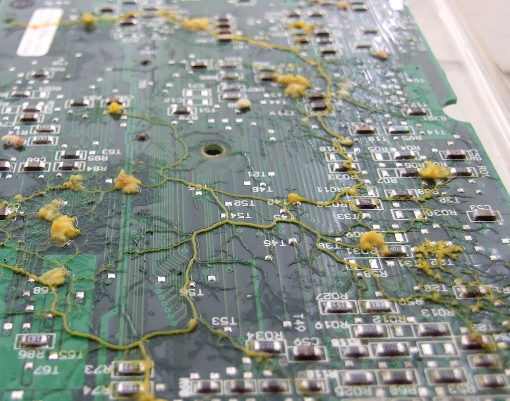
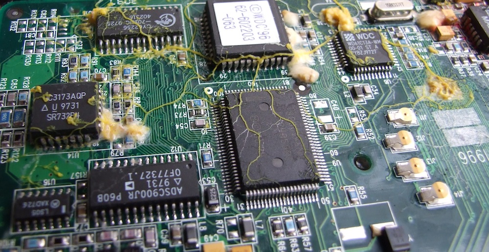
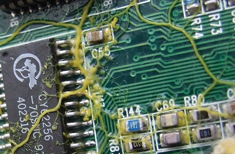
To evaluate how well Physarum propagates on a bare surface of electronic components and assemblies we conducted experiments with electronic boards (Fig. 10). Typically, a slime mould was inoculated at one edge of the board, e.g. further edge in Fig. 10a, and oat flakes were scarcely distributed on the board to attract Physarum to certain domains of the boards. The oat flakes generated chemo-attractive fields to guide Physarum wires towards imaginary pins. The boards were kept in a container, with a shallow water on the bottom to keep humidity very high. The boards with Physarum were not in direct contact with the water.
The slime mould propagated on the boards with a speed of between 1 mm to 5 mm per hour. Physarum propagated satisfactory on both sides of the boards, and usually spanned a planar set of oat flakes with networks of protoplasmic tubes ranging from spanning trees to their closures into -skeletons (Fig. 10ab). As shown in Fig. 10c a width of protoplasmic wires grown by Physarum is comparable with a width of conductive pathways on the computer boards.
7. Insulating Physarum wires
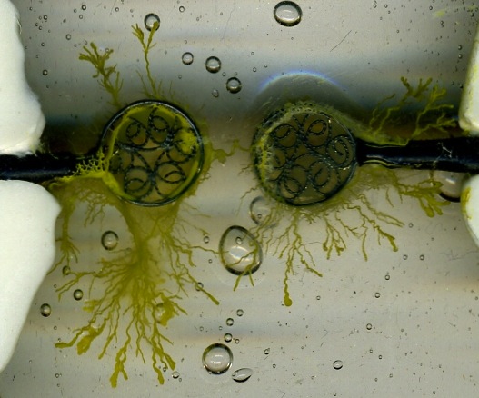
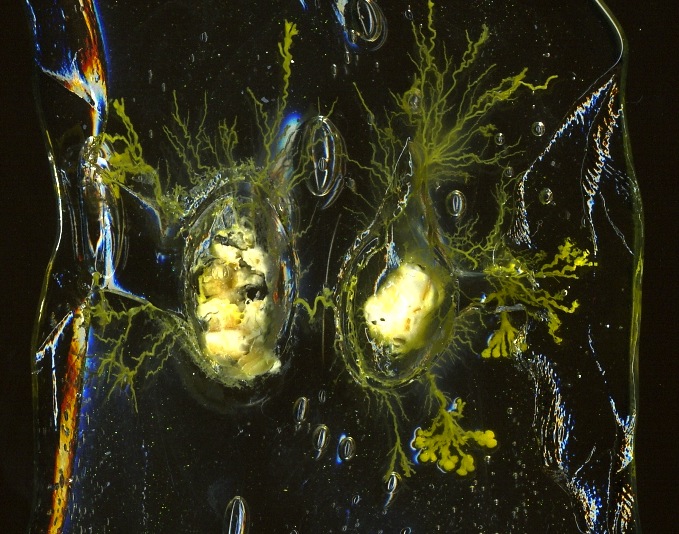
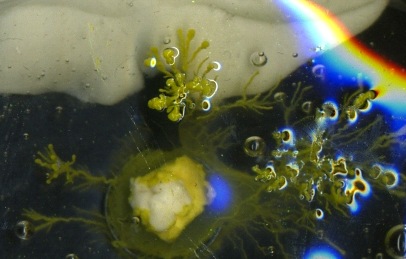
We successfully tested insulation of Physarum wires with octamethylcyclotetrasiloxane (Silastic 4-2735 Silicone Gum, Dow Corning S.A., B-7180 Seneffe, Belgium). We will use name D4 for brevity. The D4 is a liquid at normal temperature and pressure; its melting point is around 17-18 Co [36]. Its density is about 0.96 g/cm3 [36]. In experiments we gently poured D4 over Petri dish with probes and Physarum until a layer of D4 covered all objects on the bottom of the Petri dish (Fig. 11a).
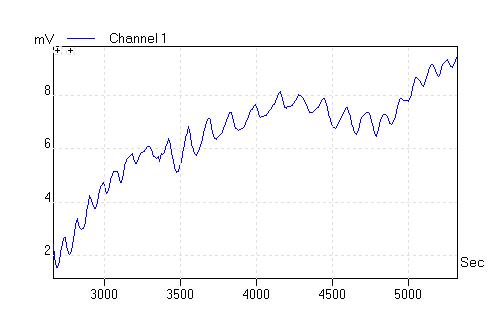
Physarum becomes completely encapsulated in the silicon insulator. When a slab of D4 removed from the Petri dish Physarum remains inside (Fig. 11b). Physarum survived inside D4 for hours. Figure 11c shows electrical activity of Physarum 2 hours after it was immersed into D4: the slime mould exhibits ’textbook classical’ oscillations of its electrical potential with amplitude around 1 mV and period 145 sec. Coincidently, the width of D4 cover is thinner on top of probes. There Physarum forms vertically ascending protrusions which reach surface of the D4 and thus Physarum is capable for intake oxygen and keeping the protoplasmic tube connecting probes alive (Fig. 11d).
8. Discussions
Growing wires from slime mould is a perspective direction of research in bio-inspired novel computing substrates. In experimental laboratory studies we shown that
-
•
Physarum’s protoplasmic tubes remains functioning and conductive under reasonable high load and capable to act as wires (Physarum wires) in electrical circuits including lightning and actuating devices.
-
•
Physarum wires can be routed on various types of substrates using chemo-attractants, chemo-repellents and electromagnetic fields.
-
•
Physarum wires can be insulated with silicon. The wires remain alive and functioning while covered in insulator for days.
-
•
Physarum can self-repair. When a Physarum wire is cut the damaged ends grow towards each other and merge, thus restoring integrity of the original wire.
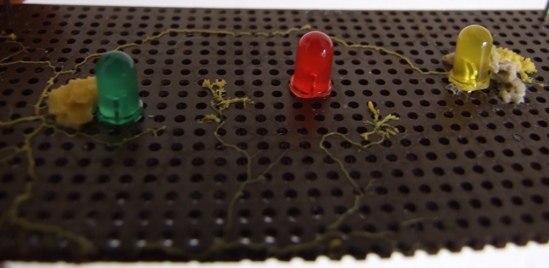
Using living protoplasmic tubes as wires suffer from few disadvantages. Physarum is always in motion and newly developed protoplasmic tubes can interfere with existing tube-wires. For example, in experiment shown in Fig. 12 Physarum grown a protoplasmic wire between two LEDs: the wire is visible near the farthest edge of the board. Few hours after the wire formed, the slime mould continued its foraging behaviour and sprouted two undesired protoplasmic tubes. These tubes grown near the closest edge of the board as seen in Fig.. 12. We should find reliable ways of inhibiting sprouting after all required wires are formed.
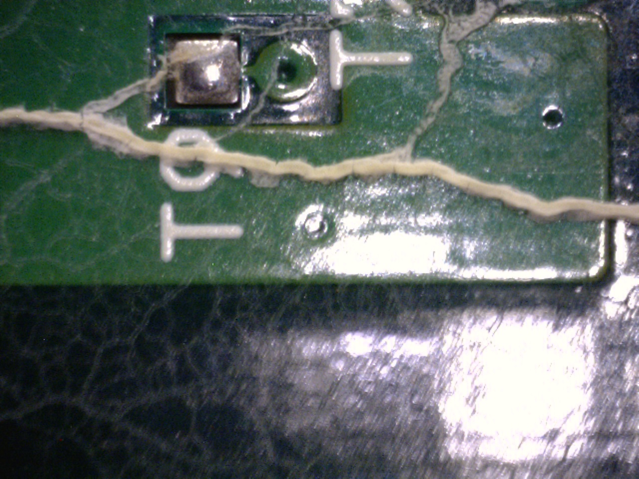
In present paper we considered living Physarum wires. Their life time is limited. Typically in 5-7 days, may be longer, depending on humidity, a protoplasmic tube becomes abandoned (Fig. 13). After becoming abandoned the tube keeps, up to some degree, its water contents, and thus conductivity, for few more days and then dries up and its walls collapse.
Physarum wires are volatile. Circuits with the protoplasmic wires can function for days but after a week at most Physarum might migrate away, or get into a sclerotium phase (if low humidity) or fructify (if exposed to light) or just get colonised by some other moulds and vanish. How to make Physarum wires long-lasting? When the slime mould develops a network of protoplasmic tubes spanning sources of nutrients, the cell maintains its integrity by pumping nutrients and metabolites between remote parts of its body via cytoplasmic streaming [50, 11, 41, 28]. The cytoplasmic streaming could be employed for the transportation of bio-compatible substances inside the protoplasmic network. In [6] we demonstrated that the plasmodium of P. polycephalum consumes various coloured dyes and distributes them in its protoplasmic network. By specifically arranging a configuration of attractive (sources of nutrients) and repelling (sodium chloride crystals) fields we can program the plasmodium to implement the following operations: to take in specific coloured dyes from the closest coloured oat flake; to mix two different colours to produce a third colour; and, to transport colour to a specified locus of an experimental substrate. Transportation of colourings per se is of little interest but shows the potential of P. polycephalum as a programmable transport medium. To employ the slime mould’s potential of internalisation and re-distribution of foreign particles for the development of a long-life wires we must consider suitable functional materials. In [35], inspired by our previous results [6] and studies on cellular endocytosis of magnetic nano-beads [34] and fluorescent nano-beads [14], and nanowire scaffolding for living tissue [51], we demonstrated internalisation and transport of two types of functional materials: magnetic nano-particles and glass spheres coated with silver. The particles could be redistributed inside the Physarum body and indeed ’spread’ along protoplasmic tubes. Initial results, rather a proof of loading, are reported by us in [35]. Final adjustments to a distribution of magnetic particles can be controlled using magnetic twizzers [15]. Mineralisation of grown Physarum wires to make them permanent wires will be a continued topic of further studies.
References
- [1] Acheubach U. and Wohlfarth-Bottermann K.E. Synchronization and signal transmission in protoplasmic strands of Physarum. Planta 151 (1981) 574–583.
- [2] A. Adamatzky, Developing proximity graphs by Physarum Polycephalum: Does the plasmodium follow Toussaint hierarchy? Parallel Processing Letters 19 (2008), 105–127.
- [3] A. Adamatzky, Developing proximity graphs by Physarum Polycephalum: Does the plasmodium follow Toussaint hierarchy? Parallel Processing Letters 19 (2008), 105–127.
- [4] Adamatzky, A. (2009). Steering plasmodium with light: Dynamical programming of Physarum machine. Arxiv preprint arXiv:0908.0850.
- [5] A. Adamatzky, Slime mould logical gates, arXiv:1005.2301v1 [nlin.PS] (2009)
- [6] Adamatzky A. Manipulating substances with Physarum polycephalum. Materials Science and Engineering: C 30 (2010) 1211-1220.
- [7] Adamatzky A. Physarum Machines (World Scientific, 2010).
- [8] Adamatzky A., Simulating strange attraction of acellular slime mould Physarum polycephalum to herbal tablets. Math Comput Modelling (2011).
- [9] Adamatzky A. Slime mould tactile sensor. Sensors and Actuators B (2013), in press.
- [10] Adamatzky A., Erokhin V., Grube M., Schubert T., Schumann A. Physarum Chip Project: Growing Computers From Slime Mould. Int J Unconventional Computing 8 (2012) 319–323.
- [11] Allen R.D., Pitts W.R., Speir D. and Brault J. Shuttle-streaming: synchronization with heart production in slime mold. Science. 1963 (13) 1485–1487.
- [12] Anderson, J. D. (1951). Galvanotaxis of slime mold. The Journal of General Physiology, 35 (1951), 1.
- [13] Anderson, J. D. Potassium loss during galvanotaxis of slime mold. The Journal of General Physiology, 45 (1962) 567–574.
- [14] Bandmann V, M ller JD, Köhler T, Homann U. Uptake of fluorescent nano beads into BY2-cells involves clathrin-dependent and clathrin-independent endocytosis. FEBS Lett. 2012 Oct 19;586(20):3626-32.
- [15] Barbic M. Magnetic wires in MEMS and bio-medical applications J. Magnetism and Magnetic Materials 249 (2002) 357–367.
- [16] Beratan D. N., Priyadarshy S., Risser S. M. DNA: insulator or wire? Chemistry & Biology 4 (1997) 3–8.
- [17] Berry V. and Saraf R.F. Self-Assembly of nanoparticles on live bacterium: An avenue to fabricate electronic devices. Angewandte Chemie 117 (2005) 6826–6831.
- [18] Cingolani E., Ionta V., Giacomello A., Marbán E., Cho H.C. Creation of a biological wire using cell-targeted paramagnetic beads. Biophysical Journal 102 (2012) 416a.
- [19] De Lacy Costello B., Adamatzky A. Assessing the chemotaxis behavior of Physarum Polycephalum to a range of simple volatile organic chemicals. Communicative & Integrative Biology 6:5, e25030; September/October 2013.
- [20] Fingerle J. and Gradmann D. Electrical properties of the plasma membrane of microplasmodia of Physarum polycephalum J. Membrane Biol. 68 (1982) 67–77.
- [21] Gale E., Adamatzky A., De Lacy Costello B. Are slime moulds living memristors? (2013) arXiv:1306.3414 [cs.ET]
- [22] Geddes L.A. and Baker L.E. The specific resistance of biological material — A compendium of data for the biomedical engineer and physiologist. Med. & BioL Engng. 5 (1967) 271–293.
- [23] Hader, D. P., and Schreckenbach, T. (1984). Phototactic orientation in plasmodia of the acellular slime mold, Physarum polycephalum. Plant and cell physiology, 25(1), 55.
- [24] Heilbrunn L. V. and Daugherty K. The electric charge of protoplasmic colloids. Physiol. Zool. 12 (1939) 1–12.
- [25] Iwamura T. Correlations between protoplasmic streaming and bioelectric potential of a slime mould, Physarum polycephalum. Botanical Magazine 62 (1949) 126–131.
- [26] Kamiya N. and Abe S. Bioelectric phenomena in the myxomycete plasmodium and their relation to protoplasmic flow. J Colloid Sci 5 (1950) 149–163.
- [27] Kashimoto U. Rhythmicity on the protoplasmic streaming of a slime mold, Physarum Polycephalum. I. A statistical analysis of the electric potential rhythm. J Gen Physiol 41 (1958) 1205–1222.
- [28] Hulsmann N. and Wohlfarth-Bottermann K.E. Spatio-temporal relationships between protoplasmic streaming and contraction activities in plasmodial veins of Physarum polycephalum. CytoViologie. 1978 (17) 317-334.
- [29] Johnsen G.K., L tken C.A., Martinsen O.G., Grimnes S. Memristive model of electro-osmosis in skin Phys Rev E Stat Nonlin Soft Matter Phys. 83 (2011) 031916.
- [30] Kosta S.P., Kosta Y.P. , Bhatele M., Dubey Y.M., Gaur A., Kosta S., Gupta J., Patel A., Patel B. Human blood liquid memristor Int. J. of Medical Engineering and Informatics 3 (2011) 16–29.
- [31] Kosta S.P., Kosta Y.P., Gaur A., Dube Y.M., Chuadhari J.P., Patoliya J., Kosta S., Panchal P., Vaghela P., Patel K., Patel B., Bhatt R., Patel V. New Vistas of Electronics Towards Biological (biomass) sensors International Journal of Academic Research 3 (2011) 511–526.
- [32] Katz E. Bioelectronics. In: Bassani F., Liedl G. L., and Wyder P. (Eds.), Encyclopedia of Condensed Matter Physics, Oxford, 2005, 85–98.
- [33] Knowles, D. J. C., and Carlile, M. J. (1978). The chemotactic response of plasmodia of the myxomycete Physarum polycephalum to sugars and related compounds. Journal of General Microbiology, 108(1), 17.
- [34] Li HS, Stolz DB, Romero G. Characterization of endocytic vesicles using magnetic microbeads coated with signalling ligands. Traffic. 2005 Apr;6(4):324-34
- [35] Mayne R., Patton D., de Lacy Costello B., Patton R. C., Adamatzky A. On loading slime mould Physarum polycephalum with metallic particles Submitted (2013).
- [36] Merck, 1996, The MERCK Index. An Encyclopedia of Chemicals, Drugs, and Biologicals, 12th Edn. Whitehouse Station, NJ. MERCK and CO. Inc.
- [37] Meyer R. and Stockem W. Studies on microplasmodia of Physarum polycephalum V: Electrical activity of different types of micro- and macroplasmodia. Cell Biol. Int. Rep. 3 (1979) 321–330.
- [38] T. Nakagaki, H. Yamada, A. Toth, Maze-solving by an amoeboid organism, Nature 407 (2000), 470–470.
- [39] T. Nakagaki, H. Yamada, and A. Toth, Path finding by tube morphogenesis in an amoeboid organism, Biophysical Chemistry 92 (2001), 47–52.
- [40] T. Nakagaki, M. Iima, T. Ueda, Y. Nishiura, T. Saigusa, A. Tero, R. Kobayashi, K. Showalter, Minimum-risk path finding by an adaptive amoeba network, Physical Review Letters 99 (2007), 068104.
- [41] Newton S.A., Ford N.C. Jr, Langley K.H. and Sattelle D. B. Laser light- scattering analysis of protoplasmic streaming in the slime mold Physarum polycephalum. Biochim Biophys Acta. 1977 (496) 212-224.
- [42] T. Nakagaki, H. Yamada, A. Toth, Maze-solving by an amoeboid organism, Nature 407 (2000), 470–470.
- [43] Palleau E., Reece S., Desai S. C. Smith M. E., Dickey M. D. Self-healing stretchable wires for reconfigurable circuit wiring and 3D microfluidics. Advanced Materials 25 (2013) 1589–1592.
- [44] Paul F. and Lapinte C. Organometallic molecular wires and other nanoscale-sized devices: An approach using the organoiron (dppe)Cp∗Fe building block Coordination Chemistry Reviews 178-180 (1998) 431–509.
- [45] Sabah A., Dakua I., Kumar P., Mohammed W. S., Dutta J., Growth of templated gold microwires by self organization of colloids on Aspergillus Niger. Digest J. of Nanomaterials and Biostructures. 7 (2012) 583–591.
- [46] Seifriz W. A theory of protoplasmic streaming. Science 86 (1937) 397–402.
- [47] T. Shirakawa, A. Adamatzky, Y.-P. Gunji, Y. Miyake, On simultaneous construction of Voronoi diagram and Delaunay triangulation by Physarum polycephalum, Int. J. Bifurcation Chaos 9 (2009), 3109–3117.
- [48] T. Shirakawa, Y.-P. Gunji, and Y. Miyake, An associative learning experiment using the plasmodium of Physarum polycephalum, Nano Communication Networks 2 (2011) 99–105.
- [49] A. Schumann and A. Adamatzky, Physarum spatial logic, New Mathematics and Natural Computation 7 (2011), 483–498.
- [50] Stewart P.A. and Stewart B.T. Protoplasmic streaming and the fine structure of slime mold plasmodia. Exp Cell Res. 1959 (18) 374-377.
- [51] Tian B, Liu J, Dvir T, Jin L, Tsui JH, Qing Q, Suo Z, Langer R, Kohane DS, Lieber CM Macroporous nanowire nanoelectronic scaffolds for synthetic tissues Nature Materials 2012, 11(11):986-994
- [52] S. Tsuda, M. Aono, and Y.P. Gunji, Robust and emergent Physarum-computing, BioSystems 73 (2004), 45–55.
- [53] Tsuda S., Jones J., Adamatzky A., Mills J. Routing Physarum with electrical flow/current. Int J Nanotechnology and Molecular Comput 3 (2011) 2.
- [54] Wang H., Wang L.=J., Shi Z.-F., Guo Y., Cao X.-P., Zhang H.-L. Application of self-assembled molecular wires monolayers for electroanalysis of dopamine Electrochemistry Communications 8 (2006) 1779–1783.
- [55] Whiting J.G.H, De Lacy Costelo B.P.J, Adamatzky A.I. Mapping chemical inputs onto electrical potential dynamics of Physarum Polycephalum (2013), submitted.
- [56] Wolf, R., Niemuth, J., Sauer, H. (1997). Thermotaxis and protoplasmic oscillations in Physarum plasmodia analysed in a novel device generating stable linear temperature gradients. Protoplasma, 197(1), 121-131.