Reducing the standard deviation in multiple-assay experiments where the variation matters but the absolute value does not
Abstract
You measure the value of a quantity for a number of systems (cells,
molecules, people, chunks of metal, DNA vectors, etc.). You repeat the whole
set of measures in different occasions or assays, which you try to
design as equal to one another as possible. Despite the effort, you find that
the results are too different from one assay to another. As a consequence,
some systems’ averages present standard deviations that are too large to
render the results statistically significant. In this work, we present a novel
correction method of very low mathematical and numerical complexity that can
reduce the standard deviation in your results and increase their statistical
significance as long as two conditions are met: inter-system variations of
matter to you but its absolute value does not, and the different assays
display a similar tendency in the values of ; in other words, the results
corresponding to different assays present high linear correlation. We
demonstrate the improvement that this method brings about on a real cell
biology experiment, but the method can be applied to any problem that conforms
to the described structure and requirements, in any quantitative scientific
field that has to deal with data subject to uncertainty.
Keywords: multiplicative systematic error, reducing standard deviation, multiple assays, inter-system variation, linear correlation, statistical significance
1 Introduction
Imagine you measure in the laboratory a given quantity for six different systems: system 1, system 2, …, and system 6 (they could be cell types, people, proteins or DNA vectors, even the same system at different times if the quantity is expected to evolve in some reproducible manner). You want to be sure that you are making no mistakes, so you repeat the whole set of six measures three times, say, in different days (you try hard so that the only thing that changes from one time to the next is the day). We will call each one of these repeated experiments an assay, in this case, assay 1, assay 2 and assay 3. At the end of the process, you are in possession of values of the quantity ; six for each assay, three for each system.
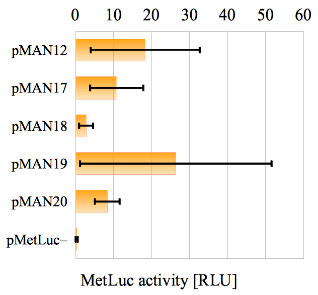
Now imagine you obtain the values in tab. 1 (the strange names for the six systems in the first column will be explained later). The first thing we can say about the results is that they do not look good at all. The standard deviation from the average is comparable to the average itself for most of the systems, and only on a couple of them you are ‘lucky’ enough so that the former is about half the value of the latter. You check the corresponding chart in fig. 1.1, and you see the same despairing situation. The error bars are humongous!
| assay 1 | assay 2 | assay 3 | ||||
|---|---|---|---|---|---|---|
| pMAN12 | 33.88 | 5.65 | 15.53 | 18.36 | 14.33 | |
| pMAN17 | 17.60 | 3.61 | 11.29 | 10.83 | 7.01 | |
| pMAN18 | 4.62 | 0.94 | 2.72 | 2.76 | 1.84 | |
| pMAN19 | 55.35 | 9.30 | 14.52 | 26.39 | 25.22 | |
| pMAN20 | 11.15 | 4.78 | 9.10 | 8.35 | 3.52 | |
| pMetLuc | 0.00 | 0.39 | 0.54 | 0.31 | 0.28 |
Before throwing in the towel, you realize two characteristics about your experiments that might save your day:
-
•
The fact is that the absolute value of for each given system is not really very important to you. What you are really interested in properly measuring is the variation in from one system to another. For example, whether or not you could safely claim that the value of corresponding to system 1 is larger than, and approximately the double of, that associated to system 5.
-
•
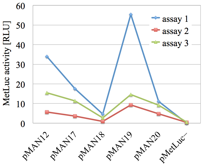
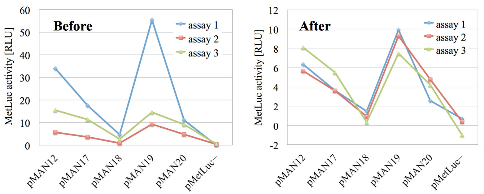
Even if you seem to be measuring huge differences in absolute value across the different assays, it looks as if the ‘tendency’ of the variations is similarly captured in all three of them. This is even more apparent in the graphical representation in fig. 1.2.
In this work, we will argue that you are right if you do not throw in the towel in such a circumstance. We will interpret the structure of the results as being caused by a multiplicative systematic error (across the different assays), and we will propose a method to correct your results in a way such that this systematic error is removed. As a consequence, the corrected numbers will not tell you anything significant about the ‘true’ absolute value of for the different systems, but, in exchange, they will maximally capture the tendency that you seemed to be correctly measuring. That is, the averages of the corrected results will present appreciable smaller standard deviations while still following the average tendency of variation.
In the next section, we will make this precise by introducing the general method of correction as well as a real experiment in cell biology which suffers from the problems (and the virtues!) that we have mentioned in this introduction. In sec. 3, we will apply the correction method to this experiment to show that both the standard deviations and the statistical significance of the results improves considerably. In sec. 4, we will discuss our interpretation of the studied situation and the proposed method, we will compare it to a simpler alternative, and we will try to explain the surprising fact that something so straightforward cannot be found (as far as we are aware) in the previous literature. Finally, in sec. 5, we will briefly summarize the main conclusions of this work, and we will outline some open questions and lines of future research
2 The method and a real example
2.1 Experimental setup
As we advanced we will have in general systems, among which a specific one may be called system , with . We now measure a quantity for each one of the systems, and we repeat times the whole set of measures. A generic repetition is termed assay , with , and each one of them is carried out under conditions that we expect to be the same. It is convenient to use to denote the value of the quantity measured for system in the -th assay (e.g., in tab. 1, ).
The different systems can be anything, from cities to DNA sequences, from people to chunks of metal. They can even be the same system at different times if the quantity is expected to evolve in some reproducible manner. The differences among the assays could be due to the experiments being performed by the same researcher on different days, by different (but in principle equally skilled) researchers using the same equipment, by the same researcher using different (but in principle equally accurate) equipment, by different (but in principle equally proficient) laboratories, etc. As long as we expect different assays to yield the same results, their definition is compatible with what we do here. For example, the different assays in table II of (Galante et al., 2012), where the production of four isoforms of Monilophthora perniciosa chitinase is presented, do not qualify as the setup described here. The reason is simple: they are knowingly carried out at different pH and temperature. Therefore, they are naturally expected to yield different results.
The experimental setup is thus very general, but we will introduce the correction method as we apply it to a specific example of a real experiment in cell biology.
2.2 The experiment
The objective of the experiment is to elucidate the regulatory network of the human protein called mitochondrial carrier homolog 1 (Mtch1), and also presenilin 1-associated protein (PSAP). Although this protein has been known for almost 15 years to be involved in apoptosis (Xu et al., 1999) and a number of studies have probed its cellular function (Lamarca et al., 2008, 2007, Li et al., 2013, Mao et al., 2008, Xu et al., 2002), not all the details are known, specially concerning its regulation, which is uncharted territory at the moment.
To identify binding sites for transcriptional regulators at the Mtch1 promoter region, different DNA vectors have been constructed and transfected into Human embryonic kidney 293T (HEK-293T) cells. Each one of the vectors contains a part of the Mtch1 promoter attached to a Metridia luciferase (MetLuc) reporter gene. When each vector is transfected into the HEK-293T cells, the MetLuc protein is produced and secreted to the medium, where its activity has been measured using the Ready-to-Glow Dual Secreted Reporter Assay Kit (Clontech). Part of this protocol involves co-transfecting each time with a vector containing the secreted alkaline phosphatase (SEAP) gene under the control of an early SV40 virus promoter. The SEAP protein is also secreted to the medium, and the measure of its activity is used to normalize the activity of MetLuc, with the objective of eliminating differences in the signal due to changes in the transfection efficiency. Hence, the activity of MetLuc is divided by that of co-transfected SEAP, and the results are reported in relative light units (RLU), which are the units used in tab. 1 and throughout this section. The complete study which will be presented elsewhere.
The example we will consider here pertains only to a small part of the data obtained for the mentioned study since it is enough for us to illustrate the correction method. We will use the MetLuc activity values corresponding to five vectors which contain incrementally deleted parts of the Mtch1 promoter (denoted by pMAN12, pMAN17, pMAN18, pMAN19 and pMAN20) as well as a control vector containing the MetLuc gene but no promoter region at all (pMetLuc). The measured MetLuc activity values (the quantity in this example) for the six vectors (the systems) in three assays are presented in tab. 1 in sec. 1. This is our starting point.
2.3 The problem with the results
As we advanced in sec. 1, the problem with the data in tab. 1 begins to emerge when we compute the average of for the system summing the results of all the assays and dividing by the total number of assays :
| (2.1) |
The corresponding standard deviation is computed as usual through:
| (2.2) |
These two values are represented for all systems in the last two columns of tab. 1, and we can see there that the standard deviations are so large that they render the results almost useless. The same problem can be appreciated if we look in fig. 1.1 (in sec. 1) at the bar chart associated to the last two columns of tab. 1.
| pMAN12 | pMAN17 | pMAN18 | pMAN19 | pMAN20 | pMetLuc | |
|---|---|---|---|---|---|---|
| pMAN12 | — | 0.475 | 0.198 | 0.662 | 0.349 | 0.161 |
| pMAN17 | — | — | 0.178 | 0.399 | 0.618 | 0.121 |
| pMAN18 | — | — | — | 0.246 | 0.077 | 0.145 |
| pMAN19 | — | — | — | — | 0.340 | 0.215 |
| pMAN20 | — | — | — | — | — | 0.050 |
| pMetLuc | — | — | — | — | — | — |
In a more quantitative way and advancing the requirement that the inter-system variation of is what really matters to us, we can calculate the probability that the observed difference between two average values, and , corresponding to two different vectors can be produced by pure chance, i.e., without the need to resort to any supplementary explanation such as the difference in the sequences of the two promoter regions in the vectors. This probability can be obtained as the so called -value associated to a two-sample Student’s -test with unequal variances (Daniel, 2009, p. 181), (Le, 2003, p. 253). One typically considers the observed difference to be statistically significant when , that is, when the probability that it can be obtained by pure chance is less than 5% (Pignatelli et al., 2003). In tab. 2, we present the -values associated to the activity measures of each pair of vectors in tab. 1, as computed by Microsoft Excel. We can appreciate that our intuition about the poor quality of our results is confirmed: Only two out of the fifteen possible pairs come close to the threshold, none is below it, and several are significantly larger.
It is at this point when we are tempted to think that everything is lost and just throw in the towel. Our results are bad. We have to dump them and perform the experiments again. Period.
However, as we advanced in sec. 1, there are two characteristics about the problem we are considering here that, when combined, can save our day.
2.4 Requirements to apply the correction method
The first one is related to the type of questions we are interested in making and answering:
We are not interested in the absolute value of for each given system (the MetLuc activity for each vector). What really matters to us is the variation in from one system to another.
For example, whether or not we could safely claim that the activity corresponding to pMAN12 is larger than, and approximately the double of, that associated to pMAN20. Indeed, if we are interested in the absolute value of MetLuc activity in RLU, the results in tab. 1 are just beyond rescue and the discussion ends here.
The second characteristic that, together with the one we just discussed, will allow us to correct the bad looking results in tab. 1 has to do with the properties of the observed measures themselves:
Even if large differences in absolute value are observed across the different assays, the ‘tendency’ of the variations is similarly captured in all three of them. Technically, the different assays present high linear correlation with one another.
This is even more apparent in the graphical representation in fig. 1.2 (in sec. 1), and without this kind of behavior in our data the correction method we will introduce next would not yield satisfactory results.
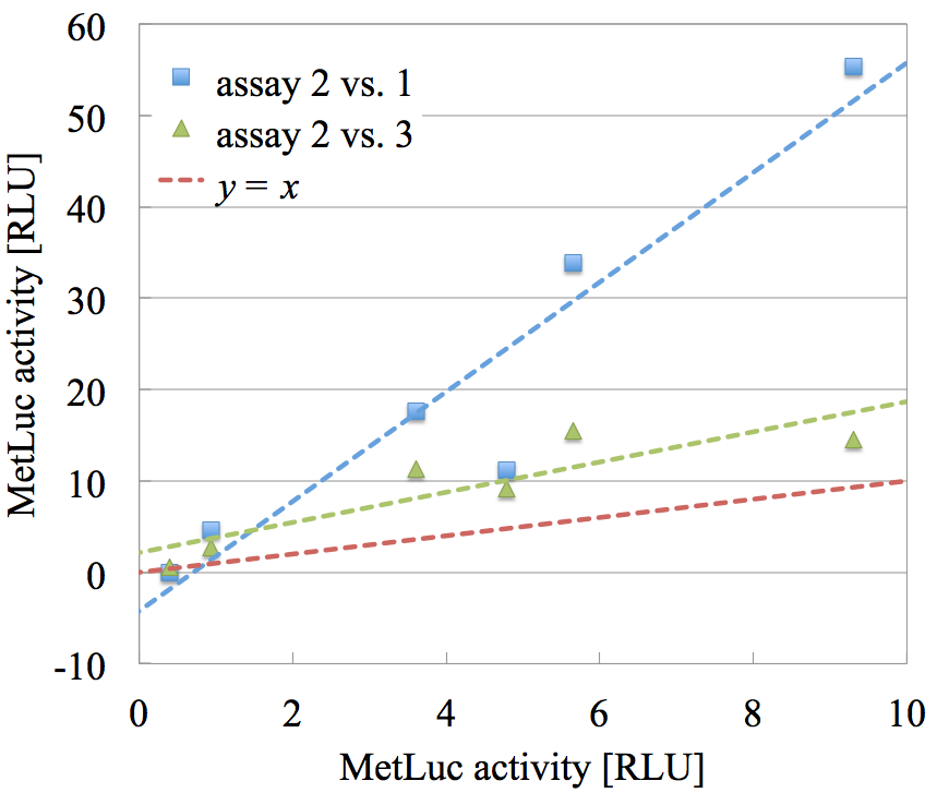
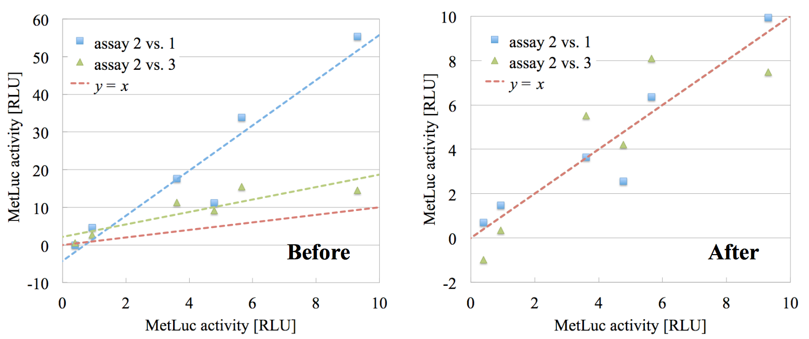
In fig. 2.1, we have represented two scatter plots: both using the values of assay 2 in the -axis, one of them using the values of assay 1 as the -coordinate (blue squares), the other using the values of assay 3 (green triangles). We have performed the two corresponding linear fits and we have depicted the corresponding tendency lines using the same color as the respective points. We also show the line in red for reference. For the reason behind the choice of these two concrete pairs of assays, see sec. 2.
Several points are worth remarking about this graph:
-
•
As we guessed, the linear correlation between the values in the different pairs of assays is high, with Pearson’s correlation coefficient for assays 2 vs. 1, for assays 2 vs. 3. This is the mathematical property that embodies the intuitive property that ‘the different assays similarly capture the tendency in the measured data’. Also, as we mentioned before, this high correlation is one of the two requirements for the method we introduce here to be applicable.
-
•
The fact that the fit lines have non-unit slope is telling us that, although the tendency is similar across the assays, the absolute value is not. The two things together mean that there is a multiplicative systematic error between the pairs of assays which is possible to remove.
-
•
The fact that the fit lines have non-zero intercept is telling us that we also have an additive multiplicative systematic error. Our method will eliminate it as well, as we shall see.
2.5 The method
To quantitatively assess the possibility that the data in tab. 1 (or the analogous one in any experiment with the structure described in sec. 2.1) satisfies the second requirement in sec. 2.4) and can therefore be corrected, we begin by performing all least-squares linear fits between all possible pairs of assays and [see, e.g., (Kirkup and Frenkel, 2006, p. 70)]. For each pair, we use the values of the first assay as the coordinate and those of the second one as the coordinate . The result of such a fit is a tendency line of the form:
| (2.3) |
where is called the slope and the intercept (or -intercept). They are computed using the following formulas:
| (2.4a) | ||||
| (2.4b) | ||||
where is the average of the measured quantity across systems and in the one single assay [not to be confused with the averages across assays for one single system computed using eq. (2.1), and presented in tab. 1 and fig. 1.1]:
| (2.5) |
Of course, is obtained just changing by in this expression.
The quantity is the standard deviation in , given by [compare now with eq. (2.2)]:
| (2.6) |
and is the covariance between the values in assay and those in assay :
| (2.7) |
With these quantities in hand, we are prepared to compute the Pearson correlation coefficient associated to the goodness of the linear fit between each pair of assays and , which is given by (Kirkup and Frenkel, 2006, eq. (5.62)):
| (2.8) |
In the first three columns of tab. 3, we present the Pearson correlation coefficients corresponding to each pair of assays in the example experiment whose results can be read in tab. 1. We can see that is close to 1.0 for all pairs, and we can therefore suspect that our correction method will produce sizable improvements in the data.
| assay 1 | assay 2 | assay 3 | ||
|---|---|---|---|---|
| assay 1 | 0.000 | 0.947 | 0.852 | 0.900 |
| assay 2 | — | 0.00 | 0.881 | 0.914 |
| assay 3 | — | — | 0.000 | 0.867 |
The first step to actually apply the method consists of selecting a reference assay. Since we do not know the ‘true’ values of the quantity (MetLuc activity) for the different systems, we will compare all the assays to the reference one and we will correct them against it.
In order to perform the selection of the reference assay with the least bias possible, we measure ‘how different’ each assay is to the rest and we choose the one that is the least different; in a sense, the most representative one. To quantify this ‘difference’ we use in fact the Pearson correlation coefficient, since it presents a property which makes it very convenient for our purposes: It discounts (is insensitive to) the possible existence of both additive and multiplicative systematic errors between the compared assays, thus measuring the difference in the variation tendency only (Alonso and Echenique, 2006); which is exactly what we need. Also notice that, as a simple consequence of its definition in eq. (2.8), is symmetric under the permutation of the indices and . This is intuitive, since it means that ‘the difference between assays and ’ is the same as ‘the difference between assays and ’.
The step that remains to be able to select the reference assay is simple: Just compute the average correlation coefficient of the -th assay with respect to all the rest of them:
| (2.9) |
and pick the one with the largest .
In the last column of tab. 3, we show the average correlation coefficient associated to each assay. We can see that it is the largest for assay 2. Therefore, we select assay 2 as our reference assay in the example we are discussing (which, by the way, explains the particular fits portrayed in fig. 2.1).
Now that the reference assay has been chosen and all the linear fits have been computed, we are ready to apply the correction to the rest of assays. If we denote by the value of the index that corresponds to the reference assay ( in our example) and we use for the corrected value associated to the original quantity (system , assay ), the correction formula reads like this:
| (2.10) |
In order to produce the whole set of corrected results, we should apply this for all assays , with , and for all systems with the index .
In order to understand the reason behind this formula, it is convenient to write the inverse transformation by solving for :
| (2.11) |
and also to notice that the systems-average of is given by:
| (2.12) |
i.e., all the averages of the corrected assays are equal to the average of the reference one. Now, if we take eq. (2.11) to the covariance in eq. (2.7) with , we obtain:
| (2.13) | |||||
where, in the last step of the second line, we have used that [as we proved in eq. (2.12)], but also that the correction in eq. (2.10) is obviously the identity for the reference assay (it suffices to notice that ), which makes , as well as all the derived quantities, such as . In the last line of eq. (2.13), we have simply used the natural notation to indicate the covariance between the corrected assays and . Finally, if we use eq. (2.13) together with the definition of the slope in eq. (2.4a) (with ), we obtain:
| (2.14) |
where we have denoted by the slope associated to the fit between the corrected assays and , and we have used that . Also, it is easy to prove that:
| (2.15) |
That is, the slope of the fits among the corrected assays is 1 and the intercept is 0. Since we argued that the first can be interpreted as a multiplicative systematic error and the second as an additive one, we have just proved that our proposed correction in eq. (2.10) has the promised effect of eliminating both errors. To see that this also has the effect of reducing the standard deviations and improving the statistical significance of our results, we turn to the next section.
But before, let us mention a final consistency property of the correction method: In mathematical jargon, it is idempotent. In plain words, applying it twice is the same as applying it once, i.e., if we apply the whole correction process to the corrected results, we find that nothing changes. The corrected-corrected results are just the corrected results.
All the formulae needed to compute the linear fits, the inter-assay correlation coefficients, as well as the correction in eq. (2.10) are provided in this section and they are very simple. The reader can choose to implement them in any spreadsheet of her liking, or she can use the Perl scripts we have written for the occasion and which can be found in the supplementary material. Also in the supplementary material, we provide a cheat sheet with the bare steps of our method, conveniently organized, briefly stated, and stripped off of all the explanatory text that surrounds the steps in this article.
3 Results
If we apply the correction in eq. (2.10) to our original results in tab. 1, we find the corrected values in the second table of tab. 4 (where we have repeated the uncorrected data to facilitate the comparison).
Before
assay 1
assay 2
assay 3
pMAN12
33.88
5.65
15.53
18.36
14.33
pMAN17
17.60
3.61
11.29
10.83
7.01
pMAN18
4.62
0.94
2.72
2.76
1.84
pMAN19
55.35
9.30
14.52
26.39
25.22
pMAN20
11.15
4.78
9.10
8.35
3.52
pMetLuc
0.00
0.39
0.54
0.31
0.28
After
assay 1
assay 2
assay 3
pMAN12
6.35
5.65
8.10
6.70
1.26
pMAN17
3.64
3.61
5.52
4.26
1.10
pMAN18
1.48
0.94
0.34
0.92
0.57
pMAN19
9.93
9.30
7.48
8.91
1.27
pMAN20
2.56
4.78
4.20
3.85
1.15
pMetLuc
0.71
0.39
0.98
0.04
0.90
At first sight, the corrected standard deviations seem much better when compared to their associated averages for each system. This impression is reinforced if we take a look at the corresponding bar charts in fig. 3.1.
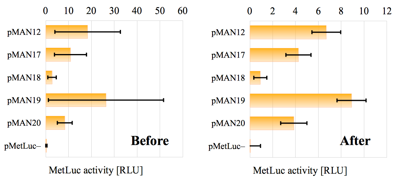
If we want to be more quantitative, and recalling that the inter-system variation of MetLuc activity is what really matters to us, we can repeat the -values calculation in sec. 2.3, this time for the corrected data. In tab. 5, we present both the original -values obtained from the uncorrected results as well as the new ones. We remind the reader that the -value’s meaning is that it quantifies the probability that the observed difference between two average values, and , corresponding to two different vectors can be produced by pure chance, i.e., without the need to resort to any supplementary explanation such as the difference in the sequences of the two promoter regions in the vectors. One typically considers the observed difference to be statistically significant when , that is, when the probability that it can be obtained by pure chance is less than 5%. As we can see in tab. 5, while the original situation was despairing, with two out of the fifteen possible pairs close to the threshold, none below it, and several significantly larger, the corrected -values show a much better behavior. For the corrected data, eleven out of the fifteen possible comparisons are below the threshold, two of them are close to it, and only two are significantly larger. This means that most of the observed differences in MetLuc activity are now statistically significant.
Before
pMAN12
pMAN17
pMAN18
pMAN19
pMAN20
pMetLuc
pMAN12
—
0.475
0.198
0.662
0.349
0.161
pMAN17
—
—
0.178
0.399
0.618
0.121
pMAN18
—
—
—
0.246
0.077
0.145
pMAN19
—
—
—
—
0.340
0.215
pMAN20
—
—
—
—
—
0.050
pMetLuc
—
—
—
—
—
—
After
pMAN12
pMAN17
pMAN18
pMAN19
pMAN20
pMetLuc
pMAN12
—
0.066
0.007
0.100
0.045
0.003
pMAN17
—
—
0.018
0.009
0.679
0.007
pMAN18
—
—
—
0.003
0.030
0.236
pMAN19
—
—
—
—
0.007
0.001
pMAN20
—
—
—
—
—
0.012
pMetLuc
—
—
—
—
—
—
In order to enrich our picture of what is going on here, we can also take a look at the corrected version of the tendency plot that we presented before in fig. 1.2 and which we now repeat here on the left of fig. 3.2. As we can see in the corrected tendency plot on the right, the fact that all three assays correctly captured the overall variation tendency of the data has been maximally leveraged by the correction in eq. (2.10). Without altering the legitimate random noise in the original results, the additive and multiplicative systematic errors have been eliminated, and the corrected tendency lines are now optimally superimposed.
Similarly, we can compare the original and corrected scatter plots in fig. 3.3. In the second one, the best fit lines corresponding to assays 2 vs. 1 and assays 2 vs. 3 have been omitted because they coincide with the zero-intercept unit-slope line. This is the precise mathematical embodiment of the fact that the correction in eq. (2.10) ‘eliminates the additive and multiplicative errors’: it transforms all the fits against the reference assay from non-zero intercept and non-unit slope to zero intercept and unit slope. The fact that the random error is unmodified can be appreciated by the remaining dispersion of the scatter plot points with respect to the line in the second graph in fig. 3.3.
4 Discussion
We have just introduced a simple method for correcting the results of multi-assay experiments which, under two very basic conditions (that only inter-system variations matter to us, and that the different assays present high linear correlation with one another), allows us to considerably reduce the standard deviation of the systems’ averages across assays, consequently increasing the statistical significance of the results. We have applied the correction method to a real experiment in cell biology where we have appreciated a great improvement.
Our interpretation of the situation is as follows: Uncontrolled differences (errors) appear when a given experiment is repeated. Some of them are random (i.e., we see no pattern in them) and cannot be eliminated. Some others are systematic and can be. If we represent a scatter plot in which the results of one assay are placed on the -axis, the results of a different one are placed on the -axis, and we perform a linear fit, we can expect to observe two different situations:
-
•
The best fit line has zero -intercept and unit slope. We interpret this as all the error being random, and no correcting action can be taken here. The data must be used ‘as is’.
-
•
The best fit line has non-zero intercept or non-unit slope or both. We interpret this as some of the error being systematic, some of it random. The non-zero intercept signals an additive systematic error; the non-unit slope a multiplicative systematic one; and the dispersion of the scatter plot points from the fit line signals the part of the error that is random. In such a case, we can apply the correction in eq. (2.10), thus eliminating both systematic components and reducing the situation to the one described in the previous point.
As it is always the case with systematic errors, one might or might not know the actual reasons behind them (we left the apparatus on too much time, the cell number was larger than usual, we inadvertently used the wrong pipette, etc.), but we do not really need to know the reasons to confidently assert that a systematic error is indeed there. If the difference between two assays is (mostly) captured by multiplying the results of one of them by a number and adding a number , we are entitled to entertain the strong suspicion that some very real causes are behind this predictable pattern. Hence, even if we do not know these causes, it would be a wasted opportunity not to apply the correction in eq. (2.10). If you do know the causes, good for you. So much the better. In fact, by applying the reasoning associated to the method described here, the presence of a non-zero intercept or a non-unit slope in the fits of the different pairs of assays (plus a high linear correlation among them) may suggest to the experimenter that some additive or multiplicative systematic error is being made from assay to assay. With this clue, she can then proceed to look for the actual experimental causes behind them (in the case that they were previously unknown).
Also notice that systematic errors might not end at the linear order. The relation between the results of two different assays could be well described for example by a quadratic relation plus some random error; or even by higher order polynomials. No a priori reason can reject this possibility, however, a treatment of these more complicated cases is outside the scope of this work.
An important part of the method introduced here is that, since we do not know the ‘real’ absolute value of the measured quantities (and in fact it does not matter to us), we have to choose a reference assay to fit all the rest of assays to. The most reasonable way to perform this choice in an unbiased manner is to select the most representative assay in the experiment, the one that is ‘most similar to all the others’. We make this condition precise by measuring the difference of every assay to all the rest of them and choosing the one that is the least different to all the others. To this end, we use the Pearson correlation coefficient associated to the goodness of the linear fit because it correctly discounts the additive and multiplicative systematic errors.
Also, it is worth mentioning that the use of the word ‘error’ for the differences between one specific assay and the rest of them might seem unorthodox at first sight. After all, the ‘error’ is ideally defined as the difference between the measured quantities and their ‘real’ values. However, we think that this apparent overuse of the term is just that: apparent. Since the ‘real’ values are never actually known, the ideal definition of ‘error’ is philosophically appealing but practically inapplicable. What researchers always do is to compare one set of measures to some more accurate ones (but not ‘real’ yet), to some theoretical prediction (not ‘real’ either), etc. In this sense, and given that the ‘real’ values of the quantity are unknown in our experimental setup in sec. 2.1 (as in all setups!), the ‘best’ guess we a priori have (before the proposed correction) of the most accurate set of measures is precisely the most representative of our assays, i.e., the one that is the least different from the rest. This is why we choose it as the reference to which all the rest of the assays are compared, and this is why the observed differences deserve to be intuitively called ‘errors’.
Although all this seems quite straightforward, we have only found in the literature one related proposal for a correction method that could be compared to the one we introduce in this work (even if the rationale is never clearly expressed as we do here). This related method readily comes to mind and it consists of dividing, in each assay, the value of for all systems by the value of one of them. For example, we could select pMAN18 as our normalizing vector, divide the activities of all the vectors in each assay by the activity of pMAN18 in the same assay, and thus obtain a new set of results now expressed as a normalized fold change in activity with respect to the pMAN18 value (which now becomes 1.0). This is used for example in (Schagat et al., 2007, Ysebrant de Lendonck et al., 2013, Matsunoshita et al., 2011, Alvarez and del Valle Loto, 2012) or (Zhang et al., 2012, fig. S3).
The result of applying this normalization to the original data in tab. 6 is presented in tab. 6. We see that the standard deviations have been reduced and in fact the overall improvement is similar to what we obtained when applying the correction method introduced in this work. However, this normalization procedure presents some drawbacks that, in our opinion, render it inferior to our method. Namely:
| assay 1 | assay 2 | assay 3 | ||||
|---|---|---|---|---|---|---|
| pMAN12 | 7.33 | 6.01 | 5.71 | 6.35 | 0.86 | |
| pMAN17 | 3.81 | 3.84 | 4.15 | 3.93 | 0.19 | |
| pMAN18 | 1.00 | 1.00 | 1.00 | 1.00 | 0.00 | |
| pMAN19 | 11.98 | 9.89 | 5.34 | 9.07 | 3.40 | |
| pMAN20 | 2.41 | 5.09 | 3.35 | 3.62 | 1.36 | |
| pMetLuc | 0.00 | 0.41 | 0.20 | 0.20 | 0.21 |
-
•
It demands an arbitrary choice (that of the normalizing system) which seems ad hoc and prevents automatization in some degree. Related to this, the fact that the corrected result for the normalizing system has zero standard deviation does not seem easy to interpret, nor completely legitimate.
-
•
If we recall the general formula for the propagation of errors (Kirkup and Frenkel, 2006, p. 50),
(4.1) where is a function of random variables with standard deviations (errors) , we can use it to compute the error in the normalized quantity , where is the measured result for the system (in a given assay) and is the quantity measured for the system chosen to normalize the results:
(4.2) We see that the error in the normalized quantity relative to the value of itself is the sum of the relative errors of and . Now, if we happen to choose a particular normalizing system with high relative error, this could spoil the whole assay when we divide all the results by , even if the rest of measures were accurate.
-
•
The described normalizing procedure seems fit to eliminate multiplicative systematic errors, but not additive ones.
Our method suffers from none of these problems:
-
•
No choice of a ‘special’ normalizing system is needed. (There is a choice of a reference assay, but it is made in a justified way, as we have explained.)
-
•
In a manner of speaking, it distributes the normalization among all the values in a given assay, thus minimizing the probability that one specially bad apple spoils the whole basket.
-
•
It eliminates both multiplicative and additive systematic errors.
If we check exhaustive textbooks in biostatistics, such as (Daniel, 2009, Vittinghoff et al., 2005, Le, 2003), or more wide ranging ones, such as (Walpole et al., 2012, Kutner et al., 2005, Mickey et al., 2004, Taylor, 1997, Mandel, 1984), we do not find any account of a correcting method that is similar to what we propose here. Some of the texts come close sometimes, but they never hit the target.
One way in which they often come close is when they discuss repeated measures. See for example (Mickey et al., 2004, chap. 9), (Kutner et al., 2005, chap. 27), or (Cnaan et al., 1997), (Vittinghoff et al., 2005, chap. 8), and (Daniel, 2009, p. 346) for detailed discussions of the concept in biosciences. ‘Repeated measures’ consists of an experimental setup very similar to the one used here and described in sec. 2.1, i.e., measuring the same quantity on systems and repeating the experiment times, but it contains a fundamental difference: it tackles measurements that are expected to change from repetition to repetition [e.g., a time series, or table II of (Galante et al., 2012) discussed in sec. 2.1]. It is a key of our setup that we expect the results of several repetitions to be the same. This is why it makes sense for us to correct them, which would be unnatural in the repeated-measures setup. Also, for repeated measures, it is not a requirement that we are not interested in the absolute value but only in the inter-system variation. In our case, this is essential.
In (Walpole et al., 2012, p. 539), another similar situation to the one we have considered here is dealt with, namely blocking, however, they do not discuss what to do if there is an obvious linear correlation between the blocks (as in their figure 13.6a). Their example in figure 13.12 also seems ripe to apply our method, but they take no correcting action on it.
One of the reasons that we imagine could be behind the fact that no precedents of our straightforward method are found in the literature (as far as we have been able to scan it) has to do with the usual interpretation of the range of application of the least-squares fit protocol. Typically, fitting some values in the -axis against those on the -axis is used to assess a possible linear relationship between two different quantities (apples and oranges, say). So much so that is typically called the independent variable, while is the dependent one. In our approach, it is a key conceptual step to realize that it actually makes sense to investigate the linear correlation of some quantity with itself (measured in two different assays), and consequently interpret any difference between the two as experimental error (in the manner we explained before).
Another reason that is possibly behind the absence of precedents is the fact that, despite being quite intuitive to us, systematic errors of the multiplicative kind are very rarely discussed in the literature. Systematic errors are normally considered to be additive.
After a thorough search we have only found anecdotal mentions in a paper that discusses the influence of natural fires on the air pollution of the Moscow area (Konovalov et al., 2011), in a proceedings paper about anticorrosion coating (Niedostatkiewicz and Zielonko, 2006), in a recent work concerned with calibration of spectrographs for detecting earth-mass planets around sun-like stars (Glenday et al., 2012), and in a similar paper focused in the detection and study of quasars (Johansson et al., 2000). In all these works the authors consider the possibility of a multiplicative systematic error in their models or measurements, but they take no action to correct it.
Something very similar happens in (Meloun et al., 1993, p. 3), where the existence of multiplicative systematic errors is acknowledged in the context of analytical chemistry, as well as the necessity to eliminate them. In (Doerffel, 1994), the possibility of both additive and multiplicative systematic errors is discussed, as well as their respective relation with non-zero -intercepts and non-unit slopes. Finally, in (Kirkup and Frenkel, 2006, p. 39), the authors not only discuss multiplicative systematic errors (which they also call gain shifts or gain errors), but they provide several examples where this multiplicative systematic error can appear. Although more space is dedicated in these last three works to the discussion of multiplicative systematic errors, the authors do not provide any method for eliminating them either.
In addition, it is worth mentioning that, in (Doerffel, 1994) and in (Kirkup and Frenkel, 2006), the authors consider the error to be defined with respect to ‘true’ (or at least more accurate) results; in the first case to calibrate experimental protocols, in the second one to calibrate measuring devices. As we explained when discussing the choice of the reference assay, our perspective on this issue is different, and so it is the approach. For example, if you want to correct your results against some ‘better’ data, you are presumably interested not only in the variations of the measured quantity, but also in its absolute value.
We have only found one work, concerned with gas electron diffraction data (Gundersen et al., 1998), in which the authors both consider the existence of multiplicative systematic errors and take actions to correct them. However, the proposed correction is particular to the concrete problem studied, and the experimental setup is different to the one described in sec. 2.1: The authors refer to systematic errors in experimental data with respect to the ‘true’ values, not to systematic errors between different measures of the same quantity as we do here.
5 Conclusions
We have introduced a method for correcting the data in experiments in which a single quantity is measured for a number of systems in multiple repetitions or assays. If we are not interested in the absolute value of but only in the inter-system variations, and the results in different assays are highly correlated with one another, we can use the proposed method to eliminate both additive and systematic differences (errors) between each one of the assays and a suitably chosen reference one. As we have shown using a real example of a cell biology experiment, this correction can considerably reduce the standard deviation in the systems’ averages across assays, and consequently improve the statistical significance of the data.
The method is of very general applicability, not only to experimental results but possibly also to numerical simulations, as long as the structure of the setup and the requirements on the data are those just mentioned and carefully discussed in sec. 2.1. This, together with its simplicity of application (the only mathematical infrastructure needed to apply it is basically least-squares linear fits), makes the method of very wide interest in any quantitative scientific field that deals with data subject to uncertainty.
Some possible lines of future work include the application of the method to a wider variety of problems, a deeper statistical analysis of its properties and the assumptions behind it, or the extension to systematic differences of higher-than-linear order that we briefly mentioned in sec. 4.
Acknowledgements
We would like to thank Professors Jesús Peña, Silvano Pino, Juan Puig, Ricardo Rosales and Javier Sancho for recommending to us the reference statistics and biostatistics textbooks that we have used in the writing of the manuscript.
This work has been supported by the grants FIS2009-13364-C02-01 (Ministerio de Ciencia e Innovación, Spain), UZ2012-CIE-06 (Universidad de Zaragoza, Spain), Grupo Consolidado “Biocomputación y Física de Sistemas Complejos” (DGA, Spain), also by grants BFU2009-11800 (Ministerio de Ciencia e Innovación, Spain), and UZ2010-BIO-03 and UZ2011-BIO-02 (Universidad de Zaragoza, Spain) to J.A.C.
References
- Alonso and Echenique (2006) J. L. Alonso and P. Echenique. A physically meaningful method for the comparison of potential energy functions. J. Comput. Chem., 27:238–252, 2006. http://bit.ly/16mmjBn.
- Alvarez and del Valle Loto (2012) A. Alvarez and F. del Valle Loto. Characterization and biological activity of Bacillus thuringiensis isolates that are potentially useful in insect pest control. In Akeem Lameed, editor, Biodiversity Enrichment in a Diverse World. InTech, 2012. http://bit.ly/1e4qFQm.
- Cnaan et al. (1997) A. Cnaan, N. M. Laird, and P. Slasor. Tutorial in biostatistics: Using the general linear mixed model to analyse unbalanced repeated measures and longitudinal data. Stat. Med., 16:2349–2380, 1997. http://bit.ly/17KzJYZ.
- Daniel (2009) W. W. Daniel. Biostatistics: A foundation for analysis in the health sciences. John Wiley & Sons, 9th edition, 2009.
- Doerffel (1994) K. Doerffel. Assuring trueness of analytical results. Fresenius J. Anal. Chem., 348:183–187, 1994.
- Galante et al. (2012) R. S. Galante, A. G. Taranto, M. G. B. Koblitz, A. Góes-Neto, C. P. Pirovani, J. C. M. Cascardo, S. H. Cruz, G. A. G. Pereira, and S. A. De Assis. Purification, characterization and structural determination of chitinases produced by Moniliophthora perniciosa. An. Acad. Bras. Cien., 84:469–486, 2012. http://bit.ly/15pgQL9.
- Glenday et al. (2012) A. G. Glenday, D. F. Phillips, M. Webber, C.-H. Li, G. Furesz, G. Chang, L.-J. Chen, F. X. Kärtner, D. D. Sasselov, A. H. Szentgyorgyi, and R. L. Walsworth. High-resolution Fourier transform spectrograph for characterization of echelle spectrograph wavelength calibrators. In Ian S. McLean, Suzanne K. Ramsay, and Hideki Takami, editors, Ground-based and Airborne Instrumentation for Astronomy IV, volume 8446, 2012. http://bit.ly/1e2fpnH.
- Gundersen et al. (1998) S. Gundersen, T. G. Strand, and H. V. Volden. Gas electron diffraction data: A representation of improved resolution in the frequency domain, a background correction for multiplicative and additive errors, and the effect of increased exposure of the photographic plates. J. Mol. Struct., 445:35–45, 1998.
- Johansson et al. (2000) S. Johansson, V. Zilio, J. C. Pickering, A. P. Thorne, J. E. Murray, U. Litzén, and J. K. Webb. Accurate laboratory wavelengths of some ultraviolet lines of Cr, Zn and Ni relevant to time variations of the fine structure constant. Mon. Not. R. Astron. Soc., 319:163–167, 2000. http://bit.ly/19oFFer.
- Kirkup and Frenkel (2006) L. Kirkup and R. B. Frenkel. An introduction to uncertainty in measurement using the GUM. Cambridge University Press, 2006.
- Konovalov et al. (2011) I. B. Konovalov, M. Beekmann, I. N. Kuznetsova, A. A. Glazkova, A. V. Vasil’eva, and R. B. Zaripov. Estimation of the influence that natural fires have on air pollution in the region of Moscow megalopolis based on the combined use of chemical transport model and measurement data. Izvestiya, Atmos. Ocean. Phys., 47:457–467, 2011.
- Kutner et al. (2005) M. H. Kutner, C. J. Nachtsheim, J. Neter, and W. Li. Applied linear statistical models. McGraw Hill, 5th edition, 2005.
- Lamarca et al. (2007) V. Lamarca, A. Sanz-Clemente, R. Pérez-Pé, M. J. Martínez-Lorenzo, N. Halaihel, P. Muniesa, and J. A. Carrodeguas. Two isoforms of PSAP/MTCH1 share two proapoptotic domains and multiple internal signals for import into the mitochondrial outer membrane. Am. J. Physiol. Cell. Physiol., 293:C1347–C1361, 2007. http://bit.ly/1bFL2HG.
- Lamarca et al. (2008) V. Lamarca, I. Marzo, A. Sanz-Clemente, and J. A. Carrodeguas. Exposure of any of two proapoptotic domains of presenilin 1-associated protein/mitochondrial carrier homolog 1 on the surface of mitochondria is sufficient for induction of apoptosis in a Bax/Bak-independent manner. Eur. J. Cell. Biol., 87:325–334, 2008.
- Le (2003) C. T. Le. Introductory biostatistics. John Wiley & Sons, 2003.
- Li et al. (2013) T. Li, L. Zeng, W. Gao, M.-Z. Cui, X. Fu, and X. Xu. PSAP induces a unique Apaf-1 and Smac-dependent mitochondrial apoptotic pathway independent of Bcl-2 family proteins. Biochim. Biophys. Acta, 1832:453–474, 2013.
- Mandel (1984) J. Mandel. The statistical analysis of experimental data. Dover Publications, 1984.
- Mao et al. (2008) G. Mao, J. Tan, W. Gao, Y. Shi, M.-Z. Cui, and X. Xu. Both the N-terminal fragment and the protein-protein interaction domain (PDZ domain) are required for the pro-apoptotic activity of presenilin-associated protein PSAP. Biochim. Biophys. Acta, 1780:696–708, 2008. http://1.usa.gov/17ThSiE.
- Matsunoshita et al. (2011) Y. Matsunoshita, K. Ijiri, Y. Ishidou, S. Nagano, T. Yamamoto, H. Nagao, S. Komiya, and T. Setoguchi. Suppression of osteosarcoma cell invasion by chemotherapy is mediated by urokinase plasminogen activator activity via up-regulation of EGR1. PLoS ONE, 6:e16234, 2011. http://bit.ly/1c2YQeN.
- Meloun et al. (1993) M. Meloun, J. Militky, and M. Forina. Chemometrics for analytical chemistry. Volume 1: PC-aided statistical data analysis. Ellis Horwood, 1993.
- Mickey et al. (2004) R. M. Mickey, O. J. Dunn, and V. A. Clark. Applied statistics: Analysis of variance and regression. Wiley, 3rd edition, 2004.
- Niedostatkiewicz and Zielonko (2006) M. Niedostatkiewicz and R. Zielonko. Time domain parameter identification of anticorrosion coating via some types of polynomial signals. In XVIII IMEKO World Congress, 2006. http://bit.ly/19oEICP.
- Pignatelli et al. (2003) M. Pignatelli, R. Luna-Medina, A. Pérez-Rendón, A. Santos, and A. Perez-Castillo. The transcription factor early growth response factor-1 (EGR-1) promotes apoptosis of neuroblastoma cells. Biochem. J., 373:739–746, 2003. http://1.usa.gov/16mm4Gv.
- Schagat et al. (2007) T. Schagat, A. Paguio, K. Kopish, and Promega Corporation. Normalizing genetic reporter assays: Approaches and considerations for increasing consistency and statistical significance. Cell Notes, 17:9–12, 2007. http://bit.ly/16mm5tS.
- Taylor (1997) J. R. Taylor. An introduction to error analysis: The study of uncertainties in physical measurements. University Science Books, 1997.
- Vittinghoff et al. (2005) E. Vittinghoff, D. V. Glidden, S. C. Shiboski, and C. E. Mcculloch. Regression methods in biostatistics: Linear, logistic, survival, and repeated measures models. Springer, 2005.
- Walpole et al. (2012) R. E. Walpole, R. H. Myers, S. L. Myers, and K. Ye. Probabilty & statistics for engineers & scientists. Prentice Hall, 2012.
- Xu et al. (1999) X. Xu, Y.-C. Shi, X. Wu, P. Gambetti, D. Sui, and M.-Z. Cui. Identification of a novel PSD-95/Dlg/ZO-1 (PDZ)-like protein interacting with the C terminus of Presenilin-1. J. Biol. Chem., 274:32543–32546, 1999. http://bit.ly/17Rm5Ud.
- Xu et al. (2002) X. Xu, Y.-C. Shi, W. Gao, G. Mao, G. Zhao, S. Agrawal, G. M. Chisolm, D. Sui, and M.-Z. Cui. The novel presenilin-1-associated protein is a proapoptotic mitochondrial protein. J. Biol. Chem., 277:48913–48922, 2002. http://bit.ly/17TjOry.
- Ysebrant de Lendonck et al. (2013) L. Ysebrant de Lendonck, F. Eddahri, Y. Delmarcelle, M. Nguyen, O. Leo, S. Goriely, and A. Marchant. STAT3 signaling induces the differentiation of human ICOS+ CD4 T cells helping B lymphocytes. PLoS ONE, 8:e71029, 2013. http://bit.ly/1c2XCzZ.
- Zhang et al. (2012) W. Zhang, D. F. A. R. Dourado, P. A. Fernandes, M. J. Ramos, and B. Mannervik. Multidimensional epistasis and fitness landscapes in enzyme evolution. Biochem. J., 445:39–46, 2012. http://bit.ly/1e4q2pT.