Symmetry breaking cilia-driven flow in the zebrafish embryo
Abstract
Fluid mechanics plays a vital role in early vertebrate embryo development, an example being the establishment of left-right asymmetry. Following the dorsal-ventral and anterior-posterior axes, the left-right axis is the last to be established; in several species it has been shown that an important process involved with this is the production of a left-right asymmetric flow driven by ‘whirling’ cilia. It has previously been established in experimental and mathematical models of the mouse ventral node that the combination of a consistent rotational direction and posterior tilt creates left-right asymmetric flow. The zebrafish organising structure, Kupffer’s vesicle, has a more complex internal arrangement of cilia than the mouse ventral node; experimental studies show the flow exhibits an anticlockwise rotational motion when viewing the embryo from the dorsal roof, looking in the ventral direction. Reports of the arrangement and configuration of cilia suggest two possible mechanisms for the generation of this flow from existing axis information: (1) posterior tilt combined with increased cilia density on the dorsal roof, and (2) dorsal tilt of ‘equatorial’ cilia. We develop a mathematical model of symmetry breaking cilia-driven flow in Kupffer’s vesicle using the regularized Stokeslet boundary element method. Computations of the flow produced by tilted whirling cilia in an enclosed domain suggest that a possible mechanism capable of producing the flow field with qualitative and quantitative features closest to those observed experimentally is a combination of posteriorly tilted roof and floor cilia, and dorsally tilted equatorial cilia.
1 Introduction
Vertebrates, from the outside, appear bilaterally symmetric. However, in many species, internal body plans are arranged asymmetrically in an organised way. For example the heart can usually be found on the left in mice, zebrafish (Ibañes & Belmonte, 2009), and humans (figures 1a). As with convention the ‘left’ of the depicted individual is on the right of figure 1a,b and in the majority of figures in this paper.
Three axes are established during early embryonic development; in order they are the dorsal-ventral, anterior-posterior and left-right axes (figure 1c) (Hirokawa et al., 2009). It is only relatively recently that the mechanisms involved in the initial establishment of left-right asymmetry have begun to be understood. Kartagener (1933), amongst others, identified a triad of conditions that included respiratory problems, male infertility and situs inversus, the lateral transposition of internal organs (figure 1b). For a detailed historical review see also Berdon & Willi (2004) and Berdon et al. (2004). In , Afzelius performed electron microscopy on the sperm of four infertile men. The flagella, which were immotile, did not possess the dynein motor protein. Three of the four men did not have normally-functioning lung cilia, moreover three of the four men were reported to have situs inversus. From this evidence, Afzelius hypothesised that, “Visceral asymmetry is determined through the movements of cilia of some embryonic epithelial tissues” (Afzelius, 1976).
Twenty years later, Sulik et al. (1994) discovered a node structure on a mouse embryo at – days post-fertilisation that expressed primary cilia. Cilia are microscopic hair-like organelles with an internal arrangement of nine pairs of microtubules arranged in a cylindrical structure. Motile ‘’ cilia, similar to the flagella of sperm, possess a central microtubule pair, and perform essential biological functions such as mucus clearance in the lung and transport of the ovum in the fallopian tube. Primary cilia by contrast are generally shorter, do not possess a central microtubule pair, and prior to the work of Sulik et al. were generally thought to be immotile. However, Sulik et al. reported that the primary cilia in the mouse node were motile and furthermore postulated that their motility may be “associated with the establishment of sidedness”, thereby providing the link between situs inversus and ciliary dysfunction in the node. Nonaka et al. (1998) then confirmed that primary cilia in the mouse node were indeed motile and in normal mice performed a clockwise rotation, when viewed from tip to base, which propels fluid. Nonaka et al. showed that knockout mice with absent cilia do not break symmetry normally and then do not survive gestation. Nonaka et al. (2002) subsequently showed that a leftward fluid flow was both necessary and sufficient for normal organ situs by placing embryos in artificial flow conditions showing that a rightward flow resulted in situs inversus. However at the time of Nonaka et al.’s work it was still unclear as to how a whirling motion could be sufficient to create a directional fluid flow.
A resolution to this question was first proposed by Cartwright et al. (2004). Representing cilia by point torque rotlet singularities in an infinite domain, Cartwright et al. showed how a clockwise rotation can generate a leftward fluid flow if the axis of rotation is tilted towards the already-established posterior direction. This theoretical prediction was verified soon after in biological observations (Okada et al., 2005) and a mechanical experimental model (Nonaka et al., 2005). Moreover, Cartwright et al.’s theoretical prediction that the tilt angle would be approximately was close to the mean value of reported by Nonaka et al. Brokaw (2005) used a computational model to show how internal chirality of dynein regulation, and internal twist, are possible mechanisms that may create the whirling motion of cilia. Furthermore Brokaw gave the first discussion of the effect of surface drag in the context of tilted rotational cilia motion, and its effect in generating a leftward flow.
Smith et al. (2007) used a computational approach to slender body theory for Stokes flow to address the roles of cilium-surface interactions and unsteadiness in the flow field, via image systems in a plane boundary (Blake, 1971). Evidence of limited chaotic advection was found and later verified experimentally (Supatto et al., 2008; Supatto & Vermot, 2011). Smith et al. (2008) also investigated the optimal tilt angle for maximum fluid propulsion by tilted rotational motion, and furthermore modelled particle drift under the action of arrays of tilted whirling cilia; results were found to lie within the bounds reported from experimental observations (Okada et al., 2005; Nonaka et al., 2005), and also the mechanical analogue model of Nonaka et al. (2005). Addressing the issue that the developing node has an enclosing upper membrane, Cartwright et al. (2007) computed flow profiles using a steady flow finite element model. It was predicted that there would exist two layers of rightward ‘return’ flow, in the ‘upper’ region away from the cilia, caused by conservation of mass in the volume, and also very close to the ciliated surface, the latter caused by the return stroke of the cilia. To investigate the influence of the enclosed fluid domain in the context of a time-dependent model, Smith et al. (2011) used the regularized Stokeslet boundary element method combined with slender body theory. The time-dependent model predicted that the leftward particle drift would occur throughout most of the ciliated layer, including very close to the ciliated surface. It was also found that the main directional flow only extended as far as the cilia array.
An important question is how a directional flow is translated into asymmetric development. Two main models have been proposed: one that the flow transports morphogen proteins to the left of the node, the other is that there are also immotile cilia in organising structures that are there to ‘sense’ the flow produced by the motile cilia. These theories are reviewed and discussed by Hirokawa et al. (2009); a detailed understanding of the flow, transport of particles, and hydrodynamic stresses produced by whirling cilia will be essential to understand this mechanism.
2 Geometry of organising structures
Many theoretical (Cartwright et al., 2004, 2007; Smith et al., 2007, 2008, 2011) and early experimental (Nonaka et al., 1998, 2002, 2005; Okada et al., 2005; Tanaka et al., 2005) studies have been conducted based on the geometry of the mouse node. This is because the mouse was one of the first species found to have cilia in its organising structure (see also reviews by Cartwright et al. (2008, 2009); Hirokawa et al. (2009) for full details).
The mouse node is a triangular depression measuring –m in width and –m in depth that forms on the ventral surface of the embryo. The mouse node is covered with a membrane, which is frequently removed for imaging purposes, and is filled with fluid. Primary cilia measuring – are expressed on the surface forming the base of the mouse node and they exhibit tilted clockwise rotations as described in the previous section.
In recent years experimentalists have turned attention to imaging the organising structure of the zebrafish (figure 1d,e), termed Kupffer’s vesicle (KV) (Kawakami et al., 2005; Kreiling et al., 2007; Okabe et al., 2008; Supatto et al., 2008). Zebrafish KV is transparent (Supatto & Vermot, 2011), meaning that internal structures can be imaged without removing any surfaces. Another distinct advantage of using KV to investigate development is that it is visible at approximately hours post-fertilisation (Kimmel et al., 1995).

Zebrafish KV is a closed spheroidal structure, measuring approximately , with cilia measuring – (Kramer-Zucker et al., 2005) expressed on the internal surfaces (figure 2) (Kreiling et al., 2007; Supatto et al., 2008). It is agreed that the cilia in KV are tilted and rotate, however there is not a consensus as to the tilt direction. Kreiling et al. (2007) report that the roof and floor cilia are posteriorly tilted; Supatto et al. (2008) report dorsally tilted side wall cilia. Both Kreiling et al. and Supatto et al. describe the same flow field, a circulation about the dorsal-ventral axis (figure 3). Supatto & Vermot (2011) suggest that dorsal tilt is necessary to create the observed flow field. Moreover, Supatto & Vermot (2011) report the directional flow magnitude in the region up to approximately above the cell surface as being in a range of approximately –.
This theoretical study is designed to investigate the flow in KV generated by these models: (1) all posteriorly tilted cilia, (2) all dorsally tilted cilia and (3) a hybrid of posteriorly and dorsally tilted cilia depending on position on the internal surface of KV. Furthermore, we shall investigate the effect of the more densely ciliated anterior-dorsal roof, as also observed in experiment.
In the next section we shall describe the mathematical theory behind our model starting with a description of the physics of a cilium beat cycle. We then formulate the regularized Stokeslet boundary integral equation because it is highly suited to modelling complex geometric problems. We will also describe our method of meshing the interior domain and cilia of KV and then solve the regularized Stokeslet boundary integral equation numerically.
3 Fluid mechanics modelling
The basic fluid mechanical mechanisms underlying how tilted cilia performing a whirling motion create a directional flow, the nodal flow, have been discussed extensively elsewhere (Smith et al., 2007, 2008, 2011), based on the theory of Blake (1971) and Blake & Chwang (1974). We briefly recapitulate these ideas before describing the computational boundary integral model of KV.
3.1 Stokes flow driven by cilia
Due to the small length scales and velocities (, ), the Reynolds number of flow near a whirling cilium is approximately . An early hypothesis regarding the generation of the nodal flow was based on the speed difference between the leftward and rightward strokes. It was known that a cilium moves more rapidly during the leftward, upright portion of the stroke than during the rightward part where the cilium moves close to the surface. The greater speed of the leftward stroke was hypothesised to produce a greater flow. This observation likely originated from intuitions regarding inertia-dominated high Reynolds number flow, however these intuitions are not accurate in the very low Reynolds number viscous-dominated regime of cilia-driven flow. Very low Reynolds number flows essentially respond ‘instantaneously’ to boundary conditions driving the flow; if the driving velocity is changed, the flow changes proportionately. If there are no other physical effects present, the lower velocity associated with the slower rightward movement is balanced by the fact that the rightward movement occurs for a longer period of time.
The physical effect producing asymmetric flow is ‘wall interaction’. Walls have very significant effects in Stokes flow, notably converting the decay of a concentrated force to an decay (Blake, 1971). The cilium moves very close to the cell surface during the rightward recovery stroke, but is fairly upright during the leftward effective stroke. As a result the amount of flow in the surrounding fluid produced by the rightward stroke is much smaller than that produced by the leftward stroke. The effect of the cell surface is to reduce the influence of the moving cilium on the bulk of the fluid; this effect is much stronger during the rightward than leftward strokes. The image systems associated with forces acting near walls in Stokes flow give further insight into the nature of the flow field (Blake & Chwang, 1974; Smith et al., 2007).
The fundamental solution of Stokes flow is the Stokeslet, defined as the solution of the Stokes flow equations driven by a point force. For a force per unit volume located at , the Stokes flow equations are,
| (3.1) |
where is pressure, is velocity, and is dynamic viscosity. The solution is given by (with summation convention), where the second rank tensor is given by,
| (3.2) |
where and , see for example Pozrikidis (1992).
Smith et al. (2007, 2008, 2011) used slender body theory to model the cilia-driven flow in the mouse node, which is based on the use of a line distribution of Stokeslets and appropriately-weighted source dipoles. The ciliated surface was modelled using the plane boundary image systems of Blake (1971). The upper membrane of the mouse node was taken into account (Smith et al., 2011) using regularized Stokeslets combined with the image system of Ainley et al. (2008).
In this study we wish to account for the curvature of the inner surface of KV, and the relatively low slenderness ratio of 1:10 of the cilia. We shall use the regularized Stokeslet boundary element method, and shall use a surface mesh of the internal cavity, and the cilia themselves. Cortez (2001) introduced the ‘regularized Stokeslet’. This is defined as the exact solution to the Stokes flow equations with smoothed point forces,
| (3.3) |
The symbol denotes a cutoff-function or ‘blob’ with regularisation parameter , satisfying . Cortez et al. (2005) showed that for the blob , the regularized Stokeslet velocity tensor is given by
| (3.4) |
Cortez et al. (2005) also showed that the resulting regularized Stokeslet boundary integral equation for flow bounded by a surface is,
| (3.5) |
for a point in the fluid, and where is a unit surface normal pointing into the fluid. The ‘single-layer’ density has dimensions of stress, so that has dimensions of force, in contrast with the localised force per unit volume of equation (3.1). The symbol is the stress tensor corresponding to the regularized Stokeslet. Since our flow domain will consist almost exclusively of rigid surfaces, we neglect the ‘double-layer potential’ arising from . The single-layer potential arising from is continuous as approaches the surface, hence we have the approximation,
| (3.6) |
3.2 Mesh generation and geometric modelling
As depicted in figure 4a, the geometry of KV is approximated as the scalene ellipsoidal surface given by
| (3.7) |
where one length unit corresponds to m. We model a vesicle with 70 cilia, one per cell, which is within the observed range of cilia reported by Kreiling et al. (2007) for 13 hours post-fertilisation.
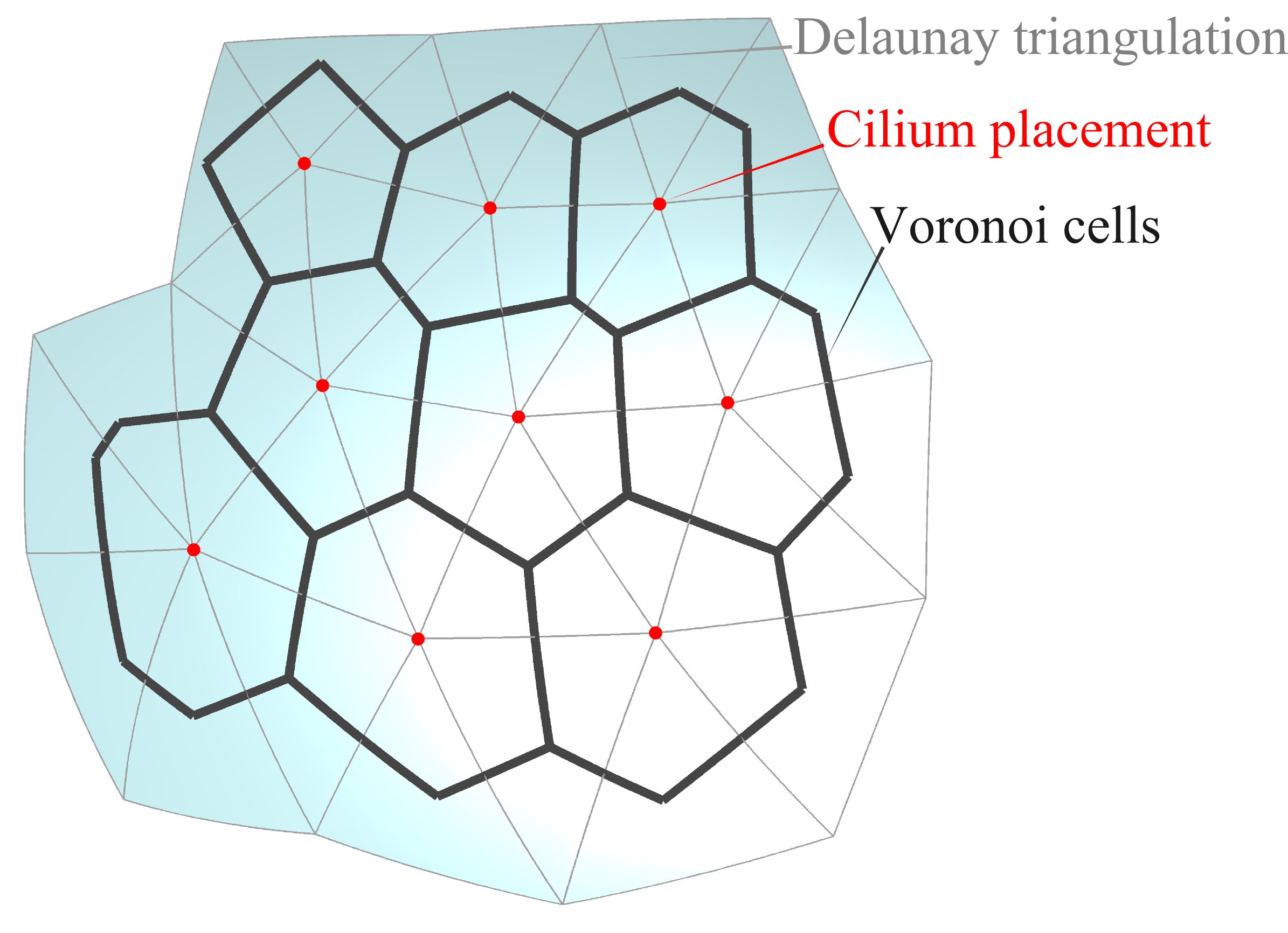
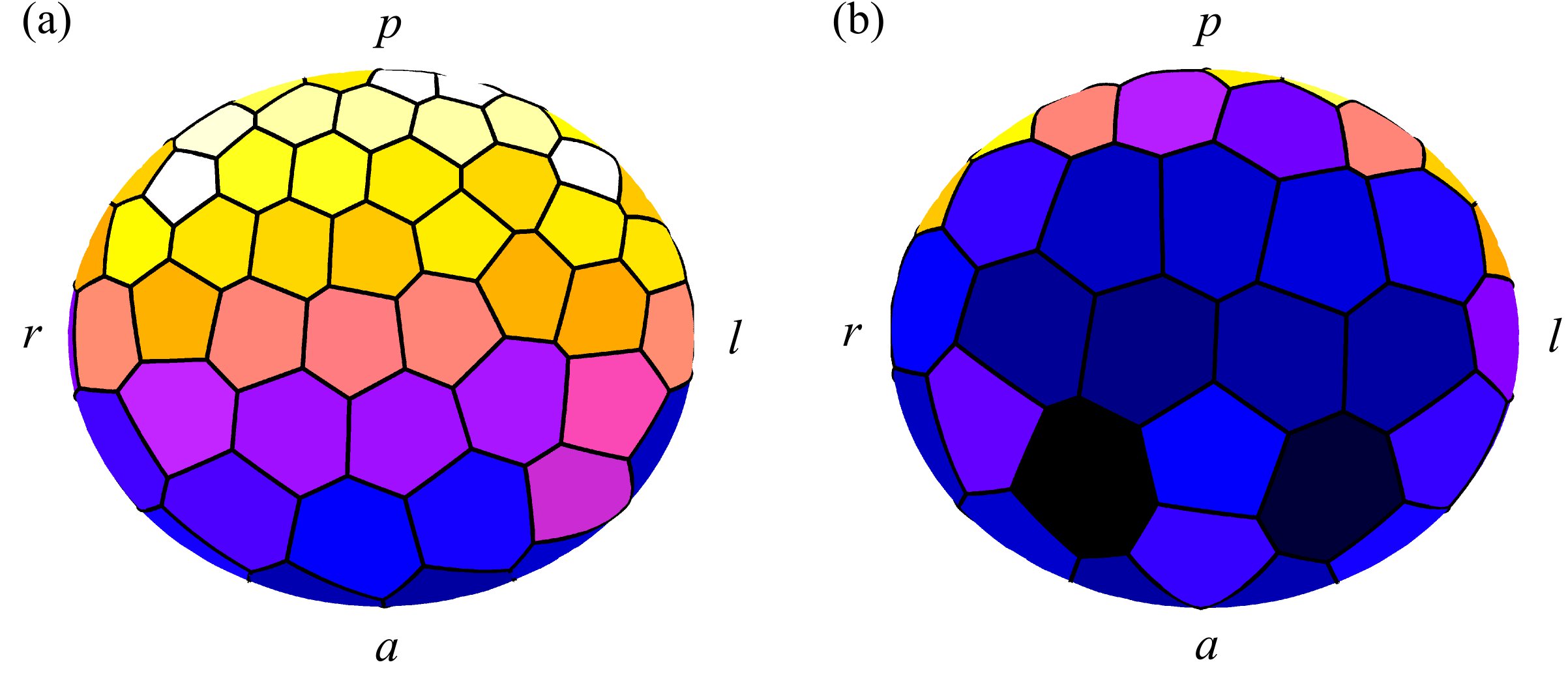
We wish to test the effect of the experimentally observed distribution of cilia, coupled with cilium tilt, on the production of flow inside KV. To achieve this, we first generate a cell-grid that defines the positions of cilia and the boundaries of each cell by creating a Delaunay surface triangulation using DistMesh (Persson & Strang, 2004) and custom MATLAB® routines. A triangulation of a set of points is said to have the Delaunay property if the circumcircle of each triangle contains no other points (Okabe et al., 1992). A cilium is located at the vertex of each triangle (figure 5) so that a triangulation of approximately uniform size yields an even distribution of cilia, whereas a varying triangle size creates a distribution closer to that reported experimentally by Kreiling et al. (2007) and shown in figure 4b. The distribution used is shown in figure 6.


The boundaries of each ciliated cell are given by linking the circumcentres of all triangles that share a given vertex, and projecting onto the curved surface defined by equation (3.7) using the MATLAB® routine fsolve. This is an approximation of the intersection of the Voronoi diagram (see also Okabe et al., 1992) of the set of cilium positions with the ellipsoidal surface, as shown in figure 4a. The cilium itself comprises a cylindrical section with diameter m and length m, and a hemispherical cap.
The cilia move through an approximately conical envelope with a semi-cone angle of . The centreline in the cilium frame at time is given by,
| (3.8) |
where is the arclength along the centreline and . The angle that the tip of the cilium makes with the vertical is given by , and the parameter places the characteristic bend in the base of the cilium shown in figure 7a. The cilium is attached to the cell smoothly via a fixed base that provides the tilt (figure 7b).

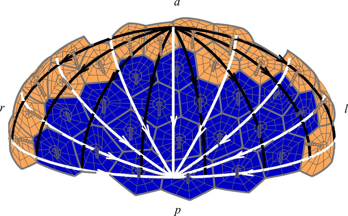
We consider three separate tilt distributions. Firstly, a purely posterior tilt (Kramer-Zucker et al., 2005), secondly, a purely dorsal tilt of (Supatto et al., 2008), and finally, a hybrid model. ‘Posterior tilt’ in our model refers specifically to tilt towards the posterior pole of the vesicle, likewise ‘dorsal tilt’ refers to tilt towards the dorsal pole. The hybrid model consists of a continuous transition from dorsal tilt close to the ‘equator’ () to posterior tilt on the dorsal roof and ventral floor (figure 8).

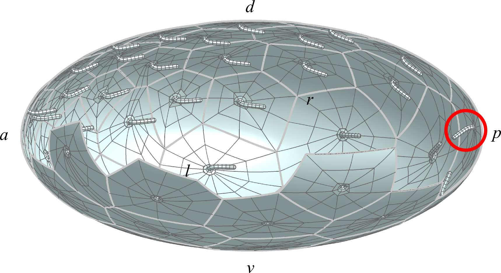
Formalised mathematically as the Hairy Ball Theorem, attempting to specify a constant magnitude direction vector over the entire surface of the ellipsoid inevitably results in a point of discontinuity. For this reason we prescribe the tilt angle to reduce smoothly to zero at the poles where this occurs, therefore we have zero tilt at the posterior pole in the posterior tilt model (figure 9, indicated with a red circle) and zero tilt at the dorsal pole in the dorsal tilt model.

3.3 Numerical implementation
The mathematical problem to be solved is the determination of the unknown stress for from the prescribed surface velocity . This is achieved by discretising the stress, and applying collocation. The stress is discretised as taking piecewise constant values on surface elements , where is the surface mesh and is the number of mesh elements. The discrete form of equation (3.6) is then given by,
| (3.9) |
Taking as the centroid of element while allowing to range over and , we then have equations for unknown scalar stress variables. The numerical solution is carried out as described in the appendix. Once the discrete approximations, , are calculated, the velocity field at any position in the domain can be found by reapplying equation (3.9). This boundary element implementation of the regularized Stokeslet method, with the variable being discretised separately from the numerical quadrature used to evaluate the regularized Stokeslet integrals, was shown by Smith (2009) to give significant advantages in efficiency, an important requirement for solving problems with complex geometry. Note that the calculation described above applies to a specific instant in time; time-dependence was kept implicit in the explanation. In order to calculate the time-averaged flow over a full beat cycle, as in the results reported in section 4, the calculation must be repeated for a sequence of timesteps, over the beat period , the flow field for each timestep being stored and then averaged.
4 Results
For comparison with the experimental results of Supatto et al. (2008) shown in figure 3, we shall examine flow in a centrally located anterior-posterior left-right (AP-LR) plane (plane in our model) at . Results will be shown viewed from the dorsal side, looking in the ventral direction. Additionally, we will compute flow in transverse () sections; we are not aware of comparable experimental data for zebrafish KV, but the results may serve as experimentally-testable predictions, and also may be compared to known flow patterns in the different but related system of medakafish KV.
There are a number of aspects of the flow that could be examined, including the oscillatory component of the flow and the advection of particles by the time-dependent flow. In the present study we shall restrict our reports to the time-average of the instantaneous flow computed by the model over discrete intervals of the cilia beat cycle. Profiles will only be reported in the bulk of the fluid.
We examine how the flow field differs between models with:
- (1)
-
(2)
all cilia tilted towards the dorsal pole (Supatto et al., 2008),
-
(3)
mixed tilt directions as described in section 3.2.
In each case, we compare flow with,
-
(i)
a spatially homogeneous cilia distribution, and
-
(ii)
a spatially varying cilia distribution with maximum density at the anterior of the dorsal roof (Kreiling et al., 2007).
4.1 All cilia tilted posteriorly
For homogeneous cilia density, with posterior tilt only, there is no evidence of an overall clockwise flow (figure 10a). The beat direction is clockwise viewed from tip to base, however the cell surfaces of the dorsal roof and ventral floor face in opposite directions. Hence dorsal roof and ventral floor cilia perform opposite motions. Because cilia density is equal on both surfaces, the overall effect is a nearly zero flow in the AP-LR midplane. A posterior tilt makes no difference to this; flow components in the AP-LR midplane are still equal and opposite. As in all results shown, the largest magnitude flow occurs close to the walls of the vesicle since this is where the cilia are located.
By comparison, inhomogeneous cilia density does produce an overall anticlockwise flow viewed from dorsal (figure 11a) because the opposing rotation directions are no longer equal; the flow produced by the dorsal roof cilia, rotating clockwise when viewed from dorsal, base to tip, predominates over that produced by the ventral floor cilia. The magnitude of the flow is however generally significantly less than the typical reported by Supatto & Vermot (2011). Our simulation results for this configuration show that the centre of the whirl in the plane is moved towards the anterior end of KV.
4.2 All cilia tilted dorsally
The cilia motion in our simulations is always clockwise about its axis of rotation, viewed from tip to base. However, examining the cilia motion about the DV () axis, a dorsally-tilted cilium located around the equatorial region () will spend half of its beat cycle moving with an anticlockwise component relative to the DV axis, and half of its beat cycle moving with an clockwise component relative to the DV axis. The dorsal tilt results in equatorial cilia consistently performing an effective stroke in the anticlockwise direction relative to the DV axis, and a recovery stroke in the clockwise direction relative to the DV axis. The anticlockwise flow relative to the DV axis will predominate, as evident in the anticlockwise pointing arrows in figure 10d (homogeneous distribution), and figure 11d (higher concentration on the dorsal roof). Comparing figures 10d and 11d, it is evident that with cilia concentrated on the anterior of the dorsal roof, the centre of the whirl is again moved to the anterior, moreover the rotational flow velocity is increased relative to the homogeneous case.
4.3 Cilia tilted dorsally and posteriorly
Results for a mixture of posterior tilt on the roof and floor, and dorsal tilt at the equator, are shown in figures 10g and 11g. For homogeneous cilia distribution, the results are very similar to the pure dorsal tilt model, however with increased cilia density on the anterior of the dorsal roof, the mixed tilt results in increased flow magnitude. This anticlockwise global vortex that is strongest from right to anterior to left is similar qualitatively to the experimental observations (Kreiling et al., 2007; Supatto et al., 2008); the flow is of similar magnitude in the region above the cilia tips, and has the largest magnitude of the models considered.
4.4 Comparison of dorsal-only and mixed tilt
While the equatorial sections shown in figures 11(d,g) do not show great differences between dorsal-only and mixed posterior and dorsal tilt, analysis of flow in a transverse section equidistant from the anterior and posterior poles, shows considerable differences. The flow field resulting from exclusively dorsally-tilted cilia is relatively small and does not show any clear directionality (figure 12a). By contrast, a mixture of posterior and dorsal tilt (figure 12b) results in a leftward flow near the dorsal roof, and rightward return flow near the ventral floor of similar magnitude.
5 Summary and discussion
Previous experimental studies of cilia-driven flow inside Kupffer’s vesicle (KV) established that the flow was a circulation about the dorsal-ventral axis from anterior to left (Kreiling et al., 2007; Supatto et al., 2008). However Kreiling et al. and Supatto et al. report different mechanisms which may be responsible for creating the flow. We have developed a theoretical model of flow in the complex geometry of KV using the regularized Stokeslet boundary integral equation. We evaluated the flow field produced by models based on the cilia position and tilt direction features reported by Kreiling et al. and Supatto et al., both separately, and in combination.
Observed profiles are most closely fitted by a model that combines the experimental observations of posterior and dorsal tilt, and moreover takes into account increased cilia density at the anterior of the dorsal roof. Analysis of flow in a transverse section suggests that the mixed posterior and dorsal tilt may more closely match experimental observations in medakafish KV than dorsal-only tilt, in particular showing a leftward flow near the dorsal roof and rightward counterflow near the ventral floor. However, medakafish KV is a different biological entity from zebrafish KV, possessing just a single population of cilia on the dorsal roof and no cilia on the ventral floor; future experimental observations may provide more information on the flow field and therefore the requirement for the mixture of dorsal and posterior tilt postulated here. An interesting finding in terms of interpreting experimental observations is that, in the dorsal-anterior corner, the posterior tilt and a dorsal tilt directions are very similar, and may not be easily distinguishable experimentally.
Our computations were restricted to a population of cilia rotating in synchrony with a frequency of Hz. The latter assumption is not a significant limitation. Reports (see for example Okabe et al. (2008)) suggest that rotational frequency lies within a very narrow range, and indeed is close to the estimated value of Hz. Moreover, the temporal invariance of the Stokes flow equations describing microscale flow entails that fluid velocity is directly proportional to beat frequency; for example that fluid velocity for a beat frequency of Hz can be calculated directly by multiplying our reported results by . The assumption of cilia synchronisation is not likely to affect time-averaged flow properties due to the relative sparsity of the cilia; particle tracking studies however may reveal significant changes to trajectories resulting from non-synchronisation. For example, even changes to cilia phase difference (see for example Smith et al. (2007) figure 12) can significantly alter the trajectories of particles released near to cilia.
There are a number of questions that remain unanswered regarding symmetry breaking flow in KV. Organising structures are growing, transient entities and the changes in size, ciliation and flow field during its existence remain to be elucidated; more extensive experimental observations and modelling will provide information on this issue. The process of how an asymmetric flow is translated into asymmetric development is still unclear. A leftward flow may allow morphogens to be transported to the left allowing a concentration gradient to be established across the left-right axis, subject to the morphogens being bound or rendered inactive before they are returned by the counterflow (Cartwright et al., 2004). If such morphogens are enclosed in micron-sized or larger lipoprotein vesicles, as reported in the mouse (Tanaka et al., 2005), the effect of diffusion may be relatively small compared with advection; we may predict the movement of such vesicles through Lagrangian particle tracking, or simulations which include finite-sized rigid or deformable vesicles suspended in the fluid. Furthermore, it will be possible to calculate the trajectories of individual particles from a range of initial positions and test for evidence of chaos in the domain, as discussed previously (Smith et al., 2007; Supatto et al., 2008) by calculating Lyapunov exponents for a group of particles initially close together (Otto et al., 2001). A related question is how finite-sized particles, including vesicles and other particles imaged experimentally, affect the flow field. The competing hypothesis of mechanical sensing by passive cilia or other mechanotranducers may also be investigated using similar approaches.
It is not clear how KV cilia ‘know’ which way to tilt. In the mouse node, the migration of the cilium basal body towards the posterior, combined with the convex cell surface, establishes the posterior tilt (Hashimoto et al., 2010). The mechanism responsible for the more complex cilia tilt distribution on the inner surface of KV is unclear. Modelling may be beneficial in helping to elucidate any possible cooperative hydrodynamic effect through which organised cilia orientation may emerge.
This study emphasises the tractability of modelling biological flow problems involving relatively complex geometries without the need for simplifying assumptions such as cilium slenderness and precisely planar/spherical boundaries. Examples of other systems which can be modelled in this more refined way include the mouse ventral node; surface features that have not previously been considered, such as the uneven ciliated surface may be taken into account. Finally, and in keeping with the theme of this special issue, we remark that an ‘inverted Kupffer’s vesicle’ bears striking similarities to more frequently-studied objects of biological fluid mechanics modelling, for example Paramecia, Chlamydomonas (Ishikawa & Pedley, 2007; Pedley & Kessler, 1992) and Volvox (Drescher et al., 2009), and therefore the theory in this paper may be generalised to the study of their swimming behaviour.
Acknowledgements
AAS and TDJ acknowledge studentships from EPSRC. DJS acknowledges funding from Birmingham Science City. The computations described in this paper were performed using the University of Birmingham’s BlueBEAR HPC service, which was purchased through HEFCE SRIF-3 funds. See http://www.bear.bham.ac.uk for more details.
JRB acknowledges the significant contributions Professor Tim Pedley has made to the development of his career over the last 40 years. The authors gratefully acknowledge comments from the anonymous referees.
Appendix: numerical implementation
Integrals were evaluated using standard numerical integration rules. Numerical tests showed that a Gauss-Legendre quadrature (Abramowitz & Stegun, 1964) for the curved quadrilateral elements and a point Fekete rule (Taylor et al., 2001) for the curved triangular elements provided satisfactory accuracy, provided the distance between the evaluation point and the element centroid is greater than (element length), where for the quadrilateral elements, for the triangular elements and element length was taken to be the longest side of a triangular element and the greatest distance between opposing corner points of quadrilateral elements. The remaining integrals were evaluated using an adaptive integration routine; briefly, integration results were compared for different rules until the difference between successive rules converged to within a specified tolerance of . These values of were decided upon by calculating each integral for one timestep using our adaptive integration routine everywhere and establishing at which distance was a suitable cut off to switch between a low-order integration rule and using the adaptive integration routine. A tolerance of was sufficient for computations; results were reported for computations performed with a tolerance of . A regularization of m was used.
Meshes contained elements, resulting in scalar degrees of freedom for the stress at each timestep. Results were reported with timesteps. Because there is no explicit time-dependence in the Stokes flow equations, calculations at each timestep were independent. Restarted GMRES was used to solve the linear system (FORTRAN library routines F11BDF/BEF/BFF, F11XAF, F11DBF, Numerical Algorithms Group, Oxford).
References
- Abramowitz & Stegun (1964) Abramowitz, M. & Stegun, I. A. 1964 Handbook of mathematical functions with formulas, graphs, and mathematical tables. Dover publications.
- Afzelius (1976) Afzelius, B. A. 1976 A human syndrome caused by immotile cilia. Science 193 (4250), 317–319.
- Ainley et al. (2008) Ainley, J., Durkin, S., Embid, R., Boindala, P. & Cortez, R. 2008 The method of images for regularized Stokeslets. J. Comput. Phys. 227 (9), 4600–4616.
- Berdon et al. (2004) Berdon, W. E., McManus, C. & Afzelius, B. 2004 More on Kartagener s syndrome and the contributions of Afzelius and A.K. Siewert. Pediatr. Radiol. 34 (7), 585–586.
- Berdon & Willi (2004) Berdon, W. E. & Willi, U. 2004 Situs inversus, bronchiectasis, and sinusitis and its relation to immotile cilia: history of the diseases and their discoverers — Manes Kartagener and Bjorn Afzelius. Pediatr. Radiol. 34 (1), 38–42.
- Blake (1971) Blake, J. R. 1971 A note on the image system for a stokeslet in a no-slip boundary. Math. Proc. Camb. Philos. Soc. 70, 303–310.
- Blake & Chwang (1974) Blake, J. R. & Chwang, A. T. 1974 Fundamental singularities of viscous flow. J. Eng. Math. 8 (1), 23–29.
- Brokaw (2005) Brokaw, C. J. 2005 Computer simulation of flagellar movement IX. Oscillation and symmetry breaking in a model for short flagella and nodal cilia. Cell Motil. Cytoskeleton 60 (1), 35–47.
- Cartwright et al. (2007) Cartwright, J. H. E., Piro, N., Piro, O. & Tuval, I. 2007 Embryonic nodal flow and the dynamics of nodal vesicular parcels. J. R. Soc. Interface 4 (12), 49–55.
- Cartwright et al. (2008) Cartwright, J. H. E., Piro, N., Piro, O. & Tuval, I. 2008 Fluid dynamics of nodal flow and left right patterning in development. Dev. Dyn. 237 (12), 3477–3490.
- Cartwright et al. (2004) Cartwright, J. H. E., Piro, O. & Tuval, I. 2004 Fluid-dynamical basis of the embryonic development of left-right asymmetry in vertebrates. Proc. Natl. Acad. Sci. USA 101 (19), 7234–7239.
- Cartwright et al. (2009) Cartwright, J. H. E., Piro, O. & Tuval, I. 2009 Fluid dynamics in developmental biology: moving fluids that shape ontogeny. HFSP J. 3 (2), 77–93.
- Cortez (2001) Cortez, R. 2001 The method of regularized Stokeslets. SIAM Journal on Scientific Computing 23 (4), 1204–1225.
- Cortez et al. (2005) Cortez, R., Fauci, L. & Medovikov, A. 2005 The method of regularized Stokeslets in three dimensions: analysis, validation, and application to helical swimming. Phys. Fluids 17 (031504), 1–14.
- Drescher et al. (2009) Drescher, K., Leptos, K. C., Tuval, I., Ishikawa, T., Pedley, T. J. & Goldstein, R. E. 2009 Dancing Volvox: hydrodynamic bound states of swimming algae. Phys. Rev. Lett. 102 (16), 168101.
- Hashimoto et al. (2010) Hashimoto, M., Shinohara, K., Wang, J., Ikeuchi, S., Yoshiba, S., Meno, C., Nonaka, S., Takada, S., Hatta, K., Wynshaw-Boris, A. et al. 2010 Planar polarization of node cells determines the rotational axis of node cilia. Nat. Cell Biol. 12 (2), 170–176.
- Hirokawa et al. (2009) Hirokawa, N., Okada, Y. & Tanaka, Y. 2009 Fluid dynamic mechanism responsible for breaking the left-right symmetry of the human body: the nodal flow. Annu. Rev. Fluid Mech. 41, 53–72.
- Ibañes & Belmonte (2009) Ibañes, M. & Belmonte, J. C. I. 2009 Left-right axis determination. WIREs: Systems Biology and Medicine 1 (2), 210–219.
- Ishikawa & Pedley (2007) Ishikawa, T. & Pedley, T. J. 2007 Diffusion of swimming model micro-organisms in a semi-dilute suspension. J. Fluid Mech. 588, 437–462.
- Kartagener (1933) Kartagener, M. 1933 Zur pathogenese der bronchiektasien. Lung 84 (1), 73–85.
- Kawakami et al. (2005) Kawakami, Y., Raya, Á., Raya, R. M., Rodríguez-Esteban, C. & Belmonte, J. C. I. 2005 Retinoic acid signalling links left–right asymmetric patterning and bilaterally symmetric somitogenesis in the zebrafish embryo. Nature 435 (7039), 165–171.
- Kimmel et al. (1995) Kimmel, C. B., Ballard, W. W., Kimmel, S. R., Ullmann, B. & Schilling, T. F. 1995 Stages of embryonic development of the zebrafish. Am. J. Anat. 203 (3), 253–310.
- Kramer-Zucker et al. (2005) Kramer-Zucker, A. G., Olale, F., Haycraft, C. J., Yoder, B. K., Schier, A. F. & Drummond, I. A. 2005 Cilia-driven fluid flow in the zebrafish pronephros, brain and Kupffer’s vesicle is required for normal organogenesis. Development 132 (8), 1907–1921.
- Kreiling et al. (2007) Kreiling, J. A., Prabhat, Williams, G. & Creton, R. 2007 Analysis of Kupffer’s vesicle in zebrafish embryos using a cave automated virtual environment. Dev. Dyn. 236 (7), 1963–1969.
- Nonaka et al. (2002) Nonaka, S., Shiratori, H., Saijoh, Y. & Hamada, H. 2002 Determination of left–right patterning of the mouse embryo by artificial nodal flow. Nature 418 (6893), 96–99.
- Nonaka et al. (1998) Nonaka, S., Tanaka, Y., Okada, Y., Takeda, S., Harada, A., Kanai, Y., Kido, M. & Hirokawa, N. 1998 Randomization of left-right asymmetry due to loss of nodal cilia generating leftward flow of extraembryonic fluid in mice lacking KIF3B motor protein. Cell 95 (6), 829–837.
- Nonaka et al. (2005) Nonaka, S., Yoshiba, S., Watanabe, D., Ikeuchi, S., Goto, T., Marshall, W. F. & Hamada, H. 2005 De novo formation of left-right asymmetry by posterior tilt of nodal cilia. PLoS Biol. 3 (8), 1467–1472.
- Okabe et al. (1992) Okabe, A., Boots, B. N., Sugihara, K. & Chiu, S. 1992 Spatial Tessellations: Concepts and Applications of Voronoi Diagrams. J. Wiley.
- Okabe et al. (2008) Okabe, N., Xu, B. & Burdine, R. D. 2008 Fluid dynamics in zebrafish Kupffer’s vesicle. Dev. Dyn. 237 (12), 3602–3612.
- Okada et al. (2005) Okada, Y., Takeda, S., Tanaka, Y., Belmonte, J. C. I. & Hirokawa, N. 2005 Mechanism of nodal flow: a conserved symmetry breaking event in left-right axis determination. Cell 121 (4), 633–644.
- Otto et al. (2001) Otto, S. R., Yannacopoulos, A. N. & Blake, J. R. 2001 Transport and mixing in Stokes flow: the effect of chaotic dynamics on the blinking stokeslet. J. Fluid Mech. 430, 1–26.
- Pedley & Kessler (1992) Pedley, T. J. & Kessler, J. O. 1992 Hydrodynamic phenomena in suspensions of swimming microorganisms. Annu. Rev. Fluid Mech. 24 (1), 313–358.
- Persson & Strang (2004) Persson, P. O. & Strang, G. 2004 A simple mesh generator in MATLAB. SIAM Rev. 46 (2), 329–345.
- Pozrikidis (1992) Pozrikidis, C. 1992 Boundary integral and singularity methods for linearized viscous flow. Cambridge Univ Press.
- Rawls et al. (2001) Rawls, J. F., Mellgren, E. M. & Johnson, S. L. 2001 How the zebrafish gets its stripes. Dev. Biol. 240 (2), 301–314.
- Smith (2009) Smith, D. J. 2009 A boundary element regularized Stokeslet method applied to cilia- and flagella-driven flow. Proc. R. Soc. Lond. A 465, 3605–3626.
- Smith et al. (2008) Smith, D. J., Blake, J. R. & Gaffney, E. A. 2008 Fluid mechanics of nodal flow due to embryonic primary cilia. J. R. Soc. Interface 5 (22), 567–573.
- Smith et al. (2007) Smith, D. J., Gaffney, E. A. & Blake, J. R. 2007 Discrete cilia modelling with singularity distributions: application to the embryonic node and the airway surface liquid. Bull. Math. Biol. 69 (5), 1477–1510.
- Smith et al. (2011) Smith, D. J., Smith, A. A. & Blake, J. R. 2011 Mathematical embryology: the fluid mechanics of nodal cilia. J. Eng. Math. 70, 255–279.
- Sulik et al. (1994) Sulik, K., Dehart, D. B., Inagaki, T., Carson, J. L., Vrablic, T., Gesteland, K. & Schoenwolf, G. C. 1994 Morphogenesis of the murine node and notochordal plate. Am. J. Anat. 201 (3), 260–278.
- Supatto et al. (2008) Supatto, W., Fraser, S. E. & Vermot, J. 2008 An all-optical approach for probing microscopic flows in living embryos. Biophys. J. 95 (4), 29–31.
- Supatto & Vermot (2011) Supatto, W. & Vermot, J. 2011 From cilia hydrodynamics to zebrafish embryonic development. Curr. Topics Dev. Biol. 95, 33.
- Tanaka et al. (2005) Tanaka, Y., Okada, Y. & Hirokawa, N. 2005 FGF-induced vesicular release of Sonic hedgehog and retinoic acid in leftward nodal flow is critical for left–right determination. Nature 435 (7039), 172–177.
- Taylor et al. (2001) Taylor, M. A., Wingate, B. A & Vincent, R. E. 2001 An algorithm for computing Fekete points in the triangle. SIAM J. Numer. Anal. 38 (5), 1707–1720.