Harnessing nuclear spin polarization fluctuations in a semiconductor nanowire
Abstract
Soon after the first measurements of nuclear magnetic resonance (NMR) in a condensed matter system, Bloch [1] predicted the presence of statistical fluctuations proportional to in the polarization of an ensemble of spins. First observed by Sleator et al. [2], so-called “spin noise” has recently emerged as a critical ingredient in nanometer-scale magnetic resonance imaging (nanoMRI) [3, 4, 5, 6]. This prominence is a direct result of MRI resolution improving to better than , a size-scale in which statistical spin fluctuations begin to dominate the polarization dynamics. We demonstrate a technique that creates spin order in nanometer-scale ensembles of nuclear spins by harnessing these fluctuations to produce polarizations both larger and narrower than the natural thermal distribution. We focus on ensembles containing phosphorus and hydrogen spins associated with single InP and GaP nanowires (NWs) and their hydrogen-containing adsorbate layers. We monitor, control, and capture fluctuations in the ensemble’s spin polarization in real-time and store them for extended periods. This selective capture of large polarization fluctuations may provide a route for enhancing the weak magnetic signals produced by nanometer-scale volumes of nuclear spins. The scheme may also prove useful for initializing the nuclear hyperfine field of electron spin qubits in the solid-state.
Spin noise is a phenomenon present in all spin ensembles that begins to exceed the mean thermal polarization as the size of the ensemble shrinks. These fluctuations have random amplitude and phase and have been observed in a wide variety of nuclear spin systems including in liquids by conventional NMR [7, 8] and in the solid-state using a superconducting interference device (SQUID)[2], by force-detected magnetic resonance [9], or by nitrogen-vacancy (NV) magnetometry [4, 5]. In addition to finding applications in nanoMRI, spin noise has been used in MRI specially adapted for the investigation of extremely delicate specimens, in which external radio frequency (RF) irradiation is not desired [10]. Through the hyperfine interaction, nuclear spin noise also sets limits on the coherence of electronic qubits in the solid state [11, 12, 13, 14]. Various efforts to mitigate these nuclear field fluctuations by hyperfine-mediated nuclear spin preparation have been developed, both in quantum dots (QDs) [15, 16, 17, 18, 19] and in NV centers in diamond [20]. We report on a manipulation and initialization technique which applies to arbitrary nanometer-scale samples, i.e. it does not require specialized structures providing a controllable electronic spin and a strong hyperfine interaction. Rather, our technique requires a detector sensitive enough to resolve nuclear spin noise and the ability to apply RF electro-magnetic pulses.
Conventional NMR and MRI techniques rely on manipulating the mean thermal polarization to produce signals. Statistical nuclear polarization fluctuations exceed the mean thermal polarization below a critical number of spins , where is the spin quantum number, is the Boltzmann constant, is the temperature, is Planck’s constant, is the gyromagnetic ratio, and is the magnetic field [21]. For 1H spins at K in an applied magnetic field T, , which in most organic samples corresponds to a critical volume . Under the ambient conditions of recent NMR experiments with NV spin sensors, statistical polarization begins to dominate for even larger samples with [4, 5]. In order to measure and initialize such ensembles, it is therefore worthwhile to consider techniques designed to both detect and control nuclear polarization fluctuations. Although nuclear magnetic signals from such small volumes are weak, if a detector is able to resolve spin noise in real-time, i.e. faster than the spin correlation time , large fluctuations can be captured. Once captured, these fluctuations can then be used to initialize the polarization of nanometer-scale spin ensembles with a fixed sign and magnitude. Such initialization schemes could provide the basis for enhancing signals from small samples and for realizing advanced pulse protocols which can be borrowed directly from conventional NMR.
In 2005, Budakian et al. first demonstrated a technique for the capture and storage of electron spin fluctuations [22]. Inspired by this work, we have adapted the protocol to nanometer-scale ensembles of nuclear spins. Given the relevance of these ensembles to nanoMRI and to the decoherence of solid-state qubits, this implementation could find wide applicability. Furthermore, the longer spin relaxation time of nuclear spins compared to electron spins – sometimes as long as several hours at cryogenic temperatures – allows captured spin order to be stored for far longer times.
We study two separate samples: an InP and a GaP NW, both grown with the vapor-liquid-solid method in a metal-organic vapor-phase epitaxy reactor using gold droplets as catalyst [23]. Using a magnetic resonance force microscope (MRFM), we measure the polarization of nanometer-scale ensembles of 31P nuclei within each NW and of 1H nuclei contained in the hydrocarbon adsorbate layer on the surface. Adiabatic rapid passage (ARP) pulses of the transverse field [24] are used to cyclically invert the polarization of nanometer-scale volumes of a particular nuclear isotope. In a magnetic field gradient, these periodic inversions generate an alternating force that drives the mechanical resonance of an ultrasensitive cantilever; the resulting oscillations, which are proportional to this force, are detected by a fiber-optic interferometer.
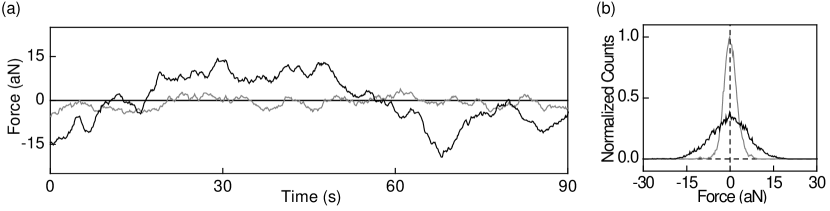
We feed the cantilever force signal to a lock-in amplifier referenced to the periodic spin inversions and monitor its in-phase () and quadrature () amplitudes. The limiting source of noise in the measurement is the thermal noise of the cantilever, which has a random phase and on average contributes equally to and . On the other hand, force fluctuations due to the nuclear spin polarization are in phase with the inversion pulses and contribute only to . Thus, as shown in Fig. 2a, we can monitor , the thermal force, and , the thermal force plus the force due to the nuclear spin inversions. is dominated by the large fluctuations and the long of the spin noise, while the thermal noise in has a smaller amplitude and a shorter correlation time set by the damped cantilever force sensor (see methods). is limited by the magneto-mechanical noise originating from the thermal motion of the cantilever in a magnetic field gradient and by the ARP pulse parameters [25]. Since the contribution of the spin signal to is large enough, we can follow – in real-time – the instantaneous nuclear spin imbalance in the rotating frame. Fig. 2b shows the variances and , which give the variance due only to the thermal noise and the variance due only to the spin noise, . is in turn related to the number of nuclear spins in the detection volume (see methods). The typical size of the spin ensembles we measure is between and for 1H and between and for 31P. Given the density of typical adsorbate layers and InP and GaP, the detection volumes discussed here are between and for 1H and between and for 31P.
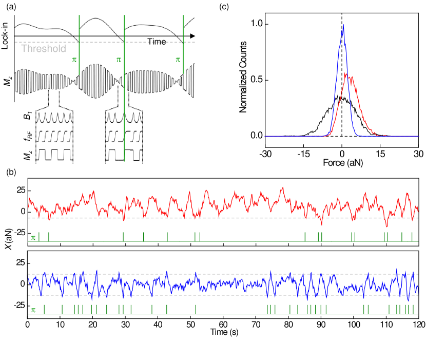
The real-time measurement of spin noise allows us to react to and control the fluctuations in polarization [22]. As a demonstration, we conditionally apply RF -inversion pulses to both rectify and narrow the naturally occurring polarization distribution. This feedback is realized via a field-programmable gate array (FPGA) in conjunction with an arbitrary waveform generator (AWG). As shown in Fig. 3a, when exceeds a predetermined threshold, the protocol applies a -inversion, by inserting an ARP pulse with a duration equivalent to a full cantilever cycle rather than the usual half-cycle. As a result, the spin ensemble’s periodic inversion at the cantilever frequency undergoes a phase shift. In this way, we can either rectify or narrow by setting appropriate thresholds as shown in Fig. 3b. Since only fluctuations due to nuclear spins are affected by the -inversions, the effectiveness of the protocol depends on the fraction of arising from spin compared to thermal fluctuations, i.e. the larger the power signal-to-noise ratio (SNR) is, the more effective the control of will be. As shown in Fig. 3c, this process can produce both hyper-polarized and narrowed nuclear spin distributions in the rotating frame.
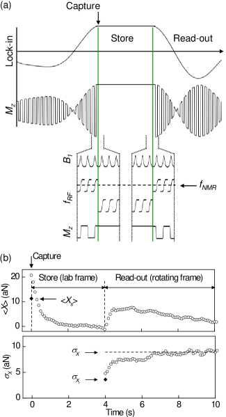
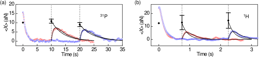
In addition to controlling an ensemble’s natural spin fluctuations, we can also capture especially large fluctuations [22]. As shown in Fig. 4, using the FPGA and AWG, we continuously measure the rotating-frame spin fluctuations by monitoring . Once reaches a predetermined threshold , the ARP pulses are tuned out of resonance with the nuclear spin ensemble, leaving the instantaneous spin polarization pointing along and transferring the transient spin order to the laboratory frame. Since the spin ensemble does not respond to the off-resonant ARP pulses, the cantilever is no longer driven by spin forces. In this way, hyper-polarized states of the nuclear spin ensemble can be captured and stored in the laboratory frame for as long as . Crucially, the time-scale required to capture the spin order – here given by the inverse of the cantilever frequency – must be much shorter than the rotating-frame correlation time . To confirm that the nuclear polarization has in fact been stored, we can reapply the resonant ARP pulses after some storage time to readout the polarization, once again cyclically inverting the polarization. These spin inversions again drive the cantilever motion, and the amplitude of reflects the size of the retrieved fluctuation.
In an idealized case, in which the spin component of the captured fluctuation is fully projected onto , the stored fluctuation will be normally distributed with a mean and a variance (see supplementary material). The fact that reflects the finite SNR of the measurement, in this case limited by the cantilever’s thermal noise. Note also that the distribution of the stored polarization is narrowed, i.e. it has a reduced variance, compared to the variance of under normal evolution (). In the limit of large SNR (), and . If during this stored polarization undergoes negligible relaxation in the laboratory frame, the corresponding retrieved fluctuation has a mean and a variance . Note that due to the finite SNR of the retrieval measurement.
In order to compare our experiments to this idealized case, we measure and for nanometer-scale 31P and 1H spin ensembles. As shown in Fig. 5, and are extracted from bi-exponential fits to during the readout sequence using our knowledge of the lock-in time constant . Deviations of from could be caused by the spin-lattice relaxation of the polarization in the laboratory frame – set by – or by the incomplete projection of the polarization onto . For both 31P and 1H in Fig. 5a and b, these deviations are negligible within our error for two values of . Limitations of the experimental hardware prevented measurements for larger , although our data show that s for 31P in InP and s for 1H on GaP at K.
Fig. 4b shows the reduced variance of the retrieved polarization , eventually approaching after a time on the order of in the rotating frame. Note that is much shorter than the time over which evolves, excluding the lock-in as a source of the behavior. This result demonstrates that the distribution of the captured fluctuations is indeed narrowed relative to the natural distribution of the nuclear spin fluctuations. The ability to prepare such distributions may find application in quantum information processing with solid-state electron spins, which is often limited by the random nuclear field distribution in the host material [15, 16, 17, 18, 19]. The nuclear spin ensemble in such a system could be initialized before each measurement by a scheme based on the capture of random polarization fluctuations, thus enhancing the electron-spin dephasing time. The degree of narrowing demonstrated in Fig. 4b represents a factor of compared to the natural distribution and is limited by the SNR of the measurement.
The size of the captured spin fluctuation is, in principle, only limited by the amount of time one is willing to wait during the capture step. For the normally distributed random variable , the average amount of wait time required to capture a fluctuation is given by , where is the average number of times crosses zero per second (see supplementary material) [26]. For example, given that Hz is a typical value in our experiments, fluctuations of are expected after just min, or alternatively for hr, a fluctuation of can be expected. For a 31P spin ensemble with at T and K, the standard deviation of the statistical polarization fluctuations is given by and its mean thermal polarization is [21]. Therefore, in the limit of large SNR where is dominated by spin fluctuations, we can expect to capture polarizations of in 15 min and in 1 hr. The polarization captured in a given could be increased even further by reducing and therefore increasing , e.g. through the periodic randomization of the spin ensemble using bursts of -pulses [27]. In principle, the nuclear spin decoherence time sets the lower limit for . As the size of the spin ensemble shrinks, the size of the achievable polarization increases as , making such a protocol increasingly relevant as detection volumes continue to shrink. Given that conventional pulse protocols based on thermal polarization require an initialization step taking at least , waiting for an extended time to capture a large spin fluctuation may be attractive – especially when the magnitude of the captured fluctuation greatly exceeds the possible thermal polarization.
Note that the crucial step in the capture protocol is that the monitoring and capture occurs in the rotating frame, where the time between statistically independent spin configurations – set by – is much faster than the equivalent time in the laboratory frame. This reduced rotating-frame correlation time, allows the system to quickly explore its spin configuration space. On the other hand, once a large fluctuation is captured and transferred to the laboratory frame, the long effectively freezes the ensemble in this rare configuration.
We therefore show the long-term storage of large polarization fluctuations arising from spin noise in nanometer-scale nuclear spin ensembles with . Storage times as long as 20 s for 31P are demonstrated, limited only by the measurement hardware. The ultimate limit to these times is set only by in the laboratory frame, which at low temperatures is typically extremely long, exceeding hours for some nuclear spins. While these results were obtained with a low-temperature MRFM, the capture and storage of spin fluctuations should be generally applicable to any technique capable of detecting and addressing nanometer-scale volumes of nuclear spins in real-time.
The ability to initialize the polarization of a small nuclear spin ensemble is important for the development of future nanometer-scale NMR techniques or possibly for the implementation of solid-state qubits and nuclear spin memory devices. Given that the mean thermal polarization is typically negligible in nanometer-scale samples, other methods to create nuclear spin order must be considered. When polarization cannot be created via effects such as Overhauser-mediated dynamic nuclear polarization or electron-nuclear double resonance, the selective capture of large statistical fluctuations provides a viable alternative. The ensembles polarized in this work, nanometer-scale volumes of 1H on an adsorbate layer and 31P in a single semiconducting NW, demonstrate the types of samples which could benefit from this technique. One could imagine, for instance, such nuclear polarization capture processes enhancing the weak MRI signals of a nanometer-scale 1H-containing biological sample on a surface or of a semiconducting nanostructure.
I Methods
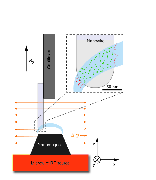
The MRFM consists of an nanomagnetic tip integrated on top of a microwire RF source, an ultrasensitive Si cantilever, and a fiber-optic interferometer to measure its displacement [28]. The entire apparatus operates in vacuum better than mbar, at temperatures down to K, and in an applied longitudinal field up to T. The NW of interest is attached to the end of the cantilever and is positioned within 100 nm of the nanomagnetic tip, as shown schematically in Fig. 1. Here the field produced by the tip, , results in a total static magnetic field , while the microwire produces a transverse RF field . ARP pulses invert the nuclear spin polarization because the polarization follows (or is “spin-locked” to) the time-dependent effective field in a frame rotating with the RF field, where is the instantaneous frequency of the ARP pulses, their amplitude, and the unit vectors are defined in the rotating frame.
The volume of inverted spins, known as the “resonant slice”, is determined by the spatial dependence of and the parameters of the pulses. This force variance is in turn related to the number of nuclear spins in this volume by the approximation, , where is the direction of cantilever oscillation [21]. This relation holds as long as the volume of spins is small enough that the gradient is nearly constant throughout; otherwise it provides a lower bound on the number of spins. From measurements of the magnetic field gradient in the vicinity of the tip, we estimate that T/m at the position of the detection volume (see supplementary material). In order to set an upper bound for the number of spins , we make a model of the magnetic field profile produced by the nanomagnetic tip and numerically integrate the spatially dependent gradient over the sample volume contained in the “resonant slice” (see supplementary material).
Ultrasensitive cantilevers are made from undoped single-crystal Si and measure 120 m in length, 4 m in width, and 0.1 m in thickness. In vacuum and at the operating temperatures, the NW-loaded cantilevers have resonant frequencies and kHz, intrinsic quality factors and , and spring constants and N/m for the InP and GaP NW experiments respectively. The InP NW is 8 m-long; its diameter shrinks from 200 nm to 60 nm along its length; and it is tipped by a 60 nm diameter Au catalyst particle, left over from the growth process. The GaP NW is 10 m-long; its diameter is 1.0 m; and it has a 1.5-m-long tapered tip which reaches 90 nm in diameter at the Au droplet. In this case we remove the Au droplet by cutting of the end of the NW with a focused ion beam, resulting in a 300-nm-diameter GaP tip. Finally, we sputter a 5 nm layer of Pt onto the NW in order to shield electrostatic interactions [29]. Each NW is affixed to the end of the cantilever with less than 100 fL of epoxy (Gatan G1). In the attachment process, we employ an optical microscope equipped with a long working distance and a pair of micromanipulators. During the measurement, the cantilever is actively damped in order to give it a fast response time of ms. Up to 50 nW of laser light at 1550 nm are incident on the cantilever as part of the fiber-optic interferometer. The nanomagnetic tips are truncated cones of Dy fabricated by optical lithography [30]. For the InP (GaP) NW experiment the tip measures 225 (280) nm in height, 270 (250) nm in upper diameter, and 380 (500) nm in lower diameter. The RF source, on which the Dy tip sits, is a 2-m-long, 1-m-wide, and 200-nm-thick Au microwire. By positioning each NW within 100 nm of the combined structure, the detection volume can be exposed to spatial magnetic field gradients exceeding T/m and RF fields larger than 20 mT without significant changes in the experimental operating temperature. We use hyperbolic secant ARP pulses with and a modulation amplitude set to 500 kHz peak-to-peak for 31P and 1 MHz for 1H [24]. These parameters, combined with the geometry of the sample and the profile of , determine the size of the detection volume [21].
Acknowledgements.
II Acknowledgments
The authors thank C. L. Degen, C. Klöffel, T. Poggio, and R. J. Warburton for illuminating discussions; Hari S. Solanki for experimental assistance; and S. Keerthana for assistance with a figure. We acknowledge support from the Canton Aargau, the Swiss National Science Foundation (SNF, Grant No. 200020-140478), the Swiss Nanoscience Institute, and the National Center of Competence in Research for Quantum Science and Technology.
III Author contributions
P.P. and M.P conceived and planned the experiments in collaboration with F.X. P.P. carried out the experiments. P.P. and M.P. analyzed the data and wrote the manuscript. P.P. and F. X. prepared the samples and devices. The nanowires were grown by H.I.T.H., S.A. and E.P.A.M.B. All authors discussed the results and contributed to the manuscript.
References
- [1] Bloch, F. Nuclear induction. Phys. Rev. 70, 460-474 (1946).
- [2] Sleator, T., Hahn, E. L., Hilbert, C. & Clarke, J. Nuclear-spin noise. Phys. Rev. Lett. 55, 1742-1745 (1985); Sleator, T. & Hahn, E. L. Nuclear-spin noise and spontaneous emission. Phys. Rev. B 36, 1969-1980 (1987).
- [3] Degen, C. L., Poggio, M., Mamin, H. J., Rettner, C. T. & Rugar, D. Nanoscale magnetic resonance imaging. Proc. Natl. Acad. Sci. USA 106, 1313-1317 (2009).
- [4] Mamin, H. J., Kim, M., Sherwood, M. H., Rettner, C. T., Ohno, K., Awschalom, D. D. & Rugar, D. Nanoscale nuclear magnetic resonance with a nitrogen-vacancy spin sensor. Science 339, 557-560 (2013).
- [5] Staudacher, T., Shi, F., Pezzagna, S., Meiger, J., Du, J., Meriles, C. A., Reinhard, F. & Wrachtrup, J. Nuclear magnetic resonance spectroscopy on a (5-nanometer)3 sample volume. Science 339, 561-563 (2013).
- [6] Nichol, J. M., Naibert, T. R., Hemesath, E. R., Lauhon, L. J. & Budakian, R. Nanoscale Fourier-transform MRI of spin noise. arXiv:1302.2977 (2013).
- [7] McCoy, M. A. & Ernst, R. R. Nuclear spin noise at room temperature. Chem. Phys. Lett. 159, 587-593 (1989).
- [8] Guéron, M. & Leroy, J. L. NMR of water protons. The detection of their nuclear-spin noise, and a simple determination of absolute probe sensitivity based on radiation damping. J. Magn. Reson. 85, 209-215 (1989).
- [9] Mamin, H. J., Budakian, R., Chui, B. W. & Rugar, D. Detection and manipulation of statistical polarization in small spin ensembles. Phys. Rev. Lett. 91, 207604 (2003); Mamin, H. J., Budakian, R., Chui, B. W. & Rugar, D. Magnetic resonance force microscopy of nuclear spins: detection and manipulation of statistical polarization. Phys. Rev. B 72, 024413 (2005).
- [10] Müller, N. & Jerschow, A. Nuclear spin noise imaging. Proc. Natl. Acad. Sci. USA 103, 6790-6792 (2006).
- [11] Merkulov, I. A., Efros, AI. L. & Rosen, M. Electron spin relaxation by nuclei in semiconductor quantum dots. Phys. Rev. B 65, 205309 (2002).
- [12] Khaetskii, A. V., Loss D. & Glazman, L. Electron spin decoherence in quantum dots due to interaction with nuclei. Phys. Rev. Lett. 88, 186802 (2002).
- [13] Childress, L., Gurudev Dutt, M. V., Taylor, J. M., Zibrov, A. S., Jelezko, F., Wrachtrup, J., Hemmer, P. R. & Lukin, M. D. Coherent dynamics of coupled electron and nuclear spin qubits in diamond. Science 314, 281-285 (2006).
- [14] Kuhlmann, A. V., Houel, J., Ludwig, A., Greuter, L., Reuter, D., Wieck, A. D., Poggio, M. & Warburton, R. J. Charge noise and spin noise in a semiconductor quantum device. arXiv:1301.6381 (2013).
- [15] Coish W. A. & Loss D. Hyperfine interaction in a quantum dot: Non-Markovian electron spin dynamics. Phys. Rev. B 70, 195340 (2004).
- [16] Reilly, D. J., Taylor, J. M., Petta, J. R., Marcus, C. M., Hanson, M. P. & Gossard, A. C. Suppressing spin qubit dephasing by nuclear state preparation. Science 321, 817-821 (2008).
- [17] Latta, C., Högele, A., Zhao, Y., Vamivakas, A. N., Maletinsky, P., Kroner, M., Dreiser, J., Carusotto, I., Badolato, A., Schuh, D., Wegscheider, W., Atature, M. & Imamoglu, A. Confluence of resonant laser excitation and bidirectional quantum-dot nuclear-spin polarization. Nature Phys. 5, 758-763 (2009).
- [18] Vink, I., Nowack, K. C., Koppens, F. H. L., Danon, J., Nazarov, Y. V. & Vandersypen, L. M. K. Locking electron spins into magnetic resonance by electron-nuclear feedback. Nature Phys. 5, 764-768 (2009).
- [19] Bluhm, H., Foletti, S., Mahalu, D., Umansky, V. & Yacoby, A. Enhancing the coherence of a spin qubit by operating it as a feedback loop that controls its nuclear spin bath. Phys. Rev. Lett. 105, 216803 (2010).¡
- [20] Togan, E., Chu, Y., Imamoglu, A. & Lukin, M. D. Laser cooling and real-time measurement of the nuclear spin environment of a solid-state qubit. Nature 478, 497-501 (2011).
- [21] Xue, F., Weber, D. P., Peddibhotla, P. & Poggio, M. Measurement of statistical nuclear spin polarization in a nanoscale GaAs sample. Phys. Rev. B 84, 205328 (2011).
- [22] Budakian, R., Mamin, H. J., Chui, B. W. & Rugar, D. Creating order from random fluctuations in small spin ensembles. Science 307, 408-411 (2005).
- [23] Assali, S., Zardo, I., Plissard, S., Kriegner, D., Verheijen, M. A., Bauer, G., Meijerink, A., Belabbes, A., Bechstedt, F., Haverkort, J. E. M. & Bakkers, E. P. A. M. Nano Lett. ASAP, DOI: 10.1021/nl304723c.
- [24] Tannús, A. & Garwood, M. Improved performance of frequency-swept pulses using offset-independent adiabaticity. J. Magn. Reson., Ser A 120, 133-137 (1996); Tomka, I. T., van Beek, J. D., Joss, R. & Meier, B. H. Spatio-chemical characterization of a polymer blend by magnetic resonance force microscopy. Phys. Chem. Chem. Phys. 15, 3438 (2013).
- [25] Degen, C. L., Poggio, M., Mamin, H. J. & Rugar, D. Nuclear spin relaxation induced by a mechanical resonator. Phys. Rev. Lett. 100, 137601 (2008).
- [26] Rice, S. O. Mathematical analysis of random noise. AT&T Tech. J. 23, 282-332 (1944); AT&T Tech. J. 24, 46-156 (1944).
- [27] Degen, C. L., Poggio, M., Mamin, H. J. & Rugar, D. Role of spin noise in the detection of nanoscale ensembles of nuclear spins. Phys. Rev. Lett. 99, 250601 (2007).
- [28] Poggio, M., Degen, C. L., Rettner, C. T., Mamin, H. J. & Rugar, D. Nuclear magnetic resonance force microscopy with a microwire rf source. Appl. Phys. Lett. 90, 263111 (2007).
- [29] Stipe, B. C., Mamin, H. J., Stowe, T. D., Kenny, T. W. & Rugar, D. Noncontact friction and force fluctuations between closely spaced bodies. Phys. Rev. Lett. 87, 096801 (2001).
- [30] Mamin, H. J., Rettner, C. T., Sherwood, M. H., Gao, L. & Rugar, D. High field-gradient dysprosium tips for magnetic resonance force microscopy. Appl. Phys. Lett. 100, 013102 (2012).