Localization phenomena in a DNA double helix structure : A twisted ladder model
Abstract
In this work we propose a model for DNA double helix within the tight-binding framework that incorporates the helicity of the molecules. We have studied localization properties of three DNA sequences,the periodic poly(dG)-poly(dC) and poly(dA)-poly(dT) sequences and the random ATGC sequence, all of which are coupled to backbone withrandom site energies representing the environmental fluctuations. We observe that due to helicity of DNA, electron transport is greatly enhanced and there exists almost a disorder-strength independent critical value of the hopping integral, that accounts for helicity of DNA, for which the electronic states become maximally extended. We have also investigated the effect of backbone energetics on the transmission and characteristics of DNA.
pacs:
72.15.Rn, 73.23.-b, 73.63.-b, 87.14.gkI Introduction
In recent years interest on DNA mediated charge migration has enhanced remarkably because its potential for the development of new generation DNA-based nanoelectronic devices and computers. Also in biology, a precise understanding of the mechanism of electron transport along DNA could play an important role for the description of the processes like damage sensing, protein binding, gene regulation, cell division, etc. However, the question that does DNA conduct electron is quite intriguing to the physicists as well as biologists, and, even today the electronic and transport properties of DNA are not well understood. Inspite of the current intense debate, the electron transport properties of DNA were being addressed soon after the discovery of the double helix structure of DNA by Watson and Crick watson . Eley and Spivey eley first suggested that DNA could behave like an electric conductor. More recently with the advent of measurements on single DNA molecule, Kelley et al. kelley showed that DNA behaves like a conducting molecular wire. But a number of conflicting experimental results fink ; porath ; cai ; tran ; zhang ; storm ; yoo ; guo and a variety of theoretical models murphy ; bixon ; berlin ; hermon ; yu ; cuni ; roche ; cuenda ; peyrard ; klotsa ; guti2 appeared in the literature. Experiments on periodic double stranded poly(dG)-poly(dC) DNA sequence reported both conducting fink as well as semiconducting porath behavior. Measurements on aperiodic -phage DNA sequence suggested that it could be metallic fink , superconducting kasumov at low temperature or insulating storm . Unambiguous and reproducible experimental results are still a great technological challenge due to the complexity of environment, thermal vibrations, contact resistance and sequence variability of the DNA molecules. On the other hand, lack of clear understanding of charge transfer mechanism in DNA leads to various phenomenological models in which charge transport is mediated by polarons conwell , solitons hermon , electrons or holes dekker ; ratner ; beratan . The electronic properties and the conducting behavior of DNA thus remain highly controversial and Ref. endres provides an excellent review on this issue.
In order to explain the diverse experimental results, one should consider three different contributions that are present in an experiment with DNA, namely, the sequence of the pairs of nucleotides(i.e., base-pairs) in DNA, the presence of backbones and the influence of environment. The environment in turn pieces out into three main parts, the substrate on which experiments are performed, the surrounding temperature guti1 and the humidity. As the sugar-phosphate backbones are negatively charged and hanging outside the double-helix structure of DNA, they can easily interact with the substrate and the effective on-site energies of the backbone get modulated to a great extent tran ; zhang ; storm ; kasumov ; lcai ; pablo . The experiments with DNA are performed mostly in two different conditions: i) in natural aqueous buffer solutions, and ii) in dry conditions. In DNA’s natural aqueous buffer solution, the backbone phosphates usually attract counter-ions and polar water molecules to neutralize the phosphates, and in the process modify their ionization potential barnett . Even for the dry case in vacuum, there are few counter-ions or water molecules that may still reside on the phosphates of the backbones even after the drying process and hence can change the effective on-site energy of the backbone sites. Though the main conducting pathway is believed to be the interaction of the stacked base-pairs in double stranded DNA and the backbones do not take part directly in the electronic transport process of DNA, one can significantly control the transport behavior of DNA just by tuning the environment which effectively modifies the backbone on-site energies introducing disorder. We have shown that this kind of control can ignite a semiconducting to metal transition in the conducting behavior of DNA.
In this paper we study the electronic conduction properties of DNA double helix within the tight-binding framework where environmental fluctuations are modelled in terms of disordered on-site potentials of the backbone sites. We propose a model that explicitly takes into account the helicity of the DNA molecule. We have observed that helicity significantly enhances electronic conduction, and, the interplayof helicity and backbone disorder has nontrivial effects on the transmission characteristics of the DNA molecules.
This paper is organized as follows. In Sec. II we introduce the model Hamiltonian and briefly describe our theoretical formulation. We analyze our numerical results in Sec. III and finally conclude in Sec. IV.
II Model and Theoretical Formulation
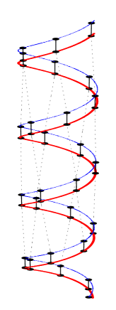
DNA, carrier of genetic code of all living organism, is a long and complex biological macro-molecule, a -stacked array of base-pairs made from four nucleotides guanine (G), adenine (A), cytosine (C)and thymine (T) coupled via hydrogen bond and forms a double-helix structure associated with sugar-phosphate backbone attached to each base-pair klotsa . In most of the theoretical models it is assumed that electronic transport cuni ; zhong ; bakhshi ; ladik is through the long-axis of the DNA molecule. In the present study, the helicity of the DNA molecule is incorporated in a tight-binding (TB) dangling backbone ladder model klotsa ; gcuni by adding hopping integrals due to the proximity of atoms in the upper strand with the corresponding atoms of the lower strand in the next pitch (see Fig. 1). The Hamiltonian for the twisted ladder model can be expressed as
| (1) |
where,
| (2) | |||||
| (4) | |||||
where and are the creation and annihilation operators for electrons in the ith Wannier state, hopping integral between neighboring nucleotides along each strand of the ladder, on-site potential energy of the nucleotides, hopping amplitude between a nucleotide and the corresponding backbone site, on-site potential energy of the backbone sites with for =I and II respectively denoting upper and lower backbone sites, interstrand hopping integral between two neighboring sites of DNA within a given pitch, interstrand hopping integral between neighboring atomic sites in the adjacent pitches which actually accounts for the helical structure of DNA. Here is the number of sites in each strand within a given pitch. For simplicity we set , and .
It is possible to obtain an analytical expression for the dispersion relation of an infinite homogeneous DNA chain (setting and ) modeled by twisted ladder in the absence backbones. Using Bloch’s theorem, the dispersion relations for the highest occupied molecular orbital (HOMO)() and the lowest unoccupied molecular orbital (LUMO)() can be expressed as , which are separated by an energy gap . Notice that for fixed and , explicitly depends on .
In order to study the transport behavior of DNA, we use semi-infinite 1D chains as leads connected cross-wise to the left (L) and right (R) ends of the DNA double helix and the Hamiltonian of the entire system is given by . The explicit form of , and are
| (5) | |||||
| (6) | |||||
| (7) |
where is the tunneling matrix element between DNA and the leads.
We use the Green’s function formalism in order to obtain transmission probability of electron datta1 ; datta2 through DNA. The single particle retarded Green’s function operator for the entire system at energy is given by , where . The transmission probability is given by , being the incident electron energy and the trace is over the reduced Hilbert space spanned by the DNA molecule. The retarded and the advanced Green’s functions in the reduced Hilbert space can be expressed as , where and , being the retarded(advanced) Green’s function for the left(right) lead. Here and are the retarded and advanced self-energies of the left(right) lead due to its coupling with the DNA molecule. It can be easily shown that .
Restricting ourselves within the linear response regime, at absolute zero temperature, the conductance is given by the two-terminal Landauer formula and the current for an applied bias voltage is given by
| (8) |
being the Fermi energy. Here we have assumed that there is no charge accumulation within the system, i.e., the voltage drop occurs only at the boundaries of the conductor.
III Results and Discussions
We first study the localization properties of the system. The localization length of the system is calculated from the Lyapunov exponent ventra
| (9) |
where = length of the system in terms of basepairs, and denotes average over different disorder configurations. In actual experimental situations there are various environmental fluctuations. We have simulated these environmental fluctuations in the model by considering the backbone site energy to be randomly distributed within the range [-w/2, +w/2], where is the average backbone site energy and w represents the disorder strength. For the purpose of numerical investigation the on-site energies of the nucleotides are taken as the ionization potentials, and the following numerical values are used through out this work: , , , . The intrastrand hopping integrals between like nucleotides are taken as while those between unlike nucleotides are taken as . We take interstrand hopping parameter to be . As all the nucleobases are connected with sugar-phosphate backbone by identical C-N bonds, the corresponding hopping parameter between the nucleobase and backbone is taken to be equal for all the cases and we take cuenda . The parameters used here are the same as those used in guo which were extracted from the ab initio calculations voit ; yan ; senth .
 |
 |
 |
 |
 |
 |
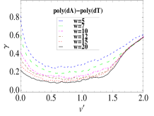
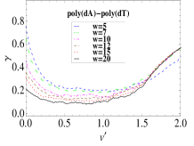
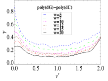
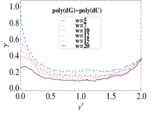
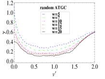
In Fig. 2 we have plotted inverse localization length of the periodic poly(dG)-poly(dC) and poly(dA)-poly(dT) sequences and the random ATGC sequence with respect to (where accounts for the helicity of DNA) for various values of the disorder strength w. It has been observed that all the curves have a general shape for the periodic as well as random DNA sequences. The variation of with is not monotonic, there exists a flat minimum of for each sequence and the position of this minimum for a specific sequence is almost insensitive to the disorder strength w. As we increase starting from zero, starts decreasing, which is quite natural because by allowing we are opening new channels for electron conduction, so the system becomes much more conductive and decreases. Now there is a flat minimum which signifies that in this region the system is most conducting as both the channels, one along the main DNA ladder and one arising due to helicity,are contributing almost equally.
Then starts increasing implying that channel starts to dominate over the other channels due to and , and as we further increase the system becomes more and more like a 1D disordered one (as the role of and becomes negligible). As in the 1D systems Anderson localization sets in if we introduce small amount of disorder into the system, the same thing is also happening here as we increase beyond the minimum, starts increasing indicating that the system effectively becoming a 1D disordered one. One may ask why these minima of versus plots are almost insensitive to disorder strength. The minimum of any versus curve is a result of the competition of two channels, the channel and resultant of and channels. Disorder can change the magnitude of but it has nothing to do with the nature of this competition, so the minima occurs almost at the same value of irrespective of the disorder strength.
 |
 |
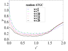
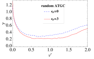 |
In Fig. 3 we have plotted versus for a fixed disorder strength (w=5) for various values of . It is to be noted that here the minima are sensitive to the average backbone site energy(), the minima get shifted as we increase . To explain this one should look at the renormalized site energies of the basepairs, as we decimate the backbone sites. Let us look at the expression for the renormalized site energy of the nucleobases , the fluctuation in due to disorder in backbone site energy becomes smaller as we increase at a given w. Thus if we consider two cases =0 and 3, we have less scattering for case, and accordingly, corresponding to its minimum has to be grater than that of the case to dominate over the other channels and hence the minimum for =3 getsshifted.
 |
 |
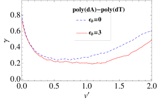
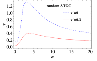 |
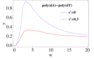
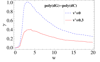
In Fig. 4 we have shown the behavior of with disorder strength for two cases with and . It is clearly visible that for the higher value of , there is a significant drop in which is obvious because by introducing we are adding other channels for conduction and the localization length of the system increases. Initially if the localization length for was a few base-pairs in presence of backbone disorder, it increases three to four times if we consider the effect of helicity of the DNA molecules. There is another thing to be noted is that there exists a maximum in each of these curves, beyond which starts decreasing even if we increase . This feature was already reported by Guo et al. for the fishbone and dangling backbone ladder models guo and the same effect is also present in our model, but this anomalous effect of backbone disorder is much smaller in our model which inturn also signify that helical nature of DNA can minimize the effect of disorder i.e., environmental fluctuations.
 |
 |
 |
 |
 |
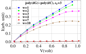 |
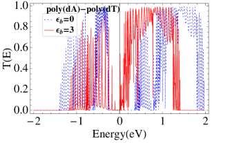
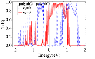
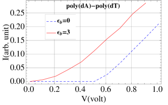
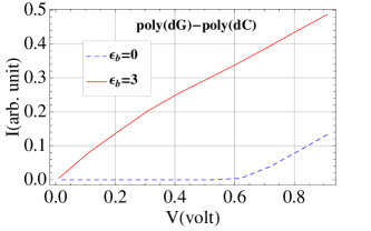
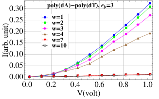
We have also investigated the response of the two periodic sequences. The temperature is set to 0 K. To minimize the contact effects we choose tunnelling parameter to be optimum , = between ds-DNA and the electrodes, where t is the hopping parameter for the electrodes macia . In Fig.5 we have shown variation of transmission probability with respect to energy for these two sequences, and it clearly shows that by tuning the backbone site energy one can control the energy gap of the system. In Fig. 6 the characteristics are shown without backbone disorder and it has been observed that for , the cut-off voltage becomes nearly equal to zero for both the periodic poly(dG)-poly(dC) and poly(dA)-poly(dT) sequences. For poly(dG)-poly(dC) case the response is almost linear which indicates that the system would undergo a transition from semiconducting to metallic phase depending on on-site energies of the backbone, not only disorder can induce that kind of transition guo . characteristics for case are in good agreement with experimental results porath . We have checked the response for different system sizes, and observed that there is no significant change in the characteristics. In Fig.7 we show the responses for the two periodic sequences for case in presence of the backbone disorder. It clearly shows that with increasing disorder the current in the system decreases.
IV Concluding Remarks
Within the tight-binding framework, the fishbone and the dangling backbone ladder models are generally used to study the transport properties of DNA like systems, but none of these models have incorporated the effect of helicity of DNA molecules. Helicity is a very fundamental aspect of the DNA structure and gives the possibility of new conduction channels in DNA. In this paper we propose a model that accounts for the helical structure of the DNA molecules and show that there is a significant change in the localization properties and response of the systems. One of the main results is that there exists a almost disorder independent critical hopping () for which localization length becomes maximum for each of the sequences that we have considered. At this critical hopping strength system is least affected by the external disturbances, i.e., environmental effects are least at this point. If one can utilize this information properly then it might be possible to minimize the environmental effects in the actual experiments. We have also shown that though backbones do not play any role directly in electron conduction but they can significantly contribute by narrowing the energy gap and reducing the cut-off voltage. It might also be possible to find DNA in a complete metallic phase even in presence of environmental fluctuations as evident from the almost linear response of poly(dG)-poly(dC) sequence for . We look forward that there might be experimental investigations as well as ab initio calculations in near future to find the exact value of the interstrand hopping integral () between nucleobases of adjacent pitches and its actual role in transport.
References
- (1) J. Watson and F. Crick, Nature (London) 171, 737 (1953).
- (2) D. D. Eley and D. I. Spivey, Trans. Faraday Soc. 58, 411 (1962).
- (3) S. O. Kelley and J. K. Barton, Science 283, 375 (1999).
- (4) H. W. Fink and C. Schönenberger, Nature (London) 398, 407 (1999).
- (5) D. Porath, S. De Vries, and C. Decker, Nature (London) 403, 635 (2000).
- (6) L. Cai, H. Tabata, and T. Kawai, Appl. Phys. Lett. 77, 3105 (2000).
- (7) P. Tran, B. Alavi, and G. Grüner, Phys. Rev. Lett. 85, 1564 (2000).
- (8) Y. Zhang, R. H. Austin, J. Kraeft, E. C. Cox, and N. P. Ong, Phys. Rev. Lett. 89, 198102 (2002).
- (9) A. J. Storm et al., Appl. Phys. Lett. 79, 3881 (2001).
- (10) K. H. Yoo et al., Phys. Rev.Lett. 87, 198102 (2001).
- (11) A-M Guo, S-J Xiong, Z. Yang, and H-J Zhu, Phys. Rev. E 78, 061922 (2008).
- (12) C. J. Murphy, M. A. Arkin, Y. Jenkins, N. D. Ghatlia, S. Bossman, N. J. Turro, and J. K. Barton, Science 262, 1025 (1993).
- (13) M. Bixon, B. Giese, S. Wessely, T. Langenbacher, M. E. Michel-Beyerle, and J. Jortner, Proc. Natl. Acad. Sci. USA 96, 11713 (1999).
- (14) Y. A. Berlin, A. L. Burin, and M A. Ratner, J. Am. Chem. Soc. 123, 260 (2001).
- (15) Z. Hermon, S. Caspi, and E. Ben-Jacob, Europhys. Lett. 43, 482 (1998).
- (16) Z. G. Yu and X. Song, Phys. Rev. Lett. 86, 6018 (2001).
- (17) G. Cuniberti, L. Craco, D. Porath, and C. Dekker, Phys. Rev. B 65, 241314(R) (2002).
- (18) S. Roche, Phys. Rev. Lett. 91, 108101 (2003).
- (19) S. Cuenda and A. Sanchez, Fluctuations and Noise Letters 4, L491-L504 (2004)
- (20) M. Peyrard, Nonlinearity 17, R1-R40 (2004).
- (21) D. Klotsa, R. A. Römer, and M. S. Turner, Biophysical Journal 89, 2187 (2005).
- (22) R. Gutiérrez, S. Mohapatra, H. Cohen, D. Porath and G. Cuniberti, Phys. Rev. B 74, 235105 (2006)
- (23) A. Y. Kasumov et al., Science 291, 280 (2001).
- (24) E. M. Conwell and S. V. Rakhmanova, Proc. Natl. Acad. Sci. USA 97, 4557 (2000).
- (25) C. Dekker and M. A. Ratner, Physics World 14(8): 29-33 (2001).
- (26) M. A. Ratner, Nature (London) 397, 480 (1999).
- (27) D. N. Beratan, S. Priyadarshy, and S. M Risser, Chem. Biol. 4, 3 (1997).
- (28) R. G. Endres, D. L. Cox, and R. R. P. Singh, Rev. Mod. Phys. 76, 195 (2004).
- (29) R. Gutiérrez, S. Mandal and G. Cuniberti, Nano Lett. 5, 1093 (2005)
- (30) P. J. de Pablo, F. Moreno-Herrero, J. Colchero, J. Gómez Herrero, P. Herrero, A. M. Baró, P. Ordejón, J. M. Soler, and E. Artacho, Phys. Rev. Lett. 85, 4992 (2000).
- (31) L. Cai, H. Tabata, and T. Kawai, Nanotechnology 12, 211 (2001).
- (32) R. N. Barnett, C. L. Cleveland, U. Landman, E. Boone, S. Kanvah, and G. B. Schuster, J. Phys. Chem. A 107, 3525 (2003).
- (33) J. Zhong, in Proceedings of the 2003 Nanotechnology Conference, Vol. 2. Edited by M. Laudon and B. Romamowicz. Computational Publications, CAMBRIDGE, MA. Nanotech 105-108 (2003).
- (34) A. K. Bakhshi, P. Otto, J. Ladik and M. Seel, Chem. Phys. 108, 215 (1986).
- (35) J. Ladik, M. Seel, P. Otto, and A. K. Bakhshi, Chem. Phys. 108, 203 (1986).
- (36) G. Cuniberti, E. Maciá, A. Rodriguez, and R. A. Römer, in Charge Migration in DNA: Perspectives from Physics, Chemistry and Biology, edited by T. Chakraborty, Springer-Verlag, Berlin (2007).
- (37) S. Datta, Electronic transport in mesoscopic systems, Cambridge University Press, Cambridge (1995).
- (38) S. Datta, Quantum Transport: Atom to Transistor, Cambridge University Press, Cambridge (2005).
- (39) M. D. Ventra, Electrical transport in nanoscale system, Cambridge University Press, Cambridge (2008).
- (40) A. A. Voityuk, J. Jortner, M. Bixon, and N. Rösch, J. Chem. Phys. 114, 5614 (2001).
- (41) Y. J. Yan and H. Y. Zhang, J. Theor. Comput. Chem. 1, 225 (2002).
- (42) K. Senthilkumar, F. C. Grozema, C. F. Guerra, F. M. Bickelhaupt, F. D. Lewis, Y. A. Berlin, M. A. Ratner, and L. D. A. Siebbeles, J. Am. Chem. Soc. 127, 14894 (2005).
- (43) E. Maciá, F. Triozon, and S. Roche, Phys. Rev. B 71, 113106 (2005)