permanent address: ]Department of Mechanical Engineering & Engineering Sciences and Applied Mathematics, Northwestern University, Evanston, IL 60208, USA
Capstan friction model for DNA ejection from bacteriophages
Abstract
Bacteriophages infect cells by attaching to the outer membrane and injecting their DNA into the cell. The phage DNA is then transcribed by the cell’s transcription machinery. A number of physical mechanisms by which DNA can be translocated from the phage capsid into the cell have been identified. A fast ejection driven by the elastic and electrostatic potential energy of the compacted DNA within the viral capsid appears to be used by most phages, at least to initiate infection. In recent in vitro experiments, the speed of DNA translocation from a phage capsid has been measured as a function of ejected length over the entire duration of the event. Here a mechanical model is proposed that is able to explain the observed dependence of exit velocity on ejected length, and that is also consistent with the accepted picture of the geometric arrangement of DNA within the viral capsid.
pacs:
87.15.hj, 87.16.ad, 82.37.Rs,87.80.Fe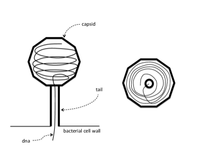
The physics of semi-flexible polymers under confinement is a subject of great interest with wide applications in biology and soft matter. Viruses are an interesting example of such an application. Bacteriophages are viruses that infect bacteria. During infection, the virus capsid itself remains outside the bacterium, only the DNA is injected into the cell hershey_independent_1952 . Several physical mechanisms by which bacteriophages inject DNA into their hosts have been identified. The specific mechanism varies by phage type and stage of the infection process molineux_fifty-three_2006 ; inamdar_dynamics_2006 ; grayson_is_2007 . A fast ejection on the timescale of seconds can be achieved simply by the release of elastic and electrostatic energy of the coiled up DNA confined within the viral capsid. It has been shown in in vitro studies that phage can eject its entire 48.5 kbp genome by this mechanism in a few seconds 111Interestingly, very recent work appears to indicate that even the phage infection mechanism may work differently in vivo vanvalen_single-molecule_2012 . In other phage species, this “syringe” mechanism may initiate the infection, which is then completed through other pathways molineux_no_2001 .
A schematic diagram of the DNA ejection process is shown in Fig. 1. If denotes the free energy of the compacted DNA, then is the force driving the ejection, where is the number of base pairs of DNA confined within the capsid and nm is the distance between base pairs. The free energy of the part of the DNA that is already outside the viral capsid is neglected as it is much smaller than the contribution from the confined part. Since inertia is completely negligible at such small scales, this driving force must be balanced by a frictional force, , where is the DNA translocation velocity and is a frictional coefficient (mobility) which may in general depend on . The linear dependence on the velocity is a consequence of the fact that at low Reynolds numbers, the fluid equations reduce to the linear Stokes equation. Thus, if the functions and were known, the translocation velocity could be determined from
| (1) |
The function can be calculated analytically purohit_mechanics_2003 ; purohit_forces_2005 on the presumption that the DNA is packed in the capsid as helical coils wound in successive layers of decreasing radii in an “inverse spool” arrangement (Fig. 1). It has been shown that such an ordered packing does indeed minimize the free energy of the system. In Brownian dynamics simulations kindt_dna_2001 , as the chain is gradually introduced into the capsid, it is observed to arrange itself in a donut shape. As becomes larger, it winds into a spool from the outside in. When the internal diameter of the spool shrinks sufficiently, the DNA stops wrapping in helical coils and simply forms loose turns parallel to the spool axis. The most direct evidence for the helical conformation of DNA within the capsid has come from imaging of DNA inside T7 cerritelli_encapsidated_1997 and T4 olson_structure_2001 phages by cryo-electron microscopy and of P22 and phages by X-ray diffraction earnshaw_dna_1977 . The images reveal multiple layers of helical coils of DNA wound around the central axis.
The analytical formula for the free energy has been subjected to direct experimental tests by a rather ingenious method. Real cells exert significant osmotic pressures that are believed to resist DNA insertion by the phage. It has been possible to actually “stall” the DNA translocation in in vitro experiments by raising the osmotic pressure in the solution outside the capsid. In such cases, the translocation stops with an amount of DNA still remaining within the capsid, where is given by
| (2) |
Here is the difference in osmotic pressures between the outside and inside the capsid and is the DNA radius. The quantities and can be directly measured for a range of values: from to a value that is high enough that none of the DNA is ejected. Thus, can be determined experimentally and compared to analytical models based on the elastic and electrostatic energy of a helically coiled semi-flexible rod. This has been done evilevitch_measuring_2004 ; grayson_effect_2006 and the theoretical models for were found to agree very well with experimental data. Further evidence for the model comes from studying an event that occurs much later in the infection process: the packaging of viral DNA into the capsid prior to lysis of the host cell and release of the phages into the environment. In an in vitro experiment, Smith et al smith_bacteriophage_2001 attached an optical bead to a DNA strand being actively packaged into the 29 bacteriophage capsid by molecular motors. By applying a variable force to the optical bead and observing the changes in the packing rate, they concluded that the elastic force against which the molecular motors must work, increases rapidly with the amount of DNA inside the capsid. This is again consistent with the rapidly decreasing nature of the function .

Unfortunately, no such theoretical result for the function in equation (1) is available. However, if is measured experimentally, then using equation (1) the function can be obtained. This was done in a recent experiment by Grayson et al. grayson_real-time_2007 where phages were immobilized on a surface and induced to eject their DNA by exposing them to cell surface proteins to which the phage would normally attach and trigger the ejection event in the native state. The ejected DNA was visualized by a fluorescent dye and the ejection process recorded by video microscopy. The data from the experiment (Fig. 5 in their paper) is reproduced in Fig. 2 from which certain conclusions may be drawn about the origin of the frictional resistance encountered by the DNA during the ejection process.
Frictional resistance to the motion of DNA can arise either from friction within the phage tail or from movement of the DNA outside or within the capsid. In the former case, would be independent of . Fig. 2 shows that becomes approximately independent of only towards the very end of the translocation process after 80 to 90 percent of the DNA has already been ejected. This is consistent with the following simple estimate of the frictional drag that is obtained if the DNA is taken as a cylinder of radius nm, concentric with a slightly larger cylinder (the phage tail) of radius nm:
| (3) |
where nm is the length of the tail and Pa-s is the viscosity of water. If we take kbp/s, the highest velocity indicated by the data, then pN may be regarded as the maximum frictional drag arising from the tail. This is much less than the pN driving force grayson_real-time_2007 ejecting the DNA from the capsid and therefore cannot be the principal source of resistance. Similarly, it is easy to show that viscous friction from the medium external to the capsid cannot be important either. The actual drag would depend on the shape of the ejected DNA coil, but, an estimate, that should be regarded as an upper bound, is obtained by regarding the entire kbp DNA to be a cylinder that translates in water parallel to its axis at a speed of kbp/s. In this case, the drag force is happel_brenner
| (4) |
which is again negligible in comparison to the driving force. However, the driving force does decay rapidly with , and for kbp the driving force is less than a pN. Thus, the hydrodynamic drag could balanced the driving force towards the very end of the translocation process, and indeed, the data shows that stops decreasing once becomes smaller than about 10 kbp (Fig. 2).
Fig. 2 also shows that as long as kbp, the experimental data can be fit very well by a function of the form
| (5) |
The fitted straight line corresponds to pN-s/kbp and kbp-1. It is significant that is hardly affected when the buffer contains the divalent Mg2+ ion which reduces the electrostatic repulsion between the DNA strands thereby reducing the driving force by almost an order of magnitude. This is consistent with our expectations, since frictional resistance is not expected to be affected greatly by interstrand repulsion.
The success of the empirical fit, equation (5), makes one wonder if there is any known physical mechanism that could lead to such an exponential friction law. We show here that if one assumes that the resistance to motion arises due to friction between the sliding DNA strand and its neighboring strands as well as with the capsid wall, then such an exponential law can be derived. For simplicity, we consider the shape of the viral capsid to be a cylinder, though bacteriophage capsids are typically polyhedral in shape and are sometimes modeled as spheres. We start from the equations of equilibrium of an elastic beam landau_theory_1986
| (6) | |||||
| (7) |
where is the arc length along the beam, is the elastic force across a cross-section, is the internal bending moment and is the external force per unit length along the beam. Since the pitch of the helix is small purohit_mechanics_2003 , we will neglect its torsion. The external force is, by Amonton’s law bowden_friction_2001 , , where and are the unit tangent and normal to the curve representing the beam centerline, points in the direction of sliding and is a friction coefficient. The force across the cross section may be resolved into a tensile () and shear () component: . The bending moment is related to the local curvature () as , where , is the Young’s modulus and is the area moment of inertia of the cross-section. Substitution in equations (6) and (7) and use of the Frenet-Serret formulas struik_lectures_1961 describing how the unit vectors change along a curve in three dimensions results in
| (8) | |||||
| (9) | |||||
| (10) |
For a helix, is a constant, thus , and therefore,
| (11) |
which may be integrated to yield
| (12) |
where is the tension in the DNA at the capsid exit and is the tension at the terminal point. By terminal point we mean the point on the DNA within the capsid marking the transition from a helix to the loosely coiled trailing end in the central core. If we express the viscous resistance on this trailing end as , then . Since the pitch of the helix is small, , so that
| (13) |
where nm, is the capsid radius. It should be noted that in equation (12), is independent of the radius of the turns but depends only on the total turn angle, . Thus, if there are multiple layers of coils, it may still be applied as long as the friction coefficient between all contacting surfaces are the same. Equation (13) is equivalent to equation (5); comparing the coefficients we determine .
It is interesting to note that equation (12) is the capstan equation (also known as Eytelwein’s formula or the Euler-Eytelwein formula) familiar from mechanics stuart_capstan_1961 . In the classical “capstan problem” a flexible line is wound around a cylinder (for example, a bollard or capstan used in ship mooring) to hold a load at one end of the line by the application of a holding force. At the point when the line is just about to slip, the relation between the load () and holding force () is described by equation (12). The exponential dependence of on explains how a very large load can be supported by a relatively modest force if the rope is wound even a few turns around the capstan. The name “capstan friction model” in the title of this paper is a reference to this correspondence. It should be noted, however, in our case there is no central cylinder. The frictional resistance arises from contact between neighboring DNA strands and possibly with the capsid wall as the tightly coiled DNA unwinds. In fact, the unfurling of a surveyor’s steel measuring tape is perhaps a better analogy.
The frictional coefficient between a pair of surfaces depends on the nature of the contact between them bowden_friction_2001 ; persson_sliding_2000 . When a lubricating fluid film is present, the coefficient is small but increases linearly with sliding speed in a manner that may be well understood from the equations of fluid flow. In the presence of a large normal load (which in the present problem would arise from the strong bending rigidity of the DNA) the fluid film thins and one enters the regime of boundary lubrication where direct molecular level contact between surfaces could occur. This is characterized by a sharp rise in the frictional coefficient. The value corresponds to this latter regime of boundary lubrication. For example, the frictional coefficient between dry highly polished metal surfaces at moderately large loads is close to this value bowden_friction_2001 . The derivation of equation (13) presumes Amonton’s law of friction which is based on the idea that contact between a pair of surfaces arise from interlocking asperities and is thus independent of the apparent area of contact but increases in proportion to the normal load bowden_friction_2001 . Such a picture is unlikely to hold for nanoscale phenomena such as the sliding of DNA strands. Nevertheless, Amonton’s law is frequently used to describe frictional effects in nanoscale phenomena even though the underlying physical models are different from the rubbing of asperities envisaged to rationalize Amonton’s law in the classical treatment of friction bhushan_springer_2004 ; gao_frictional_2004 ; mo_friction_2009-1 .
It may be shown by simple estimates that viscous friction with the fluid in the core is unimportant. Since any shear induced at the outer boundary of the fluid filled core will equilibrate on a time scale ns ( is the kinematic viscosity of water), we conclude, that, the fluid core must be in rigid rotation during most of the ejection process of duration s. The inertia of this fluid mass also plays no role. Indeed, the moment of inertia of the cylinder is and the rate of change of angular speed is where is the total length of DNA. Substituting typical values grayson_real-time_2007 we find that N-m which is many orders of magnitude smaller than the applied torque, N-m. Thus, neither the viscosity of the water within the capsid nor its inertia plays a significant role in determining the ejection velocity.
In this paper we interpret phenomena involving the translocation of single molecules
of DNA across nanometer size pores from the principles of classical continuum mechanics.
In the light of the small scale nature of the system, one might question the validity of such an approach.
However, even though the relevant length scales are approaching the limits of applicability
of the continuum description of matter, classical continuum mechanics has been applied successfully
to other problems involving translocation of single molecules across
nanopores ghosal_electrophoresis_2006 ; ghosal_effect_2007 ; luan_electro-osmotic_2008 ; van_dorp_origin_2009 .
Thus, its use in this context, for estimating forces on DNA, appears entirely reasonable.
Acknowledgement: This work was supported in part by the American Recovery and Reinvestment Act (ARRA) funds through grant number R01HG004842 to Northwestern University (USA) from the National Human Genome Research Institute, National Institutes of Health. Support from the Leverhulme Trust (UK) in the form of a Visiting Professorship is gratefully acknowledged. The author thanks the Cavendish Laboratory, Cambridge University for its kind hospitality.
References
- (1) A. D. Hershey and M. Chase, The Journal of General Physiology 36, 39 (1952).
- (2) I. J. Molineux, Virology 344, 221 (2006).
- (3) M. M. Inamdar, W. M. Gelbart, and R. Phillips, Biophysical Journal 91, 411 (2006).
- (4) P. Grayson and I. J. Molineux, Current Opinion in Microbiology 10, 401 (2007).
- (5) D. Van Valen et al., Current Biology 20(14), 1339 (2012).
- (6) I. J. Molineux, Molecular Microbiology 40, 1 (2001).
- (7) P. K. Purohit, J. Kondev, and R. Phillips, Proceedings of the National Academy of Sciences 100, 3173 (2003).
- (8) P. K. Purohit et al., Biophysical Journal 88, 851 (2005).
- (9) J. Kindt, S. Tzlil, A. Ben-Shaul, and W. M. Gelbart, Proceedings of the National Academy of Sciences 98, 13671 (2001).
- (10) M. E. Cerritelli et al., Cell 91, 271 (1997).
- (11) N. H. Olson, M. Gingery, F. A. Eiserling, and T. S. Baker, Virology 279, 385 (2001).
- (12) W. C. Earnshaw and S. C. Harrison, Nature 268, 598 (1977).
- (13) A. Evilevitch, M. Castelnovo, C. M. Knobler, and W. M. Gelbart, J. Phys. Chem. B 108, 6838 (2004).
- (14) P. Grayson et al., Virology 348, 430 (2006).
- (15) D. E. Smith et al., Nature 413, 748 (2001).
- (16) P. Grayson, L. Han, T. Winther, and R. Phillips, Proceedings of the National Academy of Sciences of the United States of America 104, 14652 (2007).
- (17) J. Happel and B. Brenner, Low Reynolds number hydrodynamics (Kluwer, Boston, 1983).
- (18) L. D. Landau, E. M. Lifshitz, A. M. Kosevich, and L. P. Pitaevski, Theory of Elasticity (Elsevier, Oxford, 1986).
- (19) F. P. Bowden and D. Tabor, The Friction and Lubrication of Solids (Oxford University Press, Oxford, 2001).
- (20) D. J. Struik, Lectures on Classical Differential Geometry (Courier Dover Publications, Mineola, 1961).
- (21) I. M. Stuart, British Journal of Applied Physics 12, 559 (1961).
- (22) B. N. J. Persson, Sliding Friction: Physical Principles and Applications (Springer, Berlin Heidelberg, 2000).
- (23) B. Bhushan, Springer Handbook of Nanotechnology (Springer, Berlin Heidelberg, 2004).
- (24) Y. Mo, K. T. Turner, and I. Szlufarska, Nature 457, 1116 (2009).
- (25) J. Gao et al., The Journal of Physical Chemistry B 108, 3410 (2004).
- (26) S. Ghosal, Physical Review E 74, 041901 (2006).
- (27) S. Ghosal, Physical Review Letters 98, 238104 (2007).
- (28) B. Luan and A. Aksimentiev, Physical Review E 78, 021912 (2008).
- (29) S. van Dorp et al., Nat Phys 5, 347 (2009).