A Developmental Network Theory of Gynandromorphs, Sexual Dimorphism and Species Formation
Abstract
Gynandromorphs are creatures where at least two different body sections are a different sex. Bilateral gynandromorphs are half male and half female. Here we develop a theory of gynandromorph ontogeny based on developmental control networks. The theory explains the embryogenesis of all known variations of gynandromorphs found in multicellular organisms. The theory also predicts a large variety of more subtle gynandromorphic morphologies yet to be discovered. The network theory of gynandromorph development has direct relevance to understanding sexual dimorphism (differences in morphology between male and female organisms of the same species) and medical pathologies such as hemihyperplasia (asymmetric development of normally symmetric body parts in a unisexual individual). The network theory of gynandromorphs brings up fundamental open questions about developmental control in ontogeny. This in turn suggests a new theory of the origin and evolution of species that is based on cooperative interactions and conflicts between developmental control networks in the haploid genomes and epigenomes of potential sexual partners for reproduction. This network-based theory of the origin of species is a paradigmatic shift in our understanding of evolutionary processes that goes beyond gene-centered theories.
Key words: Gynandromorphs, developmental control networks, cenome, CENEs, epigenomics, origin of species, evolution of species, sexual dimorphism, unisexual hemimorphism, synsexhemimorphism, hemihyperplasia, hemihypertrophy, bilateral symmetry, multicellular development, developmental systems biology, embryonic development, computational modeling, simulation
1 Introduction
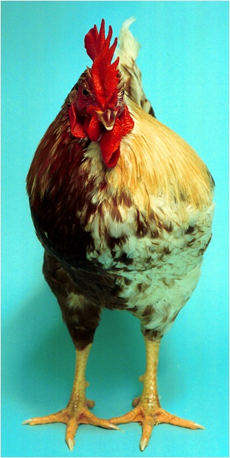
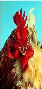
Gynandromorphs are creatures that are part female (gyn-) and part male (andro-). Bilateral gynandromorphs are half male and half female split down the middle like the lobster in Fig.1a and the Rooster-Hen in Fig.5a. Gynandromorphs occur in many species of including insects, fish, birds and mammals.


Scientists give conflicting answers to the questions of how gynandromorphs form and what makes them possible at all. The standard view used is that gynandromorphs result from an unequal distribution of sex chromosomes by way of chromosome loss or gain during cell division. Others have found the cells to have a normal complement of chromosomes, but the cells in the two bilateral halves are of different sex. Related to this is the view is that hormones determine sexual dimorphism -the differences in morphology between males and females. This paper gives an explanation of why chromosomal abnormalities and cells of different sex can lead to gynandromorphism. In addition it is shown that there is a third way for gynandromorphs to develop where all the cells of an organism are genetically normal with the normal complement of chromosomes and all the cells have the same sex, and yet they can still develop into gynandromorphs or have hemimorphic, non-bilateral bodies,
2 Networks control gynandromorph development
This essay presents a novel, general computational theory of how gynandromorphs are generated from a single fertilized egg. The theory is based on a computational theory of development, developmental control networks (CENEs) and bilateral symmetry [104, 105, 106, 108]. The standard hypotheses that an unequal chromosome distribution causes gynandromorphy are special cases of the general network theory of gynandromorphic development presented here.
What is missing in all previous accounts of gynandromorphs is a detailed explanation and theory of how gynandromorphs develop from a single fertilized egg. This paper gives an explanation of the ontogeny of gynandromorphs that is detailed enough to model the development of all known types of gynandromorphs found in multicellular organisms111When a theory cannot explain a phenomena it is simply ignored by the proponents of that theory. Research on gynandromorphs was popular in the 1930’s and later (see articles in the Journal of Heredity 1929-present). Then, with the change of biology’s scientific paradigm to molecular biology, since gynandromorphs could not be explained genetically, the area was largely ignored. However, with the developmental control network theory [106] together with the theory of bilateral symmetry [108], gynandromorphs can now be understood. . The theory also predicts a large variety of more subtle gynandromorphic morphologies yet to be discovered.
First I will give a brief introduction to developmental control networks (CENEs). Then I present some examples of types of gynandromorphs and relate them to the developmental control networks that generate them. It will be shown that each gynandromorph network has a corresponding meta-network signature that can be used to distinguish and classify gynandromorphs. The study of gynandromorph developmental networks gives fascinating new insights into how species originate, which I will discuss at the end.
3 Modeling gynandromorphs
One of the best ways of understanding the complex processes involved in the development of multicellular organisms is to computationally model the control network architecture of genomes and their interactions with cells. Cells interpret the developmental control networks in their genome and give that genome pragmatic meaning. The cell’s essential internal orientation and its external orientation in space also needs to be modeled. In addition, one also has to model the cell-cell communication and cell physics. One can then run simulations of multicellular development in space and time and observe the resulting organism. The modeling also permits making changes to the network, running the simulation again and seeing the new resulting form. Below I will describe the results of modeling gynandromorphs and the developmental control networks (CENEs) that lead to their development from starting from a single cell. This will give us a deep insight into the nature of not only gynandromorphs but into the developmental control and evolution of all multicellular life.
3.1 Developmental Control Networks or CENEs
To understand the dynamics of development I take a perspective that goes beyond the current gene-centered paradigm. My view is that the complexity of organisms and the related complexity of the ontogeny of embryos requires control information that is not in genes. Genes make up the interacting parts of the cell but they do not contain the information that orders, organizes and structures the dynamic development of multicellular systems. Genes produce parts and processes that are local to the cell. CENEs (developmental control networks) contain the global control information for multicellular development. CENEs are located in the the vast noncoding areas of genomes. CENEs are not gene regulatory networks (GRNs) [19, 18, 26, 27, 106]. Instead, CENEs subsume and control the gene regulatory networks (GRNs) that control the activation of genes. Thereby, CENEs control and organize cell actions such as division, movement and cell communication. CENEs can be linked together to form larger more complex CENEs. The complete set of all CENEs in a genome is called the CENOME. The CENOME is the global control network that directs the development of Multi-Cellular Organisms (MCOs).
3.2 The Interpretive-Executive System IES and bilateral symmetry
The cell has an interpretive-executive system (IES) that interprets the control information in the genome and executes its directives [106] to control cell actions in the dynamic system of interacting cells in a developing organism. Part of the IES is the implicit coordinate system in the cell associated with its orientation and handedness. Using cell orientation and its epigenetic interpretation by the IES, the theory of bilateral symmetry explains how bilaterally symmetric organism develop from a single cell [108]. This theory of bilateral symmetry explains the pseudo-symmetric development of gynandromorphs. Combining the developmental control theory of CENEs with the theory of bilateral symmetry, explains all the varieties of existing, possible and yet to be engineered or discovered gynandromorphs.
4 Basic gynandromorphs
Gynandromorphs found in the wild come in at least three forms: Bilateral222The term “bilateral” is used in several senses. In its usual sense all the gynandromorphs (bilateral, polar and oblique) that we are modeling are bilateral organisms in the sense that they have developed bilaterally from a single cell. In the second sense of bilateral, when used to classify gynandromorphs, it means that the male and female parts of the organism are separated into the two bilateral halves of the bilateral organism. Interestingly, there is a third sense of bilateral, that distinguishes bilateral development from the bilateral structure of an organism. While the gynandromorphs develop bilaterally from a single cell, phenotypically they are not necessarily bilateral in that the two sides of the bilaterally developing and developed organism need not be mirror images. The male parts in one bilateral half will have male characteristics different from the corresponding female structure in the other bilateral half. However, the cell orientations of the two bilateral halves will mirror each other as described in [108]. , polar, oblique [45]. Using the computational theory of developmental control networks one can model and, starting from a single cell, simulate the embryonic development all the basic gynandromorphs found in nature (see Fig. 2) as well as the all other possible forms that may exist (see Sec. 7).
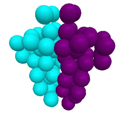


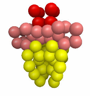
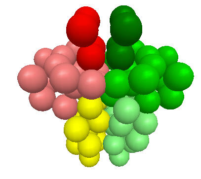
Fig. 2, illustrates the three basic forms of gynandromorphs: Bilateral gynandromorphs (bilateral body halves of opposite sex), polar gynandromorphs (anterior-posterior of opposite sex), and oblique gynandromorphs (opposite sex body sections cross the bilateral plane). The spiral gynandromorph (when the dorsal D and ventral V sections of the organism are inter folded) in Fig. 2d is a theoretical possibility I have added the category of spiral gynandromorphs . .
At the same time gynandromorphs exhibit a pseudo symmetry in that they are bilaterally split down the middle with opposite handedness (see Fig. 4b). The simulations of multicellular development of such organisms was done with software based on the theoretical framework. In the case of gynandromorphs the two bilateral founder cells have opposite orientations but now their control states activate distinct developmental control networks (CENEs). Development then proceeds as if each side developed as part of a normal bilateral organism.
5 Bilateral gynandromorphs
5.1 Cell orientation in bilateral gynandromorphs
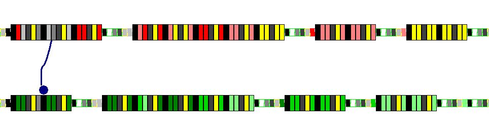
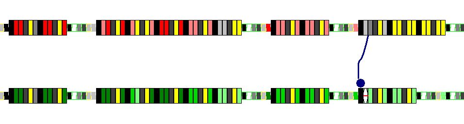
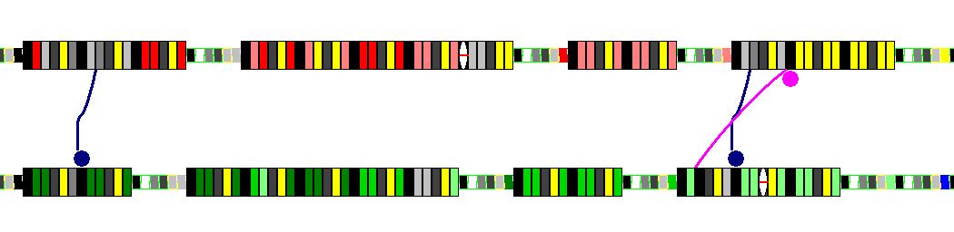
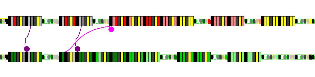
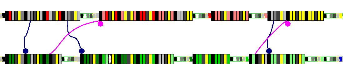
Most gynandromorphs exhibit a pseudo bilateral symmetry in that each lateral half of the organism is the bilateral symmetric half of the normal organism. Just as in the development of normal bilateral multicellular organisms, the cell orientation of gynandromorphs allows the consistent development of these pseudo bilateral halves of the gynandromorphs [108].

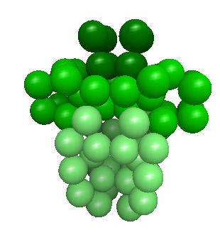
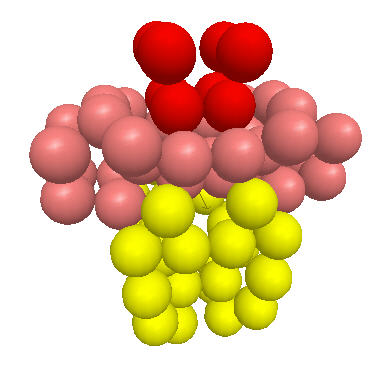
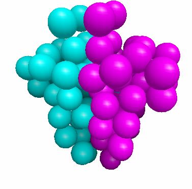
The growth of each bilateral half in Fig. 4 is based, in part, on a different developmental control subnetwork. In these multicellular organisms the two halves are morphological mirrors of one another, just the cell differentiation states are different (as indicated by the different colors in the left Fig.4a). One half is female and the other male. But, in principle, a gynandromorph-like organism could consist of halves of two different organisms with distinct genomes as long as the resulting embryo is viable. The middle Fig.4b shows the orientation states of the cells. Note the opposite orientation in the two body halves. The right Fig.4c, of stained purple and aqua marine cells, shows which parental allelic genome, paternal or maternal, is active in each cell.
5.1.1 Cell orientation in organ asymmetry
This phenomenon of autonomous development of the two body halves also relates to asymmetric development. As in symmetric development, in asymmetric development the developing cells also have a handedness and orientation that is inherited epigenetically. This means that the switch in orientation of the asymmetric body part, e.g., left side to right side, is the result of a switch in orientation and handedness of the asymmetric founder, progenitor cell. This explains the ease and consistency of the switch of an organ or limb to its mirror, because the very same developmental control network (CENE) is being used to control the development of the mirror organ or limb. All that needs to be changed is the orientation of the founder cell.
Thus, early changes in cellular orientation have major developmental effects. Hence, mutations in that lead to cell orientation switches can have vast evolutionary consequences [108].
5.2 Avian bilateral gynandromorphs
It used be thought that sexual dimorphism, the difference in morphology of male and female animals, was due to hormones. In a fundamental and important discovery it was shown that developmental differences in a chicken (gallus gallus domesticus) gynandromorph (Rooster-Hen in 5a) are cell based and not hormone based [113, 22]. The cells on the different sides of the Rooster-Hen are of the opposite sex, the rooster half being male and the hen half being female. In humans females have the homomorphic XX sex chromosomes and the males have the heteromorphic XY sex chromosomes.
Unlike humans, chicken sex chromosomes have the opposite heteromorphic sorting. The cells of female chickens have the heteromorphic ZW sex chromosomes while the males have homomorphic sex chromosomes ZZ. Almost all of cells of the male half of the Rooster-Hen were male cells having the homomorphic ZZ sex chromosomes of roosters. The cells on the female half of the Rooster-Hen were female containing the heteromorphic WZ chromosomes of hens. The same hormone has the opposite effect depending on the sex of the cells. For example it induces male progenitor cells to become testes and female progenitor cells to become the female reproductive system [113, 22]. Hence, a hormone which would be distributed evenly and effect both sides of the organism could not be responsible for the different phenotypes observed in the Rooster-Hen. As a consequence the whole view of sexual dimorphism (differences in the phenotype of males and females) and how, at least avian, gynandromorphs originate has changed.
5.2.1 Hormone information limits
While it should have been obvious, even without this result, that a hormone, which is a relatively simple molecule, cannot contain the complex control information necessary for the development of the different complex morphologies that distinguish males and females. The result of Clinton confirms the general theory that development is based on developmental control networks (CENEs) [106]. And, it confirms the network theory of gynandromorphs which states that, as is the case in the Rooster-Hen, the two dimorphic body halves are controlled in part by different developmental control networks.
5.2.2 Prediction
Since the Rooster-Hen preserves autonomous bilateral development, its cells are not only of the opposite sex and controlled by opposite sex developmental control networks, but the theory of bilateral symmetry predicts that they also are of opposite orientation and handedness [108]. Either early in development some process led to the generation of two founder cells of opposite orientation with mirror handedness. In the case of the Rooster-Hen gynandromorph these founder cells were also of opposite sex.
6 Mosquito gynandromorphs
Mosquitoes exhibit the basic palette of gynandromorphic types described in Sec. 4, namely, bilateral, polar and oblique [45]. In this section we model the mixed polar-oblique form.


In addition to sexually mixed morphology, gynandromorphs can have conflicting sex based behavior [70]. For example, polar gynandromorph mosquitos that have an anterior female half and a posterior male half. The male abdomen is much smaller than the female’s. Such a polar gynandromorph mosquito has a female brain and will behave like a female sucking blood to nourish its nonexistent eggs. It will suck blood until its small and inadequate male abdomen bursts [89].
7 Gynandromorph combinatorics
Beyond the basic gynandromorphs Sec. 4 there are many possible forms.

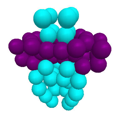


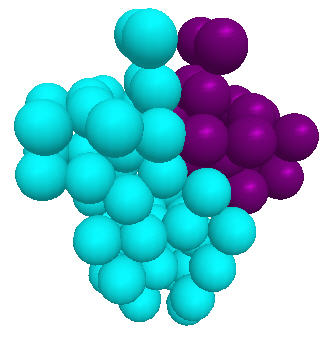
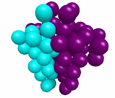
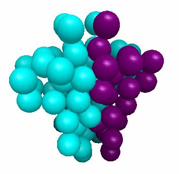
Fig. 2 and Fig. 7 illustrate different forms of an idealized, very simple 3-segmented multicellular gynandromorph consisting of only 72 cells. The interactive, transactivation of male and female chromosomes lead to the phenotypes many of which are observed in nature.
The gynandromorphs in Fig. 2 and Fig. 7 are all bilateral multicellular organisms developed from a single cell. They attain their different forms because the male (M) and female (F) chromosomes are differentially activated during development.
7.1 The possible developmental outcomes of gynandromorphs
It turns out the possible developments of gynandromorphs satisfy a combinatory logic. Given each anterior to posterior section has two bilateral halves whose sex can vary independently, each bilateral section can be in four possible states (MM, MF, FM, FF). Hence, if you just have two, anterior and posterior, body sections then if you start with MM you can get MM-MM (normal), MM-MF (polar-bilateral), MM-FM (polar-bilateral), MM-FF (polar). If you start with MF you can have MF-MM (bilateral-polar), MF-MF (bilateral), MF-FM (oblique), MF-FF (bilateral-polar), etc. However, most insects like the mosquito have 3 main sections or segments, an anterior head segment, a midsection and a posterior, tail section. Each has possibly different ventral (front) and dorsal (back) sides. Furthermore, each segment has distinct structure and possible further subsegments.
In Fig. 7, given 3 segments with 4 combinations (MM, MF, FM, FF) each, we have possible gynandromorphs. However, since we have two layers of cells, there is also a dorsal section D for each ventral section V. When these are different as in Fig.2d = (V: MF-FM-FM, D: FM-MF-MF), then, since both the ventral and dorsal sides of each section can vary independently, each section has combinations. Hence, with 3 sections, we actually get possible gynandromorphs. If we divide the midsection into two subsections, such as in the polar gynandromorph (V: MM-FF-MM-MM, D: MM-FF-MM-MM) in Fig.7a, we get more independent anterior-posterior, ventral-dorsal combinations to give a total of combinations of gynandromorphs. Many of these transformations would be rather subtle and easily missed even when explicitly looking for gynandromorphic individuals. Others are very bizarre. For example, one of these combinations is: (V: MM-FM-MF-FF, D: FM-FF-MF-MM).
7.2 Open questions
Given all these possible combinations each generating a different gynandromorph, why do we see only the major gynandromorphs? Are we missing the rest because most changes are subtle? Or is there a deeper organizational logic in the CENEs (developmental control networks) underlying the development of the sections of insects?
To what extent are the differences in sexual dimorphism determined by the sex chromosomes? Are some of the CENEs that generate the sex based morphology in CENEs located on autosomes?
8 Internetwork links between parental developmental network alleles leads to gynandromorphs
The root cause of gynandromorphism are internetwork links between allelic developmental control networks responsible for sex differences lying on allelic chromosomes. Each parent contributes a distinct set of allelic chromosomes that contain an allelic but different developmental network for an organism. The mixture of these networks leads to the ontogeny of the organism. When subnetworks responsible for the sexual dimorphism of a species are interlinked in the same developing individual organism, then that organism can exhibit aspects of both sexes. The linkage causes a jump from a developmental parental network of one sex to the opposite sex developmental parental network. This link leads from one network responsible for the morphology of one sex to the activation of the opposite sex morphology and function. The more linkages there are between opposite sex developmental networks the more variable the gynandromorph phenotype.
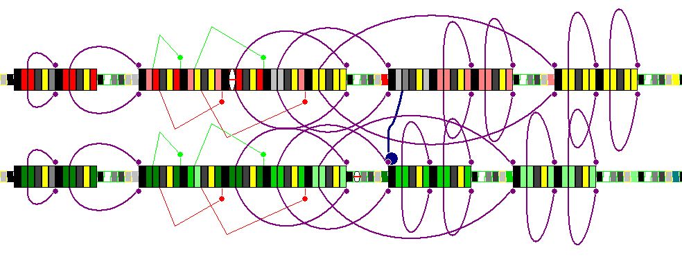
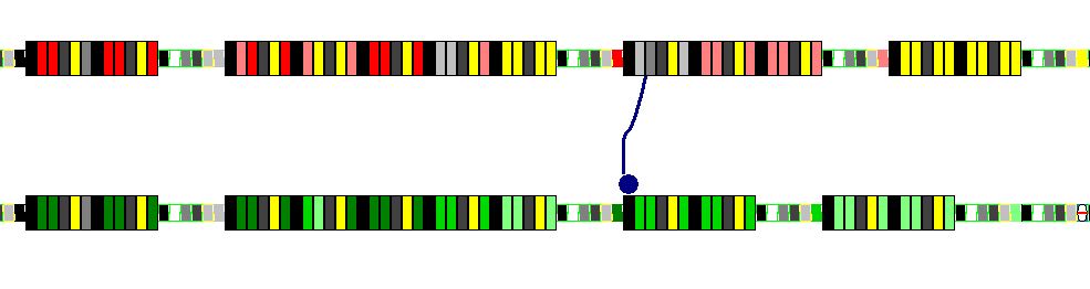
8.1 Meta-network interaction protocols between parental CENEs
Each gynandromorph has a unique signature of meta-network links between parental developmental control networks.

Irrespective of the number of combinations, what is interesting about these combinations is that they indicate an underlying global organization in the genome and its cenome. It leads me to suspect that the genome-cenome has a well organized architecture with sections of developmental control networks partitioned to be in correspondence with the major sections that vary in gynandromorphs.
8.2 Deterministic versus stochastic meta-networks
The links between opposite sex developmental control networks may be deterministic or stochastic. If they are deterministic then the same gynandromorphic phenotype will develop in all individuals whose cells are controlled by that network. If the links are stochastic and occur with low probability then we get what we see in the case of mosquitos were only a very small number of individuals appear to be gynandromorphs. However, because many gynandromorphs may be subtle variations of the millions of possibilities that are not overtly obvious, there may exist many more gynandromorphs than have been recognized as such. Hence, the chromosomal link architecture between parental CENEs may be more complex than indicated by the overt cases of insect gynandromorphs.
8.3 Bioengineering gynandromorphs
Given the existence of stochastic linkages between the developmental control networks of sex chromosomes, one should be able to engineer deterministic inter-sex developmental network links that replace stochastic links in the allelic developmental control networks of an insect like the mosquito. This would create a deterministic gynandromorphic network which if viable would lead to a specific gynandromorphic phenotype.
8.4 Genetic constraints on the space of possible gynandromorphs
The standard view is that variations in chromosome partitioning via unequal chromosomal placement in daughter cells creates gynandromorphs. However, the work of Clinton has challenged this view (see subsection 5.2). Still on both accounts, for sexually dimorphic gynandromorphs both the male and female CENEs that control dimorphic differences in development must exist in the gynandromorph’s genome. Chromosome loss or duplication can lead to inadvertent activation of the opposite sex chromosome. This is equivalent to a meta-network link being formed between the different maternal and paternal developmental networks.
We have shown above that chromosomal activation by cross chromosomal linking is an alternative way to generate gynandromorphs. The meta-network signatures that distinguish various types of gyn72 gynandromorphs assume a normal compliment of male and female CENEs, e.g., XY or ZW heteromorphic chromosomes. The gynandromorphs result from switches of control between one CENE and the sex-opposite CENE. This can be engineered into the CENEs by the creation a cross chromosomal link. It can result of a mutation that switches a link to point to the homologous area of the opposite sex chromosome. Or, it can occur by different chromosomal placement in pairs of progenitor cells as the organism develops.
Chromosomal placement can put limits on the future possible chromosomal divergence in daughter cells. For example, if XY produces an XX-cell and a YY-cell then we get one normal female cell and an abnormal male YY-cell. But neither of these cells can now generate a future mixed XY cell or cell of the opposite sex since the opposite sex complement is missing. Hence, once such a chromosomal separation occurs, if sex dimorphism is determined by CENEs on the X and Y chromosomes exclusively without the help of CENEs on autosomes then we can no longer get further gynandromorphic differences. Thus if development establishes an XX bilateral half that half can no longer generate a male Y based subsection. Whereas an XY bilateral half still has the potential to generate an XX female subsection. Therefore, chromosome placement limits the types of possible gynandromorphs.
In the network perspective, chromosome placement limits the possible meta-networks to a proper subset of the set of all possible meta-networks between sex based chromosomes. However, the early development of the embryo can influence chromosomal placement so that one may still get the basic gynandromorphs including bilateral, polar and oblique. Hence, the set of possible gynandromorphs resulting from chromosomal placement is relative to the upstream developmental network (upstream sub-CENE).
9 Synsexhemimorphism: Female and male hemimorphic organisms
In a female that contains two X chromosomes, normally one of the X chromosomes is suppressed. If not, we get abnormal development. The network theory of gynandromorphs predicts that there can exist females and males in various gynandromorph-like forms but where the bilateral, polar, or oblique versions are not governed by opposite sex chromosomes, but by different same sex chromosomes and/or autosomes from different parents. Similar can problems occur if the organism has two male Y chromosomes.
Thus, under the network theory of gynandromorphs there may exist organisms that are of the same sex (are not gynandromorphs) but that have non symmetrical or non-coherent body sections because their development was directed by distinct but same-sex developmental control networks.
Definitions: Synsexhemimorphs are same sex (male or female) organisms with asymmetric development, of normally symmetric body parts/sections, resulting from different parental allelic, developmental control networks ( CENEs). Gynohemimorphs are overtly female organisms with asymmetric body parts/sections resulting from different parental allelic, developmental control networks or CENEs. Androhemimorphs are overtly male organisms with asymmetric body parts/sections resulting from different, allelic parental developmental control networks or CENEs.
9.1 Hemihyperplasia as synsexhemimorphism
In the case of human males because they have only one Y chromosome one might think that diandromorphs cannot exist. However, homologous, allelic autosomes (and perhaps even the X chromosome) will usually contain nonidentical developmental control networks. Two homologous allelic CENEs on homologous autosomes may thus conflict in their phenotype and if asymmetrically activated this phenotypic disparity would become evident.
Examples of synsexhemimorphs (androhemimorphs or gynohemimorphs) may be humans with hemihyperplasia (hemihypertrophy) which is defined by asymmetric growth of normally symmetric body parts or sections including the cranium, face, trunk, limbs, and/or digits. This may be a form of developmental network induced male or female hemimorphism. Note, under this hypothesis hemihyperplasia need show neither chromosomal nor genetic abnormalities, because it is a result of meta-network cross talk between homologous, allelic autosomal developmental networks (CENEs).
10 Control conflicts and the origin of species
This opens up a much more general and fundamentally important question: How is the contribution of two different parents, their two different developmental control networks controlled? What determines which of the two different parental networks acts when and where?
Since these organisms are bilaterally symmetric the meta-control network that specifies which network is in charge at each point in space and time in development cannot be a random or stochastic process. Otherwise, if it were stochastic then the two bilateral sides would be morphologically dissimilar and not bilaterally symmetric. So too, identical twins show that the protocol of interaction (a meta-network) between the two parental genomes containing divergent developmental networks, cannot be random.
10.1 Sexual cooperation via meta-network protocols between parental genomes
Furthermore, the existence of gynandromorphs appears to indicate that control of which parental network is active at any given point in development cannot be a simple compromise between both networks that takes the average of two conflicting directives to a given cell at each point in time.
If this line of reasoning is correct, then it follows that there must exist a meta-control network that interlinks the two parental developmental control networks. This meta-control architecture implements a control protocol that coordinates the action of the two parental genomic contributions may be universal for a given species. Conflicts between such meta-control network linkages may be the cause of the very existence species.
Hence, the existence and origin of species is primarily the result of divergent developmental control networks and specifically divergent meta-control networks that interlink primary developmental networks. Thus, the evolution of species is the result of developmental network transformations.
10.2 Evolution of species
Since coordination conflicts would likely lead to unviable offspring, difference in the meta-control architecture, and hence, different meta-control linkages between parental networks, partition organisms into distinct classes, thereby creating different species. Hence, the origin of species results from unresolvable control conflicts between the developmental networks of different, putative species. A new species forms when the meta-control linkages between the parental networks of the new and the old species are no longer compatible resulting pathological development and/or unviable embryos.
The difference between species is a matter of degree. The greater the difference between the developmental networks of two species coordinating the networks of two divergent species becomes difficult no matter what meta-control linkages exist. Thus, if all possible meta-linkages (other than the null link and trivial links) between two parental networks fail to produce a viable offspring, then the species are very distant. Once, we decode the syntax and semantics of genomes [104, 105], we will have a new way of tracing the origin and evolution of species based on the architecture of developmental control networks.
10.3 Reverse engineering species formation
Just as it may be possible to engineer gynandromorphs, so it may be possible to engineer new species by modifying the meta-network interaction protocol or signature between parental CENEs. Also given two different species it may be possible to reverse engineer the two species by transforming their meta-network signatures to be compatible. This could make at least artificially induced fertilization between formerly distinct species possible and the resulting embryonic development viable. Hence, formerly distinct species A and B would have their meta-network signatures modified to A* and B* so that their interactions A*xB* leads to a viable embryo. However, would A*xA* or B*xB* still be viable?
Alternatively, it may be possible to reverse engineer an ancient ancestor species by transforming the meta-network signature to that of an ancestor. The case of atavisms shows that this is in principle possible. The feasibility depends on which ancient developmental control subnetworks are still there, hidden in the organism’s genome. By combining related specie’s CENEs using meta-network signature transformations one may be able to reverse engineer the common ancestor CENE taking parts from each of the two or more species CENEs. For example, could avian (bird) CENEs meta-linked to reptile CENEs by reverse species signature transformations allow the engineering of dinosaur like organisms?
11 Conclusion: How it all fits together
Gynandromorphs develop the way they do because of an interdependent cooperation between parental developmental control networks (CENEs), gene regulatory networks (GRNs), epigenetic cell orientation, cell-cell interconnection physics, and cell communication. CENEs subsume GRNs to control cell action. The cell’s interpretive-executive system (IES) interprets the control information in the genome (its cenome) relative to the cell’s orientation and executes that information using the GRNs to activate genes to perform various actions such as cell division, cell signaling, cell movement, and cell differentiation. Cell physics of intercellular connections also plays its role in the ultimate outcome. Cell communication is involved throughout development for spatial and temporal error correction, cell-cell coordination and cooperation.
Cells in the opposing symmetric body halves of bilateral organisms have opposite orientations and handedness. Handedness once established is network autonomous unless and until it is changed. Gynandromorphs show that the handedness of development of one half of the multicellular organism MCO1 is not dependent on a particular developmental control network N1 because the other oppositely oriented half MCO2 of the gynandromorph can develop according to a different developmental control network N2. Epigenetic cell polarity and not the genome is the primary cause of bilateral symmetry since cells maintain and inherit their orientation and handedness epigenetically. Cell signaling may be used to maintain polarity and orientation.
Since the same initial cell type can lead to two different morphologies in the two bilateral halves of gynandromorphs, the genome and not the containing cell is the prime driver of multicellular morphology. Gynandromorphs confirm the relative independence of morphology and cell orientation. It shows the relative independence of the morphological control information contained in the genome and the orientational information contained in the cell. Developmental control networks and not hormones determine the morphology of each bilateral half of a gynandromorph. Divergent subnetworks of the global developmental control network (the cenome) and not hormones determine the distinct morphology and function of sexually dimorphic organisms. Unequal chromosomal partitioning between cells can cause gynandromorphy because such partitioning can lead to modified meta-network linkages between allelic parental networks (whether they are located on sex chromosomes or autosomes).
The parental allelic developmental control networks must cooperate via meta-network linking protocols in order for coherent development to take place. Species form when established meta-network links change or are replaced or supplemented by new internetwork links that make sexual network cooperation between putative parental genomes unlikely or lead to unviable or infertile offsprings. Gynandromorphs led us to the insight that not genetic differences but rather developmental network divergences and incompatible meta-networks, which link the haploid developmental networks of the two parental sexes, result in the origin of species.
12 Materials and methods
Cellnomica’s Software Suite (http://cellnomica.com) was used to model and simulate gynandromorph multicellular development in space-time. The gynandromorphs in all the figures were all tested using Cellnomica’s Software Suite. Each of the concepts discussed was modeled and simulated with artificial genomes that generated multicellular bilaterally symmetric gynandromorphs starting from a single cell. Both stochastic and deterministic gynandromorph networks were developed and tested. The illustrations of multi-cellular systems are screenshots of cells that developed in virtual 4-dimensional space-time , modeled using the Cellnomica’s software. The illustrations of developmental control networks are screenshots of networks modeled and run with Cellnomica’s software.
References
- [1] A geneticist looks at human intersexes. Lancet, 273(6984):31–2, 1957.
- [2] F. Abdel-Hameed and R. N. Shoffner. Intersexes and sex determination in chickens. Science, 172(3986):962–4, 1971.
- [3] J. Adhami. Morphological abnormalities of sandflies (diptera, psychodidae) in albania. Parassitologia, 33(2-3):169–73, 1991.
- [4] S. Aw and M. Levin. What’s left in asymmetry? Developmental dynamics : an official publication of the American Association of Anatomists, 237(12):3453–63, 2008.
- [5] W. K. Baker. A fine-structure gynandromorph fate map of the drosophila head. Genetics, 88(4):743–54, 1978.
- [6] M. L. Balasov and A. V. Bgatov. Mapping of the focus of action of the lethal allele of the ecslt76 gene using genetic mosaics. Genetika, 28(11):40–7, 1992.
- [7] D. L. Barclay and W. H. Sternberg. A classification of intersexes: gynecologic considerations. Southern medical journal, 59(12):1383–92, 1966.
- [8] J. M. Belote. Male-specific lethal mutations of drosophila melanogaster . ii. parameters of gene action during male development. Genetics, 105(4):881–96, 1983.
- [9] A. S. Bernardino, T. V. Zanuncio, J. C. Zanuncio, E. R. Lima, and J. E. Serrao. Note on gynandromorphism in the eucalyptus defoliator thyrinteina arnobia (stoll, 1782) (lepidoptera: Geometridae). Anais da Academia Brasileira de Ciencias, 79(2):235–7, 2007.
- [10] D. Bodenstein. Milestones in developmental physiology of insects. Papers in development and heredity. Appleton-Century-Crofts, New York,, 1971.
- [11] S. T. Bowen and J. Hanson. A gynandromorph of the brine shrimp, artemia salina. Genetics, 47:277–80, 1962.
- [12] C. B. Bridges. Triploid intersexes in drosophila melanogaster. Science, 54(1394):252–4, 1921.
- [13] R. A. Brust. Gynandromorphs and intersexes in mosquitoes (diptera: Culicidae). Canadian journal of zoology, 44(5):911–21, 1966.
- [14] P. J. Bryant and M. Zornetzer. Mosaic analysis of lethal mutations in drosophila. Genetics, 75(4):623–37, 1973.
- [15] R. G. Bunge. What is sex?classification of intersexes and methods of diagnosis. the existence of intersexuality. Urologia internationalis, 19:165–77, 1965.
- [16] M. G. Burg and C. F. Wu. Mechanical and temperature stressor-induced seizure-and-paralysis behaviors in drosophila bang-sensitive mutants. Journal of neurogenetics, 26(2):189–97, 2012.
- [17] A. J. Campbell and M. W. Service. A gynandromorph of the mosquito aedes cantans in britain. Annals of tropical medicine and parasitology, 81(2):193–4, 1987.
- [18] S. B. Carroll. Evolution at two levels: on genes and form. PLoS biology, 3(7):e245, 2005.
- [19] S. B. Carroll. Evo-devo and an expanding evolutionary synthesis: a genetic theory of morphological evolution. Cell, 134(1):25–36, 2008.
- [20] J. E. Cilek and F. W. Knapp. Face fly (diptera: Muscidae) gynandromorph. Journal of medical entomology, 31(5):760–2, 1994.
- [21] F. C. Clarke and Y. Rechav. A case of gynandromorphism in amblyomma hebraeum (acari: Ixodidae). Journal of medical entomology, 29(1):113–4, 1992.
- [22] M. Clinton, D. Zhao, S. Nandi, and D. McBride. Evidence for avian cell autonomous sex identity (casi) and implications for the sex-determination process? Chromosome research : an international journal on the molecular, supramolecular and evolutionary aspects of chromosome biology, 20(1):177–90, 2012.
- [23] L. E. Craker, L. L. Nolan, K. Shetty, and I. S. for Horticultural Science. Proceedings of the International Symposium on Medicinal and Aromatic Plants, August 27-30, 1995, Amherst, MA USA. Acta horticulturae. International Society for Horticultural Science, Leuven, Belgium, 1996.
- [24] A. Crone-Muenzebrock and L. Leibecke. On congenital intersexes and their diagnosis and therapeutic possibilities. ii. Die Medizinische Welt, 17:924–30, 1960.
- [25] P. T. Dang and B. V. Peterson. A case of bilateral gynandromorphism in simulioum soubrense vajime and dunbar (diptera: Simuliidae). Tropenmedizin und Parasitologie, 30(4):548–50, 1979.
- [26] E. H. Davidson. The Regulatory Genome: Gene Regulatory Networks In Development And Evolution. Academic Press, 2006.
- [27] E. H. Davidson, J. P. Rast, P. Oliveri, A. Ransick, C. Calestani, C. H. Yuh, T. Minokawa, G. Amore, V. Hinman, C. Arenas-Mena, O. Otim, C. T. Brown, C. B. Livi, P. Y. Lee, R. Revilla, A. G. Rust, Z. Pan, M. J. Schilstra, P. J. Clarke, M. I. Arnone, L. Rowen, R. A. Cameron, D. R. McClay, L. Hood, and H. Bolouri. A genomic regulatory network for development. Science, 295(5560):1669–78, 2002.
- [28] I. Deak, P. R. Bellamy, M. Bienz, Y. Dubuis, E. Fenner, M. Gollin, A. Rahmi, T. Ramp, C. A. Reinhardt, and B. Cotton. Mutations affecting the indirect flight muscles of drosophila melanogaster. Journal of embryology and experimental morphology, 69:61–81, 1982.
- [29] T. Dobzhansky and B. Spassky. Intersexes in drosophila pseudoobscura. Proceedings of the National Academy of Sciences of the United States of America, 27(12):556–62, 1941.
- [30] U. Drews. Direct and mediated effects of testosterone: the development of intersexes in sex reversed mosaic mice, heterozygous for testicular feminization. Anatomy and embryology, 146(3):325–40, 1975.
- [31] E. Fekete and J. Szidonya. Abnormalities of ultrastructure and calcium distribution in the flight muscle of a flightless mutant of drosophila melanogaster. Acta biologica Academiae Scientiarum Hungaricae, 30(1):47–57, 1979.
- [32] K. F. Fischbach and G. Technau. Cell degeneration in the developing optic lobes of the sine oculis and small-optic-lobes mutants of drosophila melanogaster. Developmental biology, 104(1):219–39, 1984.
- [33] A. T. Ford. Intersexuality in crustacea: an environmental issue? Aquatic toxicology, 108:125–9, 2012.
- [34] n. Gargan, T. P., C. W. Kamau, P. C. Thande, and J. N. Wagateh. Gynandromorph of aedes mcintoshi from central kenya. Journal of the American Mosquito Control Association, 5(4):599–600, 1989.
- [35] W. J. Gehring, E. Wieschaus, and M. Holliger. The use of ’normal’ and ’transformed’ gynandromorphs in mapping the primordial germ cells and the gonadal mesoderm in drosophila. Journal of embryology and experimental morphology, 35(3):607–16, 1976.
- [36] W. H. Gerneke. Cytogenetic investigations on normal and malformed animals, with special reference to intersexes. The Onderstepoort journal of veterinary research, 34(1):219–99, 1967.
- [37] R. Goldschmidt. The interpretation of the structure of triploid intersexes in solenobia. Archiv der Julius Klaus-Stiftung fur Vererbungsforschung, Sozialanthropologie und Rassenhygiene, 21(3-4):269–72, 1946.
- [38] R. B. Goldschmidt. The beaded minute-intersexes in drosophila melanogaster meig. The Journal of experimental zoology, 112(2):233–301, 1949.
- [39] R. B. Goldschmidt. The interpretation of the triploid intersexes of solenobia. Experientia, 5(11):417–25, 1949.
- [40] I. V. Golubeva. The adaptation of intersexes to sex change. Zhurnal nevropatologii i psikhiatrii imeni S.S. Korsakova, 70(6):911–4, 1970.
- [41] M. Gonzalez-Gaitan, M. P. Capdevila, and A. Garcia-Bellido. Cell proliferation patterns in the wing imaginal disc of drosophila. Mechanisms of development, 46(3):183–200, 1994.
- [42] C. G. Goseco and V. R. Ferris. Intersexes of leptonchus obtusus thorne. Journal of nematology, 5(3):226–8, 1973.
- [43] W. G. Halina, D. W. Barrales, G. D. Partlow, and K. R. Fisher. Intersexes in swine: a problem in descriptive anatomy. Canadian journal of comparative medicine. Revue canadienne de medecine comparee, 48(3):313–21, 1984.
- [44] D. Hall. Gynondromorphism in mosquitoes’. Journal of the Florida AntiMosquito Association, 58(1):25–28, 1987.
- [45] D. Hall. Three culex salinarius gynandromorphs. JounNer, oF THE AMERTcAN Mosqurro Conrnor, AssocrATroN, 4(2):196–197, 1988.
- [46] A. Hannal-Alava and C. Stern. The sex combs in males and intersexes of drosophila melanogaster. The Journal of experimental zoology, 134(3):533–56, 1957.
- [47] J. C. Harshbarger, M. J. Coffey, and M. Y. Young. Intersexes in mississippi river shovelnose sturgeon sampled below saint louis, missouri, usa. Marine environmental research, 50(1-5):247–50, 2000.
- [48] J. L. Haynie and P. J. Bryant. Development of the eye-antenna imaginal disc and morphogenesis of the adult head in drosophila melanogaster. The Journal of experimental zoology, 237(3):293–308, 1986.
- [49] P. E. Hildreth. Doublesex, recessive gene that transforms both males and females of drosophila into intersexes. Genetics, 51:659–78, 1965.
- [50] G. W. Hinsch. Intersexes in the dog. Teratology, 20(3):463–8, 1979.
- [51] W. F. Hollander. Sectorial mosaics in the domestic pigeion: 25 more years. The Journal of heredity, 66(4):177–202, 1975.
- [52] M. J. Hollingsworth. The morphology of intersexes in drosophila subobscura. The Journal of experimental zoology, 143:123–51, 1960.
- [53] J. J. Howard, W. K. Gall, and J. Oliver. A gynandromorph of culiseta morsitans. Journal of the American Mosquito Control Association, 23(3):340–2, 2007.
- [54] J. S. Huxley. Intersexes in drosophila and different types of intersexuality. Science, 52(1333):59–60, 1920.
- [55] K. Ikeda and W. D. Kaplan. Unilaterally patterned neural activity of gynandromorphs, mosaic for a neurological mutant of drosophila melanogaster. Proceedings of the National Academy of Sciences of the United States of America, 67(3):1480–7, 1970.
- [56] W. Janning. Gynandromorph fate maps in drosophila. Results and problems in cell differentiation, 9:1–28, 1978.
- [57] R. Jimenez, M. Burgos, A. Sanchez, A. H. Sinclair, F. J. Alarcon, J. J. Marin, E. Ortega, and R. Diaz de la Guardia. Fertile females of the mole talpa occidentalis are phenotypic intersexes with ovotestes. Development, 118(4):1303–11, 1993.
- [58] J. Jones, H. W. Intersexes–surgical correction. Birth defects original article series, 13(2):137–45, 1977.
- [59] A. Kamping, V. Katju, L. W. Beukeboom, and J. H. Werren. Inheritance of gynandromorphism in the parasitic wasp nasonia vitripennis. Genetics, 175(3):1321–33, 2007.
- [60] A. Keskin, A. Bursali, and S. Tekin. A case of gynandromorphism in hyalomma marginatum koch, 1844 (acari: Ixodidae). The Journal of parasitology, 2012.
- [61] J. Kitzmiller. Genetics, cytogenetics, and evolution of mosquitoes. Advances in Genetics, 18:315–436, 1976.
- [62] R. A. Kleps, T. C. Myers, R. N. Lipcius, and T. O. Henderson. A sex-specific metabolite identified in a marine invertebrate utilizing phosphorus-31 nuclear magnetic resonance. PloS one, 2(8):e780, 2007.
- [63] M. B. Labruna, V. S. Homem, M. B. Heinemann, and J. S. Ferreira Neto. A case of gynandromorphism in amblyomma oblongoguttatum (acari: Ixodidae). Journal of medical entomology, 37(5):777–9, 2000.
- [64] M. B. Labruna, A. F. Ribeiro, M. V. Cruz, L. M. Camargo, and E. P. Camargo. Gynandromorphism in amblyomma cajennense and rhipicephalus sanguineus (acari: Ixodidae). The Journal of parasitology, 88(4):810–1, 2002.
- [65] G. Lauge. Feminization of external genitalia of triploid intersexes of drosophila melanogaster meig. under the action of elevated temperature. demonstration of a sensitive phase during development. Comptes rendus hebdomadaires des seances de l’Academie des sciences, 259:4156–9, 1964.
- [66] G. Lauge. Experimental conditions of feminization of the gonads in triploid intersexes in drosophila melanogaster meig.). Comptes rendus hebdomadaires des seances de l’Academie des sciences. Serie D: Sciences naturelles, 265(10):767–70, 1967.
- [67] P. A. Lawrence, S. M. Green, and P. Johnston. Compartmentalization and growth of the drosophila abdomen. Journal of embryology and experimental morphology, 43:233–45, 1978.
- [68] H. H. Li, C. S. Chiang, H. Y. Huang, and G. J. Liaw. mars and tousled-like kinase act in parallel to ensure chromosome fidelity in drosophila. Journal of biomedical science, 16:51, 2009.
- [69] M. Lucia, A. H. Abrahamovich, and L. J. Alvarez. A gynandromorph of xylocopa nigrocincta smith (hymenoptera: Apidae). Neotropical entomology, 38(1):901–3, 2009.
- [70] K. Maeno and S. Tanaka. Morphological and behavioural characteristics of a gynandromorph of the desert locust, schistocerca gregaria. Physiological Entomology, 32:294–299, 2007.
- [71] F. Mahmood and W. I. Bajwa. Description of a culex pipiens gynandromorph from new york city. Journal of the American Mosquito Control Association, 22(4):751–3, 2006.
- [72] R. Mann, L. Hake, and J. C. Lucchesi. Phenogenetics of triploid intersexes in drosophila melanogaster. Developmental genetics, 6(4):247–55, 1986.
- [73] K. Markopoulou and S. Artavanis-Tsakonas. Developmental analysis of the facets, a group of intronic mutations at the notch locus of drosophila melanogaster that affect postembryonic development. The Journal of experimental zoology, 257(3):314–29, 1991.
- [74] Y. Melander, E. Hansen-Melander, L. Holm, and B. Somlev. Seven swine intersexes with xx chromosome constitution. Hereditas, 69(1):51–8, 1971.
- [75] M. M. Melicow. Tumors of dysgenetic gonads in intersexes: case reports and discussion regarding their place in gonadal oncology. Bulletin of the New York Academy of Medicine, 42(1):3–20, 1966.
- [76] M. M. Melicow and A. C. Uson. Dysgenetic gonadomas and other gonadal neoplasms in intersexes; report of 5 cases and review of the literature. Cancer, 12(3):552–72, 1959.
- [77] C. J. Mitchell and T. B. Hughes. A bipolar differentiated gynandromorph of culex tarsalis coquillett, from texas. Journal of medical entomology, 6(1):78, 1969.
- [78] Y. Miyake. Cytogenetical studies on swine intersexes. The Japanese journal of veterinary research, 21(3):41–50, 1973.
- [79] J. Money. Psychology of intersexes. Urologia internationalis, 19:185–9, 1965.
- [80] B. Nilsson. A gynandromorph of the mallophagan goniodes colchici from phasianus colchicus. Angewandte Parasitologie, 17(4):223–5, 1976.
- [81] G. Paliyath, M. Bakovic, and K. Shetty. Functional foods, nutraceuticals, and degenerative disease prevention. Wiley-Blackwell, Chichester, West Sussex, UK ; Ames, Iowa, 2011.
- [82] A. R. Palmer. Animal asymmetry. Current biology : CB, 19(12):R473–7, 2009.
- [83] A. R. Palmer. Scale-eating cichlids: from hand(ed) to mouth. Journal of biology, 9(2):11, 2010.
- [84] A. R. Palmer. Developmental plasticity and the origin of novel forms: unveiling cryptic genetic variation via"use and disuse". Journal of experimental zoology. Part B, Molecular and developmental evolution, 318(6):466–79, 2012.
- [85] J. G. Rempel, J. M. Naylor, K. Rothfels, and B. Otonen. The sex chromosome constitution of chironomid intersexes parasitized by nematodes. Canadian journal of genetics and cytology. Journal canadien de genetique et de cytologie, 4:92–6, 1962.
- [86] J. L. Rodriguez Minon. Intersexes: Endocrinological aspects. Revista clinica espanola, 103(2):79–84, 1966.
- [87] E. L. Segura, Y. T. Shetty, M. Nishimizu, I. B. for Reconstruction, and Development. Fertilizer producer pricing in developing countries : issues and approaches. Industry and finance series,. World Bank, Washington, D.C., 1986.
- [88] N. Sethuraman and D. A. O’Brochta. The drosophila melanogaster cinnabar gene is a cell autonomous genetic marker in aedes aegypti (diptera: Culicidae). Journal of medical entomology, 42(4):716–8, 2005.
- [89] A. S. Shetty. Gynandromorphs of culex fatigans–the filarial mosquito. Current science, 44(19):2, 1975.
- [90] H. Stefan. Endoscopic and contrast examinations of intersexes in childhood. Ceskoslovenska pediatrie, 28(12):652–5, 1973.
- [91] J. S. Stewart. Genetic factors in intersexes. Proceedings of the Royal Society of Medicine, 52:817–8, 1959.
- [92] R. Strauss, U. Hanesch, M. Kinkelin, R. Wolf, and M. Heisenberg. No-bridge of drosophila melanogaster: portrait of a structural brain mutant of the central complex. Journal of neurogenetics, 8(3):125–55, 1992.
- [93] G. Struhl. A blastoderm fate map of compartments and segments of the drosophila head. Developmental biology, 84(2):386–96, 1981.
- [94] A. H. Sturtevant. Intersexes in drosophila simulans. Science, 51(1317):325–7, 1920.
- [95] A. H. Sturtevant. Intersexes dependent on a maternal effect in hybrids between drosophila repleta and d. neorepleta. Proceedings of the National Academy of Sciences of the United States of America, 32(4):84–7, 1946.
- [96] M. Switonski, H. Jackowiak, S. Godynicki, J. Klukowska, K. Borsiak, and K. Urbaniak. Familial occurrence of pig intersexes (38,xx; sry-negative) on a commercial fattening farm. Animal reproduction science, 69(1-2):117–24, 2002.
- [97] J. Szabad and C. Fajszi. Control of female reproduction in drosophila: genetic dissection using gynandromorphs. Genetics, 100(1):61–78, 1982.
- [98] S. Tanaka and K. Maeno. Maternal effects on progeny body size and color in the desert locust, schistocerca gregaria: examination of a current view. J Insect Physiol, 54(3):612–8, 2008.
- [99] S. Tsuchiyama, B. Sakaguchi, and K. Oishi. Analysis of gynandromorph survivals in drosophila melanogaster infected with the male-killing sr organisms. Genetics, 89(4):711–21, 1978.
- [100] G. M. Tulgetske and R. Stouthamer. Characterization of intersex production in trichogramma kaykai infected with parthenogenesis-inducing wolbachia. Die Naturwissenschaften, 99(2):143–52, 2012.
- [101] T. Uenoyama, S. Uchida, A. Fukunaga, and K. Oishi. Studies on the sex-specific lethals of drosophila melanogaster. iv. gynandromorph analysis of three male-specific lethals, mle, msl-2(27) and mle(3)132. Genetics, 102(2):223–31, 1982.
- [102] P. S. S. Veerabhadrappa. Multiple forms of carboxylesterases in the green bean (Phaseolus vulgaris L.) and pea (Pisum sativum L.). Corvallis.
- [103] H. Wallis. Psychopathological viewpoints in the treatment of intersexes. Monatsschrift fur Kinderheilkunde, 109:156–8, 1961.
- [104] E. Werner. In silico multicellular systems biology and minimal genomes. DDT, 8(24):1121–1127, 2003.
- [105] E. Werner. Genome semantics, in silico multicellular systems and the central dogma. FEBS Letters, 579(7):1779–1782, 2005.
- [106] E. Werner. On programs and genomes. arXiv:1110.5265v1 [q-bio.OT], http://arxiv.org/abs/1110.5265, 2011a.
- [107] E. Werner. Cancer networks: A general theoretical and computational framework for understanding cancer. arXiv:1110.5865v1 [q-bio.MN], http://arxiv.org/abs/1110.5865, 2011b.
- [108] E. Werner. The origin, evolution and development of bilateral symmetry in multicellular organisms. arXiv:1207.3289v1 [q-bio.TO], http://arxiv.org/abs/1207.3289, 2012a.
- [109] E. Werner. How to grow an organism inside-out: Evolution of an internal skeleton from an external skeleton in bilateral organisms. arXiv:1207.3624v1 [q-bio.TO], http://arxiv.org/abs/1207.3624, 2012b.
- [110] B. Wilczynski and E. E. M. Furlong. Challenges for modeling global gene regulatory networks during development: Insights from drosophila. Developmental Biology, 340(2):161–169, 2010.
- [111] A. S. Yang and E. Abouheif. Gynandromorphs as indicators of modularity and evolvability in ants. Journal of experimental zoology. Part B, Molecular and developmental evolution, 316(5):313–8, 2011.
- [112] J. Yoshizawa, K. Mimori, K. Yamauchi, and K. Tsuchida. Sex mosaics in a male dimorphic ant cardiocondyla kagutsuchi. Die Naturwissenschaften, 96(1):49–55, 2009.
- [113] D. Zhao, D. McBride, S. Nandi, H. A. McQueen, M. J. McGrew, P. M. Hocking, P. D. Lewis, H. M. Sang, and M. Clinton. Somatic sex identity is cell autonomous in the chicken. Nature, 464(7286):237–42, 2010.
- [114] D. Zhao, D. McBride, S. Nandi, H. A. McQueen, M. J. McGrew, P. M. Hocking, P. D. Lewis, H. M. Sang, and M. Clinton. Somatic sex identity is cell autonomous in the chicken. Nature, 464(7286):237–42, 2010.