Anomalous diffusion of phospholipids and cholesterols in a lipid bilayer and its origins
Abstract
Combining extensive molecular dynamics simulations of lipid bilayer systems of varying chemical composition with single-trajectory analyses we systematically elucidate the stochastic nature of the lipid motion. We observe subdiffusion over more than four orders of magnitude in time, clearly stretching into the sub-microsecond domain. The lipid motion delicately depends on the lipid chemistry, the lipid phase, and especially on the presence of cholesterol. We demonstrate that fractional Langevin equation motion universally describes the lipid motion in all phases including the gel phase, and in the presence of cholesterol. The results underline the relevance of anomalous diffusion in lipid bilayers and the strong effects of the membrane composition.
pacs:
87.16.dj,87.10.Mn,05.40.-a,02.50.-rRecent advances in single molecule spectroscopy unveil anomalous diffusion of microscopic tracers in the crowded environment of living cells, starting to reshape our views of molecular cell biology and underlining the role of modern statistical physics pt . Extensive experimental studies show subdiffusion in terms of the non-linear scaling in time of the mean squared displacement (MSD) ralf1
| (1) |
where is the anomalous diffusion exponent and the generalized diffusivity of physical dimension . Subdiffusion (1) was reported for various microscopic tracers under the densely crowded conditions inside living cells golding ; weber ; jeon ; seisenhuber ; garini and in control experiments weiss ; pan , as well as for proteins in cell membranes weiss1 ; weigel . These experiments demonstrate the ubiquitous presence of subdiffusion of a large variety of tracers and crowded environments over many orders of magnitude in time, but see note1 . Subdiffusion alters significantly the diffusion control of biochemical reactions, and its effects are therefore far-reaching for a wide range of molecular cellular processes minton . While subdiffusion (1) slows down long-distance diffusional exchange and may affect surface-bulk exchange irwin , it may indeed be beneficial for local interactions in cells golding ; guigas ; leila . Depending on the magnitude of the exponent anomalous diffusion may effect the localization of objects such as chromosomes or membrane channels garini ; weigel , and impact on the formation and dynamics of membrane domains.
Here we study in detail the diffusive behavior of lipids in bilayer systems through trajectory analysis from extensive molecular dynamics simulations. We find that in all investigated bilayers the lipids exhibit subdiffusion up to a few nanoseconds, before a crossover to either normal diffusion or to persistent anomalous diffusion with a larger exponent. The observed behavior depends strongly on the phospholipid chemistry, their mixture with cholesterol, and the bilayer phase (liquid/gel). Subdiffusion ranges at least up to several hundreds of nanoseconds in the presence of cholesterols. Our analysis shows that the lipid motion is consistent with viscoelastic subdiffusion driven by correlated Gaussian noise in both liquid and gel phases, and thus provides a unified physical framework for lipid diffusion in membranes.
Subdiffusion (1) is described by several prominent models based on different physical mechanisms ralf1 ; stas2 . In continuous time random walks (CTRWs) jumps are separated by random waiting times with heavy-tailed distributions scher . CTRW motion was identified for microbead motion in reconstituted actin networks wong , lipid granules in cellular cytoplasm jeon , and protein channels in plasma membranes weigel . In contrast fractional Brownian motion (FBM) and fractional Langevin equation (FLE) produce ergodic subdiffusion (1) with long-ranged anti-correlation () lutz
| (2) |
of spatial displacements . FBM is defined by an overdamped Langevin equation driven by athermal, external Gaussian noise with power-law correlation. FLE is a generalized Langevin equation driven by the same noise. Due to its memory kernel the FLE describes thermal motion of a particle in viscoelastic media igor . While FLE in the overdamped limit produces FBM-like subdiffusive motion, below the momentum relaxation time, FLE motion is ballistic. FBM/FLE motion describe subdiffusion in living cells of mRNA magdziarz , chromosomal loci weber , and of lipid granules at longer times jeon , as well as the motion of macromolecules in a crowded dextran solution weiss . While sharing the scaling form of the MSD (1), CTRW and FBM/FLE lead to completely different dynamics of diffusion-control. Knowledge of the subdiffusion mechanism in membranes is therefore vital to advance our understanding of their physical and biochemical properties.
Lipid bilayers are quasi two-dimensional, highly packed systems made up of phospholipid molecules, which undergo thermally driven lateral diffusion and thus constantly reorganize the membrane. The lateral MSD of membrane lipids typically spans three distinct regimes: short-time ballistic (), intermediate subdiffusive (), and long-time Brownian motion () flenner ; armstrong . The long-time diffusive motion of various kinds of phospholipid molecules in lipid bilayers has been extensively studied vaz2 ; almeida . Diffusion of lipids in pure bilayers occurs both in the liquid disordered and the gel phases below the melting temperature, the latter with decreased diffusivity. Moreover, in bilayers mixed with cholesterols, the diffusivity of the lipids tends to decrease with higher cholesterol concentration.
Lipid subdiffusion at shorter time scales is comparatively poorly understood. In the traditional microscopic picture, the lateral movement of lipid molecules is assumed to occur through jumps when sufficient void space is thermally activated at nearest sites almeida ; vaz . Between jumps the molecule, caged by its neighbors, undergoes rattling motion. This CTRW-type jump-diffusion model has been used to estimate the diffusivities of lipids in the liquid-disordered phase and in bilayers containing cholesterol vaz2 ; almeida . However, atomistic simulations falck and quasi-elastic neutron scattering experiment busch showed that such jump-like displacements rarely occur, and the lipids move concertedly with their neighbors as loosely defined clusters. Moreover, conflicting results were reported on the stochastic nature of the lipid diffusion: Refs. flenner ; kneller demonstrated that the lipid motion is consistent with FLE dynamics, whereas Ref. akimoto claimed to observe CTRW-type motion governed by non-Gaussian fluctuations and scale-free rattling dynamics.
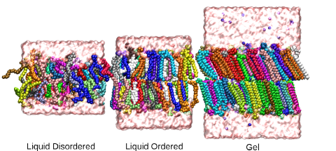
Lipid bilayers of 128 phospholipid molecules were studied by molecular dynamics simulations under periodic boundary conditions, for details see the Supplementary Material (SM) supp . We used three pure single component lipid bilayers composed of DSPC, SOPC, and DOPC phospholipids in the liquid disordered phase REM . We also studied these systems with additional 32 cholesterols (20% molar concentration) in the liquid ordered phase. A pure membrane of 288 DSPC molecules was also simulated in the gel phase. Fig. 1 shows typical snapshots in the three phases. In this work we focus on the characterization of the lipid diffusion. To that end we note that during the simulation the center of mass of the upper and lower lipid layers undergo free, independent translational motion (Fig. S1), as reported previously akimoto ; edholm . Free center of mass diffusion causes apparent normal diffusion of individual lipid molecules at longer times, irrespective of their actual diffusion characteristics. To avoid this we analyze the relative motion from the center of mass of lipids and cholesterols. Figs. S2, S5, and S11 in SM show sample trajectories.
From individual trajectories we obtained the time-averaged (TA) MSD of lipids typically defined as golding ; weber ; stas2
| (3) |
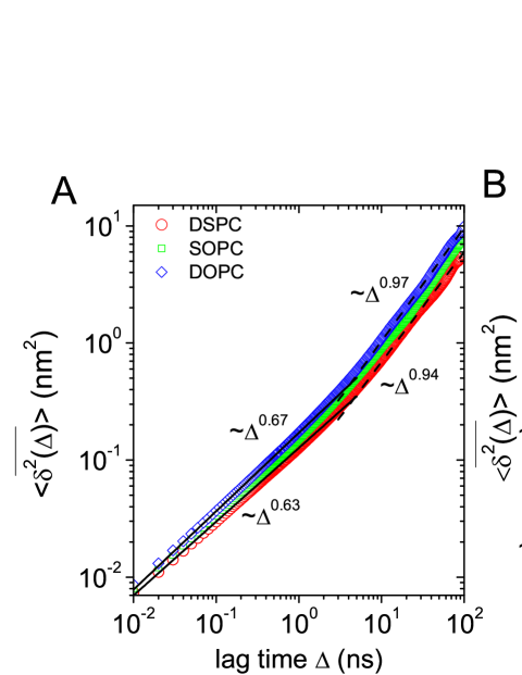
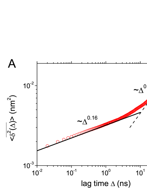
where is the lag time and the length of the trajectory (measurement time). Fig. 2 shows the mean taken over the trajectories of all phospholipids, for the cases of DSPC, SOPC, and DOPC in absence and presence of cholesterol. In each case, the result was fitted by at short and long times, respectively. The corresponding diffusion exponents and diffusivities are summarized in Tab. 1. In Fig. 2, the scaling behaviors for pure DSPC and DOPC at short (solid line) and long (dashed line) times are indicated. In absence of cholesterol, all three types of lipid molecules show similar behavior: anomalous diffusion with exponent below a crossover time ns, and normal Brownian motion beyond . The crossover time roughly corresponds to the diffusion time of a lipid molecule needed to span its nearest-neighbor distance. The structural difference in the tails of the lipids affects somewhat the long-time diffusion, in particular, the values of .
| DSPC | 0.63(0.06%) | 0.032(2%) | 0.94(0.9%) | 0.020(5%) |
| SOPC | 0.66(1%) | 0.038(2%) | 1.00(16%) | 0.020(60%) |
| DOPC | 0.67(2%) | 0.043(2%) | 0.97(13%) | 0.028(16%) |
| DSPC | 0.52(1%) | 0.013(2%) | 0.82(4%) | 0.0076(6%) |
| SOPC | 0.58(0.4%) | 0.019(0.5%) | 0.87(5%) | 0.012(4%) |
| DOPC | 0.61(0.5%) | 0.023(0.4%) | 1.04(22%) | 0.0098(41%) |
Fig. 2B shows that cholesterol significantly affects both the short and long time diffusion of the lipids. Especially for saturated DSPC with the smallest cross section area hector2 , we observe that below 10 ns decreases to about 0.5 and a new subdiffusion regime emerges with up to 100 ns. An additional 1 s-long simulation confirms that this new regime above is in fact a slow transition toward normal diffusion that lasts over hundreds of nanoseconds (Fig. S10). In the displayed time window the behavior is well fitted by the scaling exponents listed in Tab. 1. For unsaturated DOPC, the effect of cholesterols is small, albeit is significantly reduced, and no second subdiffusion regime occurs.
The observed subdiffusive behavior of lipids is mainly attributed to the unique structural complexity of the lipid molecule. Spherical-shaped hard particle systems cannot have such a long subdiffusion regime and values of as small as 0.5-0.6 (Fig. S18). To gain additional physical insight into the lipid motion we now check the detailed stochastic properties of the lipids.
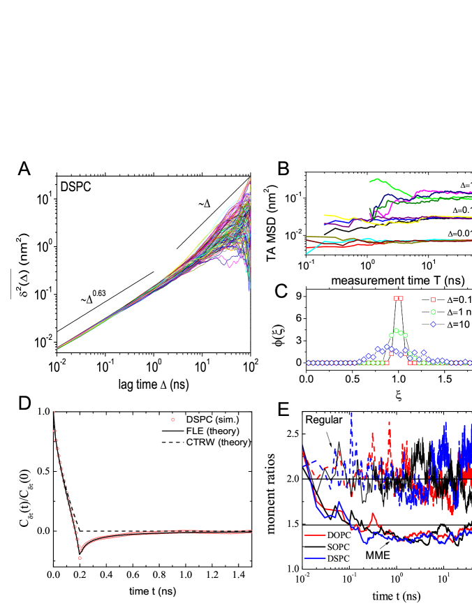
Time-averaged observables obtained from single trajectories provide information on the ergodic properties of a stochastic motion, and thus about the physical nature of the underlying dynamics. We call a process ergodic when the long time average of a quantity (e.g., the MSD) equals the corresponding ensemble average stas2 ; stas1 . For free CTRW subdiffusion free grows like , while the ensemble average (1) scales sub-linearly He ; stas2 . Free FLE motion is ergodic deng ; jae , and . The observation of a sublinear slope in Fig. 2 already indicates that the observed motion is not of CTRW type. This is further confirmed by the independence of of the measurement time , Fig. 3B, in contrast to the scaling of CTRW stas2 ; He . Fig. 3C shows the distribution of trajectory-to-trajectory amplitude variations of for the 128 individual molecules of Fig. 3A, as function of the dimensionless variable . All curves are centered around the ergodic value . The broadening of with increasing mirrors large fluctuations of at long due to insufficient statistics when calculating . also narrows at fixed when is increased (Fig. S3). These properties demonstrate that the lipid molecules perform ergodic motion different from CTRW.
We obtained the displacement autocorrelation function
| (4) |
for arbitrary time step for several diffusion models in SM III supp . Normalized, is a fit free function for given and . Fig. 3D shows at ns of DSPC lipids from simulations in the subdiffusion regime, along with theoretical results for CTRW and FLE motion supp . We find excellent agreement with FLE motion. Here was taken from the TA MSD. The lipid motion is thus anti-correlated, in line with Eq. (2). The behavior in Fig. 3D differs distinctly from free CTRW motion stas2 where for (dashed line). Fig. 3E also shows the moment ratios and , where denotes the maximal distance of a given particle from its initial position reached up to time tejedor . Moment ratios have unique values depending on the stochastic process tejedor , as summarized in SM supp . Fig. 3E shows that fluctuates around 2, and decreases from to stay , as predicted for FLE and violating CTRW.
We conclude that the subdiffusive behavior shown above is robust, all analysis tools convincingly point to FLE motion. Analogous results were obtained for SOPC and DOPC molecules. The results are preserved at varying temperature: temperature increase only leads to an increase of in the Brownian regime (Fig. S4). Consistent with previous studies falck ; busch the collective motion of lipids exhibit a flow-like pattern (Fig. S16).
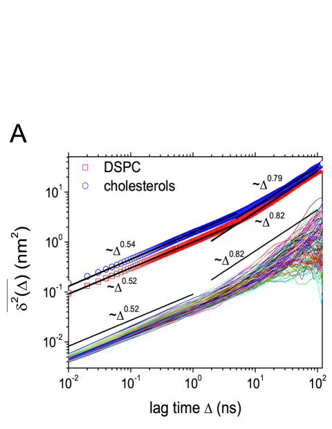
As shown in Fig. 2, the diffusion of the lipid molecules is drastically changed by the presence of cholesterols. Is the stochastic nature also affected by cholesterols? We find that ergodicity of the motion is preserved, while cholesterols significantly affect the distribution of . Comparing with the pure bilayer (Fig. 3C), with cholesterol noticeably broadens (Fig. 4B). Individual lipids thus undergo considerable variations while diffusing in the presence of cholesterols, as seen in Fig. 4A. Moreover, with cholesterols the displacement autocorrelation has a slightly deeper well, consistent with a stronger anti-correlation of the displacement (Fig. S6).
For the motion of the cholesterol molecules themselves we find that their behavior is almost identical to the lipids’, of FLE-type anomalous diffusion. Fig. 4A compares the TA MSD averaged over all trajectories separately for DSPC and cholesterol molecules. While cholesterols universally diffuse faster than the lipids, their scaling behaviors are almost the same. Meanwhile, the scatter distributions of cholesterols are sharper than that of the lipids, implying that cholesterol diffusion is more uniform than that of lipids. This may be related to the fact that at any moment only some lipids are in direct contact with cholesterols which modifies their behavior from those of the remaining lipds. The displacement autocorrelation of cholesterols is hardly different from that of the lipids in the lipid-cholesterol bilayer (Fig. S9), and the moment ratios agree with FLE motion (Fig. S7).
To obtain a full picture of the diffusive motion in lipid bilayers, we also studied the gel phase. For DSPC molecules we obtain: (i) Fig. 5A shows that scales with at short times and is thus remarkably smaller than the value in the liquid phase. Moreover, in the gel phase the TA MSD remains subdiffusive with beyond the crossover. (ii) The scatter distribution of individual shows that the lipid motion remains ergodic in the gel phase (Fig. S12) jae1 . (iii) The results for the gel phase autocorrelation are consistent with FLE motion with exponent (Fig. 5B), as are the moment ratios (Fig. S13). (iv) Contrasting recent claims akimoto , the rattling dynamics of lipids is consistent with FLE motion (Figs. S14, S15).
In summary, we here report extensive molecular dynamics simulations of lipid bilayer systems and the analysis of individual trajectories using stochastic analysis tools. While we find a moderate dependence on the lipid chemistry, the effect of cholesterols is striking. Cholesterols effect more pronounced and persistent subdiffusion. FLE motion is identified as the unifying process for the motion of both phospholipids and cholesterols in liquid and gel phases. Our study thus provides an integral picture of lateral motion of lipids by showing the compatibility of FLE-type stochastic motion of individual molecules and their flow-like collective motion.
Cholesterols significantly affect the phospholipids diffusion via increasing membrane packing and inducing 2D ordering hector_new (Fig. 1). is lowered significantly to below ns. In agreement, recent experimental and computational studies show that decreases with increase of the concentration of proteins in the bilayer horton ; matti . Interestingly we observe a pronounced variation between individual lipid’s motion, likely due to the asymmetric disturbance caused by cholesterols hector_new . While the slowing down of lipid diffusion by cholesterols is known from experiment almeida ; lindblom and simulations edholm , the dramatic effects of cholesterols on intermediate-time lipid diffusion have not been reported to our best knowledge.
Given above results we speculate that in biomembranes, whose complexity is higher than the bilayers’ studied here (e.g., larger number of lipid moieties, proteins, and higher cholesterol concentration), subdiffusion may range to macroscopic times, thus altering our current view of membrane dynamics. Single particle tracking together with advanced simulations techniques and stochastic analysis tools are promising methods to explore this intriguing possibility.
Acknowledgements.
We thank Otto Pulkkinen and Ilpo Vattulainen for discussions. We acknowledge financial support from the Academy of Finland (FiDiPro scheme) and the CSC–IT Center for Science in Finland for computing resources.References
- (1) E. Barkai, Y. Garini, and R. Metzler, Physics Today 65, 29 (2012).
- (2) R. Metzler and J. Klafter, Phys. Rep. 339, 1 (2000); R. Metzler and J. Klafter, J. Phys. A 37, R161 (2004).
- (3) I. Golding and E. C. Cox, Phys. Rev. Lett. 96, 098102 (2006).
- (4) S. Weber, A. J. Spakowitz, and J. A. Theriot, Phys. Rev. Lett. 104, 238102 (2010).
- (5) G. Seisenberger et al., Science 294, 1929 (2001).
- (6) J.-H. Jeon et al., Phys. Rev. Lett. 106, 048103 (2011).
- (7) I. Bronstein et al., Phys. Rev. Lett. 103, 018102 (2009).
- (8) J. Szymanski and M. Weiss, Phys. Rev. Lett. 103, 038102 (2009).
- (9) W. Pan et al., Phys. Rev. Lett. 102, 058101 (2009).
- (10) M. Weiss, H. Hashimoto, and T. Nilsson, Biophys. J. 84, 4043 (2003).
- (11) A. V. Weigel et al., Proc. Nat. Acad. Sci. USA 108, 6438 (2011).
- (12) Note that there also exist reports on normal diffusion, especially for small tracer particles: J. Elf, G.-W. Li, and X. S. Xie, Science 316, 1191 (2007); J. A. Dix and A. S. Verkman, Annu. Rev. Biophys. 37, 247 (2008).
- (13) A. P. Minton, J. Cell Sci. 119, 2863 (2006); D. Ben-Avraham and S. Havlin, Diffusion and Reactions in Fractals and Disordered Systems (Cambridge University Press, Cambridge UK, 2005); E. Abad, S. B. Yuste, and K. Lindenberg, Phys. Rev. E 81, 031115 (2010); D. Froemberg and I. M. Sokolov, Phys. Rev. Lett. 100, 108304 (2008).
- (14) M. A. Lomholt, I. M. Zaid, and R. Metzler, Phys. Rev. Lett. 98, 200603 (2007); I. M. Zaid, M. A. Lomholt, and R. Metzler, Biophys. J. 97, 710 (2009).
- (15) G. Guigas and M. Weiss, Biophys. J. 94, 90 (2008).
- (16) L. E. Sereshki, M. A. Lomholt, and R. Metzler, EPL 97, 20008 (2012).
- (17) S. Burov, J.-H. Jeon, R. Metzler, and E. Barkai, Phys. Chem. Chem. Phys. 13, 1800 (2011); I. M. Sokolov, Soft Matt. DOI: 10.1039/c2sm25701g.
- (18) H. Scher and E. W. Montroll, Phys. Rev. B 12, 2455 (1975).
- (19) I. Y. Wong et al., Phys. Rev. Lett. 92, 178101 (2004).
- (20) E. Lutz, Phys. Rev. E 64, 051106 (2001).
- (21) I. Goychuk, Adv. Chem. Phys. 150, 187 (2012).
- (22) M. Magdziarz, et al., Phys. Rev. Lett. 103, 180602 (2009).
- (23) E. Flenner, J. Das, M. C. Rheinstadter, and I. Kosztin, Phys. Rev. E 79, 011907 (2009).
- (24) C. L. Armstrong et al., Soft. Matt. 7, 8358 (2011).
- (25) W. L. C. Vaz, R. M. Clegg, and D. Hallman, Biochem. 24, 781 (1985).
- (26) P. F. F. Almeida, W. L. C. Vaz, and T. E. Thompson, Biochem. 31, 6739 (1992).
- (27) W. L. C. Vaz and P. F. Almeida, Biophys. J. 60, 1553 (1991).
- (28) E. Falck, T. Rog, M. Karttunen, and I. Vattulainen, J. Am. Chem. Soc. 130, 44 (2008).
- (29) S. Busch, C. Smuda, L. C. Pardo, and T. Unruh, J. Am. Chem. Soc. 132, 3232 (2010).
- (30) G. R. Kneller, K. Baczynski, and M. Pasenkiewicz-Gierula, J. Chem. Phys. 135, 141105 (2011).
- (31) T. Akimoto et al., Phys. Rev. Lett. 107, 178103 (2011).
- (32) Supplementary Material.
- (33) Their chemical notation is 1,2–distearoyl–sn–glycero–3–phosphatidylcholine (DSPC), 1,2–dioleoyl–sn–glycero–3–phosphatidylcholine (DOPC), and 1–stearoyl–2–oleoyl–sn–glycero–3–phosphatidylcholine (SOPC).
- (34) C. Hofsäß, E. Lindahl, and O. Edholm, Biophys. J. 84, 2192 (2003).
- (35) H. Martinez-Seara et al., Biophys. J. 95, 3295 (2008).
- (36) S. Burov, R. Metzler, and E. Barkai, Proc. Nat. Acad. Sci. USA 107, 13228 (2010).
- (37) Free CTRW and FLE motions refer to the processes in free space, without external confining potential or obstacles that could constrain the motion.
- (38) Y. He, S. Burov, R. Metzler, and E. Barkai, Phys. Rev. Lett. 101, 058101 (2008).
- (39) W. Deng and E. Barkai, Phys. Rev. E 79, 011112 (2009).
- (40) J.-H. Jeon and R. Metzler, Phys. Rev. E 81, 021103 (2010); ibid. 85, 021147 (2012).
- (41) V. Tejedor et al., Biophys. J. 98, 1364 (2010).
- (42) J.-H. Jeon and R. Metzler, J. Phys. A 43, 252001 (2010).
- (43) H. Martinez-Seara et al., PLoS One 5, e11162 (2010).
- (44) M. R. Horton et al., Soft Matt. 12, 2648 (2010).
- (45) M. Javanainen et al., Faraday Discuss. DOI: 10.1039/C2FD20085F
- (46) A. Filippov, G. Oradd, and G. Lindblom, Biophys. J. 84, 3079 (2003).