Induction of a photostationary ring-opening/closing state of spiropyran monolayers on the semi-metallic Bi(110) surface
Abstract
Molecular switches on metal surfaces typically show very little photoreactivity. Using scanning tunneling microscopy we show that the ring-opening/-closing switch nitrospiropyran thermally and optically isomerizes to the open merocyanine form on a Bi(110) surface. Irradiation by blue light of a monolayer spiropyran molecules leads to mixed domains of the two isomers. At large illumination intensities a photostationary state is established, indicating the bidirectional ring-opening and closing reaction of these molecules on the bismuth surface. The enhanced photoactivity contrasts with the case of adsorption on other metal surfaces, probably due to the low density of states at the Fermi level of the semi-metallic Bi(110) surface.
The combination of organic molecular switches with inorganic materials foresees extending their switchable functionality to a large variety of physical and chemical processes. Several interesting perspectives arise from such hybrid systems like, for example, using the different conductance of two isomers to act as an electronic switch in molecular based devices Kudernac09 ; vanderMolen10 , or the different optical adsorption properties to produce tunable coatings FeringaBook ; Rosario04 . The application of molecular switches in devices, however, relies on the persistence of their switching ability by external stimuli when interacting (mechanically and electronically) with a metal electrode or a surface. The presence of a metal surface may introduce alternative excitation routes in the switching mechanism: photoexcited electrons/holes from the substrate may be transferred to the molecule enabling an isomerization in the anionic/cationic states, respectively Hagen08a . In spite of that, it is found that the switching processes can be irreversible or even fully suppressed for molecules in direct contact with a metallic substrate. The origin is attributed to the fast quenching of excited states due to the presence of the metal; the coupling of molecular states with metallic electronic bands allows dumping the excitation energy into a continuum of substrate excitations. The strong reduction of excite states’ lifetime down to time scales much shorter than the isomerization process Dulic03 ; Henningsen08 ; Tegeder11 drastically decreases the quantum yield many orders of magnitude with respect to electronically isolated molecules Crommie07 .
Spiropyran based molecules are prototype molecular switches. The 1,3,3-Trimethylindolino-6-nitrobenzopyrylospiran (spiropyran, SP) isomer is a three-dimensional, inert, and colorless molecule. Cleavage of the central C-O bond leads to the planar, chemically active, and colored merocyanine isomer (MC) (Fig. 1). In solution, the ring-opening reaction is induced by ultra-violet light, while the back-reaction is triggered by visible light or temperature Rumpf53 ; Hirshberg52 . Multilayers of these molecules are also reversibly photo-isomerizable Karcher07 . In contrast, monolayers of SP on a Au(111) surface behave completely different; the only switching event observed was the thermally activated ring-opening reaction Piantek09 . The inversion of the thermodynamic stability and inhibition of photoreactive switching emphasized the influence of the substrate Tegeder11 . One strategy to reduce the role of the surface could be to employ materials which weakly couple electronically with adsorbates.
Here, we investigate the response to illumination of spiropyran molecules adsorbed on Bi(110), a semi-metallic surface with low density of states at the Fermi level Hofmann01 ; Hofmann06 . We find that, contrary to other metal surfaces, spiropyran exhibits here photo-reactive activity; illumination with monochromatic (blue) light produces increasing amounts of the open isomer with the applied power, until reaching a photostationary state consisting of both closed (SP) and open (MC) isomers with a ratio reflecting their difference in photoisomerization cross-section. The existence of this state implies that photoisomerization occurs in both directions, i.e. the C-O bond is cleaved and formed by the same external stimuli.
The bismuth surface was prepared by successive ion sputtering and annealing (420 K) cycles in ultra-high vacuum. The spiropyran molecules were evaporated using a Knudsen cell onto the bismuth sample held at room temperature. The sample was then cooled down and transferred into a custom-made low temperature scanning tunneling microscope (STM), at K, for inspection. For the illumination experiments, the sample was removed from the STM and irradiated with a blue laser diode (wavelength = 445 nm, photon energy Eph= 2.8 eV, total power P= 45 mW) in the preparation chamber with an angle of 45∘ and at 300 K. The samples were afterwards cooled and inspected again at K.
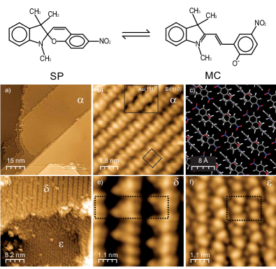
Deposition of spiropyran molecules onto the Bi surface at RT leads to self-assembled extended islands (Fig. 1 a) composed of molecular features arranged in densely packed rows with a rectangular unit cell ( nm2). The structure of the islands resembles in shape and size spiropyran islands grown on a Au(111) surface at temperatures below K (see inset of Fig. 1 b) Piantek09 . From the striking resemblance of STM images on the two surfaces we conclude that also on bismuth the ring-closed spiropyran conformation (SP) can be stabilized on the surface. The corresponding structural model of the molecular arrangement is shown in Fig. 1 c). The structure is stabilized by -H bonds and H-bonds between the molecules. This structure is labeled as “” in the following.
The structure of the molecular layer changes drastically when the bismuth sample is annealed to K. While the initial pattern vanishes, two new ordered phases appear on the surface, plus some disordered regions (Fig. 1 d). The first of the two novel phases, labeled (Fig. 1 e), is characterized by broad stripes (unit a cell nm2). Here, the STM images do not provide any concluding fingerprint about the isomeric composition of the structure. The second phase () has a smaller unit cell ( nm2) and clearly shows inside a double-lobe molecular feature repeated 4 times with different orientations (Fig. 1 f). This homogeneous phase is the only one observed on the sample after annealing to higher temperatures ( K). As for the case on Au(111) Piantek09 , we expect that at sufficiently high temperatures the complete molecular layer undergoes a thermally-activated ring-opening reaction. Hence, we identify the high-temperature phase as being composed exclusively of the ring-opened isomer merocyanine (MC). Correspondingly, the intermediate phase is formed by a mixture of both isomers within its unit cell.
To investigate the photoisomerization ability of the molecules on the Bi(110) surface, a pristine SP layer was exposed to blue laser light (Eph= 2.8 eV) for minutes, while keeping the sample temperature at 300 K, below the temperature for thermal isomerization note1 . The laser spot was not focused on the surface; instead, it had an oval shape of about mm mm size. Since the photon intensity is not homogeneous across the laser beam, we expect that different areas of the sample were exposed to different light intensity. This allows us to investigate in a single experiment the effect of different photon fluences on the isomerization simply by exploring with the STM different areas within the laser spot. To obtain a function describing the photon fluence across the surface, we approximate the total power distribution across the laser spot with a Gaussian function and consider that it integrates to the calibrated value of 45 mW. In this way, we obtain that the laser fluence during the experiment gradually decreased from kJ/cm2 at the center of the spot to zero, at the boundaries.
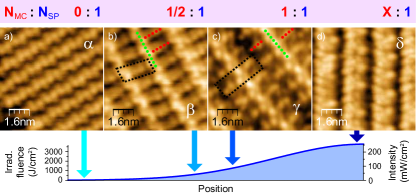
After irradiation of the SP molecular layers, a set of new structurally different phases appear on the surface, generally coexisting, and with some disordered areas of molecules in between. Fig. 2 summarizes the different phases and the position where they are most frequently observed with respect to the center of the laser spot. The drastic change in molecular structure with respect to the initial phase is an indication that they do not consist of pure SP molecules, but of a mixture with the other isomer (MC). The type of phases most frequently observed in a region (i.e. their relative area) depends on the position within the broad laser spot. This suggests that there is a correlation with the light distribution across the laser spot.
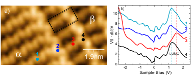
On positions with low irradiation power, the initial pure SP phase can still be observed (Fig. 2 a). However, in regions with moderate irradiation two novel grid phases, labelled and , appear (Fig. 2 b and 2 c). These phases combine structures with two different shapes in the STM images, forming ladder-like arrangements. Scanning tunneling spectroscopy (STS) measurements allows us to identify the corresponding isomers (Fig. 3). The spectra on the higher species show an unoccupied molecular resonance at V, characteristic of molecules in the phase. We thus identify them as spiropyran isomers. Correspondingly, the lower features are identified as the open isomer merocyanine, with spectral features consisting of an empty state shifted to higher values and the onset of an occupied resonance below V. Careful analysis of the shape and size of the patterns’ unit cell allows us to conclude molecular ratios MC:SP of 1:2 and 1:1 for the and phases, respectively note2 . The different composition of the two phases correlates with different photon dosage ( kJ/cm2 for and kJ/cm2 for ). This is in agreement with an increase of the MC fraction for larger radiation doses.
At the center of the laser spot the photon fluence reaches its maximum value, more than kJ/cm2. There, all former patterns are transformed into a single phase: the phase (the same as found after annealing a SP monolayer to K (Fig. 1f). However, even after extended irradiation periods of more than 8 hours, the phase was the only one observed; no hint of the full MC phase (phase in Fig. 1) was ever found. Following the tendency of an increase in the MC fraction with the irradiation intensity, this structure is probably composed of a larger fraction of MC isomers. The structure of the large unit cell appears rather inhomogeneous, suggesting a varying ratio between the two isomers and/or involving various conformers of the ”flexible” merocyanine backbone. Unfortunately, our STM measurements are not clear enough to reveal the precise isomer composition of this phase.
The experiments thus show that illumination of a pure spiropyran monolayer on Bi(110) results in pronounced changes in the molecular self-assembled structures. The structural changes reflect the isomerization of large fractions of the spiropyran molecules towards their merocyanine form. We can rule out a thermal-activated cleavage of the C-O bond in spiropyran: persistent monitoring of the sample temperature and heat conduction calculations ensure that the surface temperature does not rise significantly during the light exposure. Thus, we attribute the isomerization to a photon-induced process.
The large-scale molecular reorganization implies that the two molecular species are highly mobile on the surface during irradiation. Most probably the system can be described as a two dimensional molecular “liquid”; only in this way, a gradual increase of the fraction of MC isomers with light would cause an increase of MC-rich phases. The various mixed MC/SP structures are then crystallized upon cooling. Their relative area changes gradually with the position, reflecting the local composition of the isomeric mixture. At the center of the laser spot, the mixed phase is always observed, even after much larger illumination times (same light intensity but much larger fluence). The stabilization of a MC:SP equilibrium structure independent of the photon fluence is a fingerprint of the system being driven into a photostationary state through SP MC bidirectional photo-isomerization processes. This contrasts with a situation where the backswitching channel is blocked, what would inevitably lead to a saturation of the pure reaction product MC note3 .
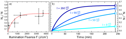
In order to get a quantitative insight into the kinetics of the reaction, we have estimated the fraction of MC isomers as a function of the position within the laser spot. In each site, we used wide range STM images to determine the relative area occupied by each phase and, using their MC composition obtained above, the fraction of MC isomers in sample regions exposed to a certain illumination. The resulting values are represented as a function of the fluence as points in Fig. 4a. The saturation of the system into a photostationary mixed phase is clear from these results.
We further analyze the observed behavior by fitting these data to a rate equation model, typically used to describe equilibrium states in chemical reactions. The number of MC isomers per unit of area, , follows the rate equation:
| (1) |
where and (in mJcm) are the probabilities for photoexcitation of the SP MC and MC SP reactions, respectively, is the density of SP isomers, and is the photon intensity. The quantities and are treated as average densities for sufficiently large microscopic areas. Density variations caused by diffusion through borders of this area can be assumed to be negligible. Solving this differential equation leads to the expression
| (2) |
describing the fraction of MC molecules after irradiation with a given fluence . A fit of the data points of Fig. 4 (a) using the derived function Eq. (2) provides an estimation of the photoexcitation rates of the two isomerization processes: mJ cm and mJ cm note4 .
In our experiment, the different photon fluence in every sample position stems from illuminating with different laser intensities during a fixed exposition time. Using the results of the rate equations modelled above it is also possible to monitor the time evolution of the fraction of the MC isomers in sample areas exposed to different laser intensities. The results, plotted in Fig. 4 (b), show that the higher is the photon intensity in a region of the laser spot, the earlier the photostationary state is reached. Furthermore, the whole sample would reach this state at large enough irradiation time scales.
From the obtained switching rates, we can derive the corresponding set of photon cross sections cm2 and cm2 note5 . These values are orders of magnitude lower than the typical photon cross sections of SP MC isomerization in solution, ranging between cm2 and cm2 Goerner00 . On the other hand, the efficiency on bismuth is at least two orders of magnitude larger than on a gold surface, where the absence of observable switching events after similar exposure as here imposes an upper limit to the cross section of cm2 Tegeder11 . Both in gold and bismuth the employed photon energy is smaller than the HOMO-LUMO energy difference, and larger than either the HOMO or the LUMO alignment with respect to EFnote6 . Since an identical adsorption state is observed for SP layers on both surfaces, similar cross-sections for photo-excitation are expected. The differences in photo-isomerization activity are then ascribed not to the excitation process itself, but to differences in the isomerization dynamics in the excited state.
Metal surfaces provide a continuum of electron-hole excitations that favors the fast quenching of molecular excited states, thus drastically reducing the excitation lifetime PerssonJPC78 and, consequently, the switching efficiency. Bismuth and gold differ in their density of electronic states in the energy region around the Fermi level: bismuth, as a semimetal, has a lower electron/hole density of states that could resonantly couple with the photo-excited molecular resonances. The photo-excitations are then expected to live longer, in agreement with the larger isomerization yields observed in our experiments. In a similar way, photoactivity is also enhanced on insulating surfaces or when the molecular species are functionalized with bulky endgroups Hagen08a ; Crommie07 ; these decouple the molecule from the metal surface and confine excitations in the molecule for larger time-scales. From our results, the surface electronic structure thus appears as an additional crucial parameter to steer the functionality of a molecular layer TrumpPRL04 .
A further intriguing aspect from the photon induced switching on bismuth is the bidirectionality of the isomerization process with monochromatic light. In fact, further experiments have shown that irradiation with photon energies from the red-visible to the UV range lead to the phase as the final product of the reaction. This apparent independence on photon-energy contrasts with the behavior in solution, where either of the two reactions can be activated by selecting light with photon energy matching the corresponding absorption transitions. We note that the photon energy of our experiments is too low to induce a direct HOMO-LUMO transition. Instead, we consider that Bi(110) has a narrow surface band around EFHofmann that is probably involved in the excitation process. The continuum source of electronic states of this substrate’s band allows photon activated electron/hole transfer into molecular states Hagen08b ; Tegeder09 , supporting the bidirectional switching with a single photon energy in a broad spectral range.
In summary, we have reported a photo-stationary ring-opening-closing state of spiropyran on a bismuth surface. This state is based on the enhanced photoactivity of these molecular switches respect to other metal surfaces. We attribute the origin of this enhancement to the the semimetallic character of bismuth. The presence of the surface enables activation mechanisms using photo-excited electrons from a surface band.
We thank C. Bronner, A. Krüger, W. Kuch, F. Leyssner, and P. Tegeder for fruitful discussions. Financial support by the DFG through Sfb 658 and SPP 1243 is gratefully acknowledged.
References
- (1) T. Kudernac, N. Katsonis, W.R. Browne, B.L. Feringa, J. Mater. Chem. 19, 7168 (2009).
- (2) S.J. van der Molen, P. Liljeroth, J. Phys.: Condens. Matter 22, 133001 (2010).
- (3) B. L. Feringa, Molecular Switches, Wiley-VCH (2001).
- (4) R. Rosario, et al., J. Phys. Chem. B 108, 108, 12640 (2004).
- (5) S. Hagen, J. Chem. Phys. 129, 129, 164102 (2008).
- (6) N. Henningsen, et al.,J. Phys. Chem. C 2007 111, 14843
- (7) C. Bronner, G. Schulze, K.J. Franke, J.I. Pascual, P. Tegeder, J. Phys.: Condens. Matter 23, 484005 (2011).
- (8) D. Dulic, et al., Phys. Rev. Lett. 91, 207402 (2003).
- (9) M.J. Comstock, et al., Phys. Rev. Lett. 99, 038301 (2007).
- (10) O. Chaud\a’e, P. Rumpf, C. R. Hebd. Seances Acad. Sci. 236, 697 (1953).
- (11) E. Fischer, Y. Hirshberg, J. Chem. Soc., 4522, (1952).
- (12) M. Karcher, C. Rüdt, C. Elsässer, P. Fumagalli, J. Appl. Phys. 102, 084904, (2007).
- (13) M. Piantek, et al., J. Am. Chem. Soc. 131, 131, 12729 (2009).
- (14) S. Agergaard, et al., New J. Phys. 3, 15 (2001).
- (15) Ph. Hofmann, Prog. Surf. Sci. 81, 191 (2006).
- (16) Additional blank tests without illumination exclude that the observed reactions were caused by external influences other than the laser illumination
- (17) Although the size of the unit cell and appearance of the molecular features in the STM images suggests a MC/SP ratio of 1:2 for phase , we cannot completely exclude a 1:1 ratio, instead. The quantitative analysis described later in the text yields similar conclusions for both assumptions.”
- (18) We can discard that an eventual high thermodynamical stability of the mixed-phase could be the cause of its saturation upon irradiation because such stable phase should have been observed in more or less amount all over the sample. Besides, thermal full isomerization to MC films is observed at slightly higher temperatures.
- (19) mJ cm and mJ cm with standard error of the mean (SEM).
- (20) , cm-2 and cm-2 with SEM.
- (21) H. Görner, Phys. Chem. Chem. Phys. 3, 416 (2001).
- (22) The HOMO-LUMO energy gap of SP in the phase is 3.9 eV, following the results on Au(111) Tegeder11 , where SP is physisorbed in an identical phase. For MC, the gap is 3.4 eV, based on the spectra of Fig. 3. Test measurements reproduced the reported isomerization with photon energies below 2.1 eV, supporting isomerisation through ionic transition states, rather than a HOMO-LUMO transition.
- (23) B. N. J. Persson, J. Phys. C: Solid St. Phys. 11, 4251 (1978).
- (24) G. E. Thayer, J T. Sadowski, F. Meyer zu Heringdorf, T. Sakurai, R. M. Tromp, Phys. Rev. Lett. 95, 256106 (2005).
- (25) Ph. Hofmann, Prog. Surf. Sci. 81, 191 (2006).
- (26) S. Hagen, P. Kate, F. Leyssner, D. Nandi, M. Wolf, and P. Tegeder, J. Chem. Phys. , 129 (2008).
- (27) M. Wolf, P. Tegeder, Surf. Sci. 603, 1506 (2009).
- (28) I. Horcas, et al., Rev. Sci. Instrum. 78, 013705 (2007).