Sorption of proteins to charged microgels: characterizing binding isotherms and driving forces
Abstract
We present a set of Langmuir binding models in which electrostatic cooperativity effects to protein sorption is incorporated in the spirit of Guoy-Chapman-Stern models, where the global substrate (microgel) charge state is modified by bound reactants (charged proteins). Application of this approach to lysozyme sorption to oppositely charged core-shell microgels allows us to extract the intrinsic, binding affinity of the protein to the gel, which is salt-concentration independent and mostly hydrophobic in nature. The total binding affinity is found to be mainly electrostatic in nature, changes many orders of magnitude during the sorption process, and is significantly influenced by osmotic deswelling effects. The intrinsic binding affinity is determined to be about 7 for our system. We additionally show that Langmuir binding models and those based on excluded-volume interactions are formally equivalent for low to moderate protein packing, if the nature of the bound state is consistently defined. Having appreciated this, a more quantitative interpretation of binding isotherms in terms of separate physical interactions is possible in future for a wide variety of experimental approaches.
I Introduction
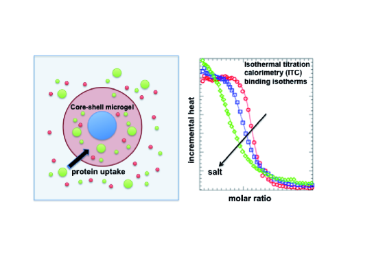
Functionalized colloids and nanoparticles play an increasingly prominent role in the development of biomaterials and imminent biotechnological applications, for example, drug delivery, enzyme biocatalysis, or control of gene expression. Peppas et al. (2000); Caruso (2001); Alarcon et al. (2005); Bajpai et al. (2008); Levental et al. (2006); Cedervall et al. (2007a); Lindman et al. (2007); Calderón et al. (2010); Walczyk et al. (2010) In particular, multiresponsive hydrogels are of great interest due their biocompatibility, resemblance to biological tissue, and tunable viscoelastic properties. Peppas et al. (2000); Alarcon et al. (2005); Bajpai et al. (2008); Levental et al. (2006); Eichenbaum et al. (1999a, b); Sassi et al. (1996); Khoury et al. (2003); Bridges et al. (2008); Cedervall et al. (2007b) Dispersed in water these colloidal microgels create an enormous surface that may be taken as a model for soft biological interfaces. Hence, protein storage, activity, and uptake properties may be changed by physiological stimuli, such as pH, salt concentration, and temperature. Moreover, colloidal hydrogels qualify for a number of applications, e.g., as drug carrier devices. Blackburn et al. (2009); Smith and Lyon (2012); Ghugare et al. (2009) However, the detailed control of functionality requires a quantitative understanding of the underlying physical interactions between microgels and biomolecules in the aqueous environment.
Recent studies have demonstrated that protein sorption to nanoparticles is mostly driven by global, nonspecific electrostatic interactions and more local, probably hydrophobic interactions. Cedervall et al. (2007a); Lindman et al. (2007); Sassi et al. (1996); Kabanov et al. (2004); De et al. (2007); Cai et al. (2008); Jung et al. (2002); Welsch et al. (2012a); Johansson et al. (2007, 2010); Li et al. (2010) The balance between those two is highly system-specific and can be manipulated by chemical functionalization or copolymerization. Charged microgels, for instance, can be used to favor or disfavor the sorption of net-charged proteins, while their osmotic swelling and storage volume can be tuned by pH, salt and charge density Eichenbaum et al. (1999a, b); Longo et al. (2012); Johansson et al. (2010) essentially via the Donnan equilibrium. Flory (1953) However, during the uptake of the charged, polyionic proteins, swelling and Donnan equilibria are typically changing in an interconnected fashion. Kabanov et al. (2004); Johansson et al. (2007, 2010); Li et al. (2010) These highly cooperative effects render the interpretation of binding isotherms, and thus the separation and quantification of global electrostatic and local hydrophobic contributions to binding, a difficult task. Moreover, binding affinities in these systems depend on protein load which presents an additional complication when modeling the adsorption isotherm.
Up to now, the modeling of protein uptake has been done often by using the standard Langmuir isotherm,Volmer and Mahnert (1925); Masel (1996) in particular when evaluating protein adsorption as measured by isothermal titration calorimetry (ITC). T. Wiseman and Lin (1989); Cedervall et al. (2007a); Lindman et al. (2007); De et al. (2007); Jung et al. (2002); Welsch et al. (2012a) In the standard Langmuir approach, however, protein association with single, independent binding sites is assumed which neglects electrostatic cooperativity effects and volume changes during sorption. Additionally the term ’binding’ of proteins to soft polymeric layers and hydrogels is somewhat ill defined, as the system may remain in a fluid-like state where proteins are still mobile on average, albeit slower than in bulk. Khoury et al. (2003); Li et al. (2011) Consequently the stochiometry and binding affinities to ’sites’ in the gel obtained from Langmuir fitting are not so easy to interpret.
Alternatively, the hydrogel matrix may be viewed as a homogeneously charged background to the mobile proteins where saturation of sorption may set in due to excluded-volume (EV) packing. In recent binding models based on such a view, Biesheuvel et al. (2006); de Vos et al. (2008); Biesheuvel and Wittemann (2005); Johansson et al. (2010) it is typically assumed that proteins can be treated as simple charged hard spheres, i.e., hard polyions. The electrostatic problem can then be tackled by approximative Poisson-Boltzman (PB) ’cell’ or ’box’ models, Gunnarsson et al. (1980); Jönsson and Wennerström (1987); Alexander et al. (1984); Denton (2010); Biesheuvel et al. (2006); de Vos et al. (2008); Biesheuvel and Wittemann (2005); Tamashiro et al. (1998); Deserno et al. (2000); Allen and Warren (2004); Hansson (2009) where electrostatic cooperativity and osmotic ion effects to deswelling can be included. So far it has not been attempted, however, to separately treat more specific effects in EV-based binding models such as hydrophobic interactions or restraints of the configurational protein degrees of freedom in the bound state. Also the relation between Langmuir and EV models, if any, is unclear. However, modeling protein adsorption by a meaningful and physically sound isotherm is the prerequisite for a quantitative understanding of the driving forces.
Here we present an in-depth discussion of the isotherms suitable for modeling protein sorption into microgels and soft polymeric layers in general. This discussion will then provide the basis for a detailed investigation of the driving forces of protein binding. We demonstrate that electrostatic cooperativity and the effects of microgel volume changes can be introduced into standard Langmuir models following the spirit of Guoy-Chapman-Stern theory for binding of charged molecules to charged surfaces. Aveyard and Haydon (1973); Seelig (2004); McLaughlin (1989) We test the performance of the binding models by fitting to binding isotherms obtained previously from ITC of chicken egg white lysozyme sorption onto a negatively charged core shell microgel.Welsch et al. (2012a) The core-shell particles consist of a polystyrene core onto which a charged poly (N-isopropylacrylamide-co-acrylic acid) (NiPAm) network is attached. Previous characterization of this system shows that it is an ideal model system as it reaches full equilibrium with high binding affinities, and the globular lysozyme maintain its folded state in the microgel with even enhanced activity. Welsch et al. (2012a) We then demonstrate that we can separate out the global electrostatic contribution leading to consistent values for the salt concentration independent binding affinity. Finally we show that EV-based binding models are formally equivalent to the Langmuir approach in the low-packing regime if the bound state is consistently defined. Consequences to the interpretation of Langmuir models are discussed.
II Experiments: Materials and Methods
II.1 Materials
In this study the same batch of microgel dispersion was used as in previous work. Welsch et al. (2012a) In brief, the polystyrene core was synthesized by emulsion polymerization in the first step. After purification of the core particles, the microgel shell, containing 5 mol-% N,N -methylenebisacrylamide (BIS) crosslinkers and 10 mol-% acrylic acid with respect to the amount of NiPAm, was polymerized on the polystyrene core by seed polymerization. After purification the particles were transferred into buffer solution by ultrafiltration against 10 mM MOPS buffer at pH 7.2. In this preparation state the gel is swollen and strongly hydrated with more than 90% volume fraction of water. Dynamic Light Scattering (DLS, Malvern Instruments) was applied to determine the hydrodynamic radius of the polystyrene core to nm and the radius of the total core-shell microgel to to nm depending on the solution condition.Welsch et al. (2012a)
The ITC experiments were performed using a VP-ITC instrument (Microcal) as described previously. Welsch et al. (2012a) Briefly, a total of 300 ml of lysozyme solution (0.695 mM) was titrated into the sample cell filled with 1.4 ml of buffer-matched microgel dispersion at a mM concentration. The pH-value was held constant at pH=7.2. The experiments were performed at 298 and 303 K. The following three buffer systems were used: (i) 10 mM MOPS, 2 mM NaN3 (7 mM ionic strength); (ii) as (i) with additional 10 mM NaCl (17 mM ionic strength); (iii) as (i) with additional 25 mM NaCl (32 mM ionic strength). The incremental heat changes were measured during the course of the titration experiment where the protein solution is stepwise injected into the microgel dispersion. The integrated heat change after each injection was corrected by the heat of dilution.
III Theory
III.1 Basic model
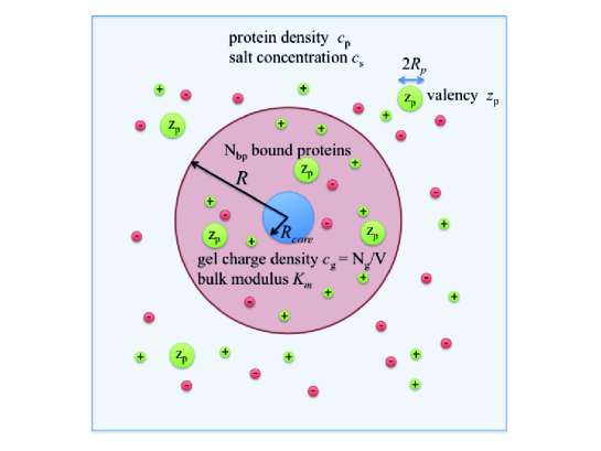
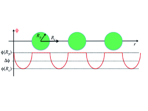
In our model one core-shell hydrogel particle is modeled as a perfect sphere with radius having a core with radius as depicted in Fig. 1. The net gel volume is therefore . Potentiometric measurements show that a total number of charged monomers are present in the gel. The network monomers are assumed to be homogeneously distributed in the gel as justified by small angle scattering. Seelenmeyer et al. (2001) The mean gel charge density is , where is the monomer charge valency. Since we work at a pH , much larger than the pKa-value of of polyacrylic acid, and deal with weakly charged gels (with a charge fraction ), we can safely assume that charge regulation plays a minor role and in the following. The salt is monovalent with a bulk concentration , where and are the concentrations of cations and anions, respectively. The Bjerrum length is , where is the elementary charge, and the vacuum and bulk permittivities, respectively, and the thermal energy. In our systems nm, while the electrostatic Debye-Hückel screening length in bulk is between 3.7 nm (7 mM salt), 2.4 nm (17 mM salt), and 1.7 nm (32 mM salt).
The lysozyme proteins are modeled by monopolar, homogeneously charged spheres with diameter , assumed to have a valency (i.e., a net charge of +7) estimated from titration. Kuehner et al. (1999) Hence, multipolar contributions, e.g., dipolar or quadropolar or charged-patch interactions to sorption are neglected. The proteins are found in bulk solution with density . No dispersion or other attractive nonelectrostatic interactions between the proteins are considered, and we assume a homogeneous distribution of proteins in the gel region. Welsch et al. (2012a) Aggregation of proteins in the gel is unlikely because at full load the system is still below the solubility threshold of a bulk system at comparable pH and electrolyte concentration. Retailleau et al. (1997) The number of bound protein inside the gel is denoted by giving rise to an internal protein packing fraction . Finally, the gel and the aqueous buffer are modeled as a continuum background with elastic, dielectric, and osmotic properties as detailed in the following sections. The introduced quantities and variables are summarized in Tab.1.
| variable | meaning |
|---|---|
| gel radius | |
| gel volume | |
| charged monomer concentration | |
| bulk salt concentration | |
| inverse Debye length in gel | |
| inverse Debye length in bulk | |
| bulk protein concentration | |
| total protein concentration | |
| molar ratio | |
| number of proteins in the gel | |
| number of Langmuir binding sites | |
| protein load | |
| effective protein diameter | |
| effective protein radius | |
| protein packing fraction in gel | |
| Donnan potential | |
| binding constant | |
| gel modulus | |
| constants | meaning |
| nm | core radius |
| number of charged monomers | |
| monomer charge valency | |
| protein charge valency | |
| M | microgel concentration |
| nm | Bjerrum length |
| =l/mol | standard volume |
III.2 Electrostatic contributions to the interactions of the proteins with the charged gel
III.2.1 Donnan equilibrium
Homogenization of the charged gel by salt ions leads to a electrostatic potential difference between bulk and gel, the Donnan potential. The latter can be derived by assuming electroneutrality in the gel for monovalent, ideal ions leading to Flory (1953)
| (1) |
where is the charge ratio between gel and bulk charge densities and the tilde in denotes the dimensionless potential scaled by .
We estimate a mean separation of about nm between two charged monomers on the same polymer within the gel, considerably larger than the Bjerrum length nm. Thus, charge regulation effects by counterion (Manning) condensation or inhomogeneity effects can be safely neglected. Devore and Manning (1978); Buschmann and Grodzinsky (1995); Zeldovich and Khokhlov (1999); Dobrynin and Rubinstein (2005) For our microgel we further estimate the charged monomer concentration to be mM, depending on the swelling state. Given bulk salt concentrations between 7 and 32 mM, thus is on the order of unity ( 25 mV) and significant for the interaction with charged macromolecules.
The Donnan equilibrium leads to an osmotic pressure difference between gel and bulk ions and, in the ideal gas limit, is given by the difference of internal and external ionic concentrations Flory (1953)
For the salts and concentrations considered in this work corrections due to nonideal activity are small Robinson and Stokes (2002) and can be safely neglected.
III.2.2 Electrostatic protein-gel interaction
The transfer of a charged protein from the bulk solution with salt concentration into the charged gel with monomer charge concentration is accompanied by a (Gibbs) transfer free energy change per particle (or chemical potential). If we assume the gel monomers to be mobile, this can be modeled by the transfer of a spherical polyion with radius and valency from one bath at salt concentration and zero potential to another at mean potential and salt concentration . The difference in solvation free energies on a Debye-Hückel level is then
| (3) |
where we defined the inverse screening length in the gel, characterizing approximately the screening by mobile polyelectrolyte charges and their neutralizing counterions (see Appendix A). The Donnan potential (1) must now be corrected for the change of total net charge by the charge of bound protein via
| (4) |
The first, purely entropic term in eq. (3) simply expresses the electrostatic transfer energy of a charge from bulk to a region at potential . The second term reflects the difference in the Born solvation free energies in a salty medium. McQuarrie (2000) The first term is attractive if microgel and protein have opposite net charge, otherwise repulsive, while the second one is attractive if , otherwise repulsive. Since we deal with a weakly charged gel additional entropic effects from the release of condensed counterions can be neglected in our system. Henzler et al. (2010)
In Appendix A a more rigorous derivation of eq. (3) is presented in the framework of a linearized PB cell model Gunnarsson et al. (1980); Jönsson and Wennerström (1987); Alexander et al. (1984); Denton (2010); Biesheuvel et al. (2006); de Vos et al. (2008); Biesheuvel and Wittemann (2005); Tamashiro et al. (1998); Deserno et al. (2000); Allen and Warren (2004); Hansson (2009) corrected for particle-particle interactions. While eq. (3) is valid strictly only in the limits of small salt concentrations and small protein charges, its quantitative validity seems to cover a wide range of protein load as long as the protein valency is not too high, as discussed in Appendix A. For our system with lysozyme (surface charge density 0.17e/nm2) in monovalent salt and the weakly charged gel (charge fraction 1/10), Debye-Hückel level assumptions are justified.
III.3 Osmotic and elastic swelling equilibrium
In equilibrium the gel size is determined by a mechanical balance between osmotic and elastic pressures in the gel via Flory (1953); Dubrovskii et al. (2001); Horkay and Zrinyi (1982); Rubinstein et al. (1996); Buschmann and Grodzinsky (1995); Overbeek (1956)
| (5) |
The osmotic term has two contributions one from the solvent, the other from the ions. The first is often expressed by a de Gennes-like scaling type of relation , where typically for neutral polymers under good solvent conditions. de Gennes (1979); Horkay and Zrinyi (1982) For charged networks corrections may arise, Rubinstein et al. (1996) while for our case of weakly charged and weakly stretched gels the 9/4 exponent likely to be valid. Horkay and Zrinyi (1982); Hu et al. (1993); Dubrovskii et al. (2001) (According to definitions by Dobrynin and Rubinstein, Dobrynin and Rubinstein (2005) we estimate an elecrostatic blob size of about 1.3 nm using a monomer length 0.35 nm and our charge fraction 1/10. This is indeed smaller than the correlation length of 3.2 nm given by our mean mesh size, estimated from the linker density.)
The ionic osmotic pressure is dominated by the ideal gas pressure of ions in the gel as given by eq. (III.2.1). This is counterbalanced by the elastic pressure as represented essentially by the shear modulus . Weakly charged networks obey Gaussian chain statistics Rubinstein et al. (1996); Dubrovskii et al. (2001), and scales via the power law with . Various experiments corroborate that scaling. Horkay and Zrinyi (1982); Hu et al. (1993); Skouri et al. (1995); Nisato et al. (1995); Dubrovskii et al. (2001); Dubrovskii and Rakova (1997) The expression for the total pressure is
where and (or ) are volume-independent constants. For high salt concentration the ionic contribution vanishes, , and the equilibrium gel is in the ’neutral’ reference state with volume . The bulk modulus of the gel in this state is defined via . By fitting eq. (III.3) to a variety of salt concentrations (without proteins) we will obtain both the unknowns and and thus .
In first order the effect of protein addition to the gel is lowering of the gel net charge and the inhomogenization of the charge and electrostatic potential distribution inside the gel. As shown in the PB cell model in Appendix A those effects can be approximately included in (III.3) by replacing with
| (7) |
with . The potential is the electrostatic potential at the cell boundary in the PB cell model (Appendix A).
Estimating other protein-induced contributions to the pressure from more local effects on elasticity, such as cross-linking by local binding, Borrega et al. (1999) conformational restraints of the polymer network, or possible contributions from the protein osmotic pressure is challenging due to the lack of precise knowledge of the nature of the bound state and is out of scope of this paper.
III.4 Binding isotherms
The transfer of one protein into the gel will be accompanied by a release of the binding enthalpy . If proteins bind and we assume that the heat per protein does not change with load, the total heat released is
| (8) |
where is the total titration volume. In the ITC experiments is measured vs. the total protein concentration . Introducing the molar ratio we can write . The incremental heat per molar concentration of protein is more sensitive to fitting and reads
| (9) |
For large binding constants and small , almost all of the proteins immediately get sorbed, and , and a plateau of heigh is expected, independent of the binding constant. For large , typically saturates and . Thus we recognize that fitting to binding models is most sensitive to intermediate values of the molar ratio , in the pre-saturation regime, near the inflection point of .
III.4.1 Standard Langmuir Binding Model
The standard Langmuir model is based on identifying the association reaction A+BAB, with a binding constant K = [AB]/[A][B], where the square brackets denote concentrations. The basic assumptions in the Langmuir model are that ideal particles are binding to a fixed number of identical and independent bindings sites. The equilibrium constant in the Langmuir framework is usually written as Volmer and Mahnert (1925); Masel (1996)
| (10) |
where denotes the fraction of bound protein and total sites (see also derivation in Appendix B for the canonical ensemble). The protein density outside the gel can be expressed by the total protein density in the sample minus bound protein density by . If it is assumed that , , and are protein concentration independent, solving (10) for gives the total heat
| (11) |
with . The fitting of to the experimental data then yields the unknown constants , , and . Typically a sigmoidal curve is measured for , where describes the plateau for the first injections (small ), the inflection point, and the sharpness of the transition at . Fitting to Langmuir isotherms is thus most sensitive close to , when the molar ratio equals the number of available binding sites.
The Gibbs binding free energy in the Langmuir model is defined by
| (12) |
where is the ’standard volume’ which describes the ’effective’ configurational volume in one of the binding boxes (see also Appendix B). Thus the absolute value of depends on the magnitude of the standard volume , an often overlooked fact in literature. Zhou and Gilson (2009) Typically nm3 is chosen which is a reasonable choice for molecular binding where spatial fluctuations are on a nanometer length scale. While for quantitative estimates thus precise knowledge of the nature of the bound state is necessary, we will satisfy ourselves in this work with the standard choice .
III.4.2 Extended Langmuir Binding: Separating Nonelectrostatic and Electrostatic Contributions
When many charged entities bind to charged regions a cooperativity effect comes into play due to the change of the global electrostatic properties during loading. This has been appreciated in the Guoy-Chapman-Stern theory for the binding of charged ligands to charged surfaces. Aveyard and Haydon (1973); Seelig (2004); McLaughlin (1989) Consequently, the total binding constant has to be defined more generally and split up into an ’intrinsic’ part and an electrostatic part via
| (13) |
i.e., . In our work we assume to be given by eq. (3). The intrinsic, -independent binding constant defines the intrinsic adsorption free energy
| (14) |
which only contains contributions from specific interaction between the protein and the gel environment, and the nonspecific, leading order electrostatic effect has been separated out. Specific interaction may include local solvation effects – where the hydrophobic effect is naturally one of the biggest contributors – and possibly specific local interactions, such as salt bridges.
III.4.3 ’Excluded Volume’ (EV) Binding Isotherms
An alternative binding isotherm that may be used for evaluating the ITC data is based on the equivalence of chemical potentials in bulk and inside the gel, where in addition to electrostatic and intrinsic binding effects the EV interaction between the (hard spherical) proteins inside the gel are taken into account. Biesheuvel et al. (2006); de Vos et al. (2008); Biesheuvel and Wittemann (2005); Johansson et al. (2010) One assumption is here that in the very contrast to Langmuir models, the proteins are not ’condensed’ to fixed sites in the gel but are able to freely move around, under the restraint only that their translational freedom is confined by packing (excluded volume). This ansatz yields the Boltzmann-like equation for the density of hard spheres in the gel
| (15) |
where is the partition function of the bound state more precisely defined later, and is the Carnahan-Starling (CS) excess chemical potential Hansen and McDonald (2006)
| (16) |
representing the free energy of transferring one hard sphere to a solution with the packing fraction . The effective diameter of the sphere is expected to be close to the diameter of gyration or hydrodynamic diameter of the protein (i.e., nm for lysozyme) Hamill et al. (2005) and will serve as a fitting parameter in the following. Since packing effects play a role in the EV approach we correct the gel volume by the polymer volume fraction. A PNIPAM monomer has an excluded volume roughly of nm3. We estimate 106 monomers in one microgel what makes 106 nm3. This corresponds to a polymer volume fraction in the range of 5% (pure, unloaded gel) to 12% (fully loaded gel in 7mM salt).
In (15) we have also separately described the nonspecific electrostatic part () and the intrinsic part () to binding as in the Langmuir model above. In the EV model, the contribution to can be still thought of being induced by hydrophobic interactions or salt bridges but only in a weak-interaction sense, such that the particles in the gel are still translationally free. However, we additionally consider in eq. (15), the partition function of the protein in the bound state, which can include contributions from vibrational and orientational restrictions of the protein’s degrees of freedom within the gel, e.g., by partial sliding on polyelectrolyte chains. Henzler et al. (2008)
III.4.4 Equivalence Between the Langmuir and the EV Approach
For small protein packing fractions we may linearize the CS chemical potential in eq. (15). We obtain
| (17) |
where we identified the second virial coefficient of hard spheres . Physically describes the volume excluded to the centers of the other spheres taken by one sphere. We rearrange to obtain
| (18) |
If we now make the substitution and as in the Langmuir model above, we end up with the standard Langmuir form (10)
| (19) |
If , thus the EV treatment is equivalent to a Langmuir picture where a bound ideal gas particle has a configurational freedom (volume) of , i.e., it can freely move around in one of the binding boxes. However, as discussed above, in the Langmuir-type bound state the configurations are restricted to an effective configurational volume with respect to , such that the partition function . With that definition we exactly end up with the standard Langmuir model eq. (10).
Thus, in the approximation of small protein packing , the EV approach and the standard Langmuir model, eq. (10), are formally equivalent and are allowed to be compared, if in the Langmuir picture and . The parameter , the number of fixed binding sites, can then be interpreted as the maximum number of binding spots available for hard spheres simply due to packing in the available volume . In both models, Langmuir and EV, the binding constant is referenced with respect to a standard volume .
III.5 Numerical evaluation including volume change
In the Langmuir approach the derivative of eq. (11) is fitted to the experimental data by scanning through , , and values until the least square deviation (LSD) to the experimental data is minimized. The radius is fixed in the Langmuir fittings to nm. In the EV approach, eq. (15) is solved numerically and , as more generally defined in eq. (9) is fitted to the experimental data by minimizing the LSD. Here the fitting parameters are , , and .
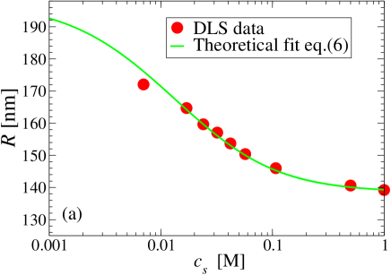
One challenge arises because also the gel volume is in general a function of , the protein concentration. The electrostatic transfer energy defined by eq. (3), in turn, depends on the gel volume. Thus, for separating out the electrostatic contributions to binding, the fitting of binding isotherms needs take into account the volume change. Since a predictive theory for is out of scope of this paper, we satisfy ourselves in the following by employing experimental DLS data for and at 7 mM salt concentration as shown in Fig. 2. We fit by the empirical function
| (20) |
with and being the maximum and minimum gel radius at and , respectively, is the location of the inflection point, and the distribution width. For K and 7 mM salt we find nm, nm, , and can be identified with . For the other salt concentrations, where no DLS data are available, is obtained by using the known nm (17 mM) and 157.1 nm (32 mM) (cf. Fig. 2a) and (cf. Tables II-IV), while nm is used from the 7 mM fit. The now only free variable is employed as an additional parameter obtained by least square fitting the ITC data. We find = 31000 ( 17 mM) and 42000 ( 32 mM) which appears correlated with the change of the sharpness of the binding isotherms with .
IV Results and Discussion
IV.1 Gel shrinking by salt and proteins
In agreement with previous observations, Eichenbaum et al. (1999a); Kabanov et al. (2004); Johansson et al. (2010) DLS shows that the charged PNIPAM microgel shrinks upon the systematic addition of salt as summarized in Fig. 2 (a) up to a concentration of M. The gel radius decreases monotonically and saturates at molar concentrations at a radius close to nm. The best fit of our mechanical balance approach eq. (III.3), also shown in Fig. 2 (a), yields very good agreement in almost the whole range of . From the fit we obtain nm and a bulk modulus kPa. Assuming Poisson’s ratio to be 1/3, valid for neutral polyacrylamide or poly-NiPAm gels, Li et al. (1993); Yoon et al. (2012) then Young’s modulus . Our determined value for is thus fully consistent with recently measured Young moduli investigated for varying BIS cross linker density at similar temperatures. Burmistrova et al. (2011) In the latter was measured between kPa and kPa for BIS contents of 2% and 10%, respectively, compared to 5% in this work. The good agreement supports our considerations leading to eq. (III.3).
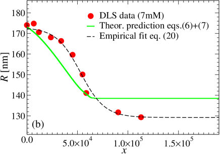
In Fig. 2(b) we show the measurement of the hydrogel radius vs. the molar ratio for the system at 7 mM salt concentration. Analogously to the salt-only case, shrinking of the gel is now observed with increasing protein concentration, i.e., with increasing protein load. At the highest investigated ratio the radius is notably smaller than pointing to binding-related network tightening. By fitting to the the empirical function (20), we find nm. Thus the gel volume under full protein load is times smaller than in the neutral reference state. If we assume that the charged proteins under full load lead to complete charge neutralization and thus , then the bulk modulus induced by protein binding scales and is times larger than without proteins, i.e., the gel is roughly 2 times stiffer due to protein sorption. This has to be considered a lower bound as it is likely that ionic contributions to the osmotic pressure still play a role, and maybe protein osmotic effects due to EV interactions need to be considered.
However, in order to check to what amount purely electrostatic effects by proteins induce gel shrinking, we plot the description by eqs. (III.3) and (7) also in Fig. 2 (b) using the experimental ITC binding isotherm as input. This description is now a prediction and no fitting is involved. While the overall shrinking of the gel is reasonably captured, the model yields a too fast decrease for small . This may point to shortcomings of the mean-field cell model (Appendix A), where no ion and protein fluctuations are included. For large , the experimental saturation of nm is not reached indicating that nonelectrostatic effects to gel elasticity play a role. Similar unsatisfying performances of simple Donnan models have been observed also in a recent work. Johansson et al. (2010) However, from our comparison it is quite reasonable to judge that the dominant effect to gel shrinking by protein uptake is of ion osmotic origin.
IV.2 Characterizing experimental binding isotherms
IV.2.1 Langmuir models
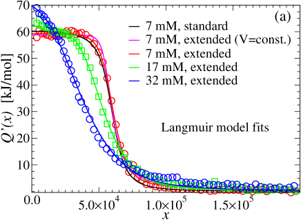
The evaluation of the ITC data by the Langmuir-type binding models is presented in Fig. 3 (a). First we compare the results of different model assumptions for 7 mM ionic strength: i) standard Langmuir model vs. ii) extended Langmuir (including electrostatic cooperativity) with constant volume vs. iii) extended Langmuir including the DLS-measured volume change. From looking at the fits by eye and judging from the overall least square deviation (LSD) to the ITC data, cf. Tab. II, all fits look comparably well and can only be distinguished in the presaturation region around . As discussed above fitting is most sensitive to this transition region. The fitting parameters are summarized in Tab. II: while the heat of binding kJ/mol and total number of binding sites are relatively insensitive to the model assumptions, the changes in the binding constant are big. From the standard model kJ/mol. In the extended model without volume change ii), kJ/mol., i.e., more than 12 kJ/mol correspond to -dependent electrostatic contributions. Including the volume change, however, has a considerable effect on the electrostatic contribution which grows by 18 kJ/mol. The latter trend is understood by the fact that the monomer charge density increases with shrinking and the contributions in as given by eq. (3) rise. Thus considering volume changes in charged systems is important for quantitative fitting, especially in those systems where deswelling is significant. The value of the intrinsic binding free energy is kJ/mol for 7 mM ionic strength.
In the next step the extended Langmuir model including volume change has been applied to the other ionic strengths 17 and 32 mM as also shown in Fig. 3(a) and summarized in Tab. II. Note again that the starting gel volumes at decrease for increasing ionic strengths, see the values for in Tab. II. Here we first observe that the number of Langmuir binding sites decreases with higher ionic strength. The reason is a priori unclear as the Langmuir model assumes a fixed number of binding sites independent of ionic strength. We further notice that the heat of adsorption slightly increases with ionic strength. More importantly, however, stays fairly independent of . Thus, in contrast to the standard Langmuir model, the nonspecific electrostatic contributions have been consistently separated out and the remaining becomes salt concentration independent. On average we find kJ/mol that might be attributable to hydrophobic interactions or possibly other local binding effects.
| LSD | ||||||
| [nm] | [kJ/mol] | [l/mol] | [kJ/mol] | |||
| 7 (i) | – | 60100 | 61 | 2.6 | -36.6 | 132 |
| 7 (ii) | 172.1 | 65500 | 59 | 1.9 | -24.5 | 89 |
| 7 | 172.1 | 63000 | 59 | 1.6 | -18.3 | 44 |
| 17 | 165.7 | 57500 | 62 | 2.0 | -18.9 | 57 |
| 32 | 157.1 | 42500 | 72 | 1.3 | -17.8 | 130 |
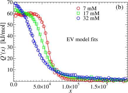
IV.2.2 Excluded Volume model
The fitting of the ITC data by the EV model is presented in Fig. 3 (b). Corresponding fitting parameters , , and are summarized in Tab. III. As in the Langmuir models the binding isotherms are described very well by the fits judging from the overall LSD to the experimental data. The results for the heat are also consistent with the Langmuir fits. This is not surprising as this value is determined by the plateau in for small far away from the saturation regime. The fitting parameter is now replaced in the EV model by the parameter , the effective hard-core radius of the protein. The analysis yields values of between to nm, see Tab. III, weakly depending on ionic strength. Those numbers are indeed very close to the hydrodynamic radius of lysozyme of about nm. Hamill et al. (2005) This good agreement is actually remarkable and justifies the assumptions leading to the EV model, i.e. corroborates well with a packing picture of globular proteins.
The magnitude of the intrinsic adsorption energy in Tab. III are between 15 and 18 kJ/mol matching closely the ones from the Langmuir model in Tab. II. A very small salt dependence of remains indicating a slightly less accurate subtraction of the nonspecific effects in this model. However, the salt concentration dependence is quite small and on average we find kJ/mol that has to be attributed to specific local binding effects. In Tab. III we also show the results of EV model fitting to experimental data gathered at a higher temperature, K. The intrinsic binding affinity increases slightly to an average kJ/mol which corroborate with the typical thermodynamic signature of increasing hydrophobic association at enhanced temperatures. Dill and Bromberg (2003)
| LSD | ||||||
| [nm] | [nm] | [kJ/mol] | [kJ/mol] | [K] | ||
| 7 | 172.1 | 1.77 | 59 | -14.9 | 30 | |
| 17 | 165.7 | 1.83 | 62 | -17.9 | 33 | 298 |
| 32 | 157.1 | 2.01 | 71 | -18.4 | 25 | |
| 7 | 172.1 | 1.78 | 70 | -15.1 | 111 | |
| 17 | 165.7 | 1.79 | 70 | -18.9 | 43 | 303 |
| 32 | 157.1 | 1.85 | 65 | -19.6 | 75 |
IV.2.3 Donnan potentials and the binding constant
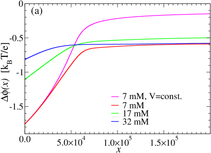
The Donnan potential , the electrostatic binding energy , and so the total binding constant depend on protein load, and thus, in turn, on the molar ratio . The Donnan potential as resulting from the fitting to the extended Langmuir model is plotted in Fig. 4(a) for all ionic strengths. In the case of 7 mM, at the beginning of the titration, , the potential magnitude is a considerable 1.77 /e ( mV). The potential quickly decreases with protein load due to charge neutralization, c.f. eq. (4), and saturates for due to saturation of . The decrease of gel volume in the adsorption process has a notable effect on the potential at large due to the accompanying increase in monomer charge density. Increasing the ionic strength, on the other hand, considerably lowers the potential at small load, while surprisingly remains roughly independent of for high load. The reason for the latter is that less proteins bind for higher ionic strengths, i.e., there is less charge neutralization. The Donnan potential is expected to be highly correlated with the surface potentials of the microgels which govern colloidal stability in solution. Johansson et al. (2010)
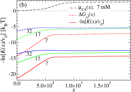
The total electrostatic contribution to the binding constant is plotted in Fig. 4(b) together with the total binding free energy . The curves and are parallel with an offset given by the intrinsic part , as defined in eq. (13). The electrostatic contribution for very small load () is big and contributing a favorable 15 ( kJ/mol) for 7 mM and 6 ( kJ/mol) for 32 mM to the total binding affinity of about 22 and 14 , respectively, at the beginning of the titration. We find that the Born contribution (second term in (3)) constitutes a significant (favorable) 2-3 to the total electrostatic energy and is thus not negligible. All curves decrease in their absolute values and saturate for values , when the molar ratio exceeds the number of binding sites in the Langmuir model. Note that for 7 mM the saturation value of is close to the value of obtained from standard Langmuir fitting, c.f., Tab. II, because fitting is most sensitive to the presaturation region above which remains constant. This statement holds also for the other salt concentrations when is compared to previous standard Langmuir fitting. Welsch et al. (2012a)
Also shown in Fig. 4 (b) is the Carnahan-Starling contribution in the EV model. This entropic penalty due to hard sphere packing slowly rises with in the regime until quickly increasing to unfavorable 3 in the saturation regime .
IV.3 Interpretation of Langmuir vs. EV model results
In section III.4.4 we argued that the EV model is equivalent to the Langmuir approach in the low packing regime (), if , the number of binding sites, is equated to the number of free spots just by packing. Indeed if one now considers a mean gel radius of nm and protein diameter nm obtained from fitting above, we end up with , totally consistent with numbers obtained from fitting to the Langmuir models above, cf. Table II. This agreement implies that the strict Langmuir assumption of a fixed set of binding sites can still be considered an interpretable quantity, even if the nature of the bound state is ill-defined. From that point of view, the decrease in in the Langmuir models with increasing ionic strengths can be understood: for increasing , the gel volume decreases and packing penalties are becoming more important for a smaller number of bound protein. Hence, in standard Langmuir fitting of ITC data for sorption to soft materials, T. Wiseman and Lin (1989); Cedervall et al. (2007a); Lindman et al. (2007); De et al. (2007); Jung et al. (2002); Welsch et al. (2012a) where it is likely that not a condensation-like binding of proteins takes place, the stochiometry of binding may be interpretable by packing effects.
As we demonstrated in Fig. 4 the electrostatic energies and therefore the binding affinity are functions of the molar ratio . How do we interpret a constant as deduced from standard Langmuir fitting? T. Wiseman and Lin (1989); Cedervall et al. (2007a); Lindman et al. (2007); De et al. (2007); Jung et al. (2002); Welsch et al. (2012a) As argued in the Methods section fitting is most sensitive to the presaturation region . Indeed the data in Fig. 4 (b) shows that equals the values of standard Langmuir fitting, see Tab. II. Thus, we can conclude that a binding constant obtained from a standard Langmuir fit is a reasonable number which can be interpreted as binding affinities in the presaturation regime, i.e., in the intermediate to high protein load regime, where also volume changes are not so large anymore. However, for small (small protein load) our separation into electrostatic and hydrophobic contributions shows that binding affinities can be about 1000 times larger than in the presaturation regime. This may have implications for the modeling and interpretation of protein binding kinetics. Kabanov et al. (2004); Dell’Orco et al. (2010); Casals et al. (2010); Welsch et al. (2012b)
It may be on first glance remarkable that in both models, extended Langmuir and EV, the consistent separation of electrostatic and hydrophobic effects yields the same number for the hydrophobic (intrinsic) binding affinity. In our system we find which must be attributed to nonelectrostatic binding effects and constitutes roughly 1/3 or 1/2 of the total binding affinity in the small and high load regimes, respectively. The very weak dependence of with salt concentration in both models indicates a successful separation of nonspecific electrostatic and intrinsic effects in our treatment. However, note that the agreement can only be established if the nature of the bound state is identically defined in both models as discussed in section III.4.4. Since at low packing we exemplified total equivalence of both models, the agreement for is thus not unexpected. Probably for higher packing densities than observed in this study (where ), the results from both models may disagree stronger. Thus for not too high packing, one model does not seem superior over the other in describing experimentally measured binding isotherms, if correctly interpreted.
Furthermore we would like to comment on the magnitude of for the instrinsic interaction of lysozyme with the PNIPAM network. If methane-methane interactions are taken as reference with attractions on the order of 2-3 , then 7 correspond to 2-3 hydrophobic protein-PNIPAM contacts on average which seems reasonable. Increasing temperature led to an increase in conform with the signature of hydrophobic interactions. Dill and Bromberg (2003) A recent study on a similar system showed hardly uptake of lysozyme by an uncharged PNIPAM microgel. Johansson et al. (2010) Reasons for this discrepancy may be the different gels preparation state, e.g., larger pore sizes within the gel. Alternatively, maybe we overestimate the effects of hydrophobicity and other local effects, such as salt bridges, play a more important role as expected.
Finally, we note that effective net charge of chicken egg white lysozyme as used in this work may be
slightly larger on average due to the protonation effects ( to 8)
within the gel. Welsch et al. (2012a) However, using = +7.5 or +8 in our analysis we end up
with a similar ; the reason is that while the prefactor in the
electrostatic contribution (3) rises, the Donnan potential (2) decreases quicker
with load. These effects roughly cancel each other for our particular system.
V Concluding Remarks
In summary we have systematically introduced binding models to characterize the physical interactions in the process of equilibrium protein sorption to microgels. The binding models separate out electrostatic cooperativity and include deswelling effects. The analysis yields a hydrophobic, salt-independent interaction of around for our core-shell system and hen-egg white lysozyme.
The found constitutes roughly 1/3 or 1/2 of the total binding affinity in the small and high load regimes, respectively. For small protein load, binding affinities can be about 1000 times larger than in the presaturation regime due to high net charge of the protein which is gradually neutralized upon binding. This may have implications for the modeling of protein binding at early stages of sorption, especially to understand kinetic rates. Kabanov et al. (2004); Dell’Orco et al. (2010); Casals et al. (2010); Welsch et al. (2012b) Moreover, the Donnan potential is expected to be highly correlated with the surface potentials of the microgels which govern colloidal microgel stability in solution. Johansson et al. (2010)
We find gel deswelling to be mostly of electrostatic origin. However, the gel becomes at least 2 times stiffer at high load pointing either to more specific effects at high load (e.g., cross links) or electrostatic correlations not accounted for in mean-field PB cell model approaches. The change and control of material properties upon protein load is essential for functionality, Levental et al. (2006) and suggest challenging investigations in future.
In two complementary models, extended Langmuir and EV, the consistent separation of electrostatic and hydrophobic effects yields the same number for the hydrophobic (intrinsic) binding affinity. The agreement can only be established if the nature of the bound state is identically defined in both models. Details of the bound state can be inferred, for instance, from small angle scattering. Henzler et al. (2008) We work at low protein packing; hence, one model does not seem superior over the other in describing experimentally measured binding isotherms, at least for not too high loads (packing). With that a more quantitative interpretation of binding data T. Wiseman and Lin (1989); Cedervall et al. (2007a); Lindman et al. (2007); De et al. (2007); Jung et al. (2002); Welsch et al. (2012a); Johansson et al. (2010); Li et al. (2010) in terms of separate physical interactions is possible in future. Especially we have shown that fitting based on standard Langmuir models yields interpretable binding affinities and stochiometry. T. Wiseman and Lin (1989); Cedervall et al. (2007a); Lindman et al. (2007); De et al. (2007); Jung et al. (2002); Welsch et al. (2012a) More challenges arise, however, if the systems of practical interest exhibit microscopic irreversibiltiy and the final protein binding cannot be considered in equilibrium terms. Walczyk et al. (2010); Cedervall et al. (2007b)
Acknowledgements.
Financial support by the Deutsche Forschungsgemeinschaft (DFG), Schwerpunkt Hydrogele , and by the Helmholtz Virtual Institute is gratefully acknowledged.Appendix A Linearized Poisson-Boltzmann (LPB) cell model
To obtain the electrostatic contributions to the transfer of charged hard spheres from bulk to gel we make use of the Poisson-Boltzmann cell model. While often numerical solutions have been employed, Marcus (1955); Buschmann and Grodzinsky (1995); Gunnarsson et al. (1980); Jönsson and Wennerström (1987); Alexander et al. (1984); Tamashiro et al. (1998); Deserno et al. (2000); Allen and Warren (2004); Hansson (2009) for weak perturbations the linearized form can be treated analytically. Denton (2010); Biesheuvel and Wittemann (2005)
In the cell model each protein is represented by a homogeneously charged sphere with radius and valency centered in a spherical cell with radius and volume . The gel is assumed to be made up of such cells whose dimensions are determined by the number of proteins in the volume, i.e.,
| (21) |
where is the total number of proteins in the volume . Each cell is in contact with a reservoir with monovalent salt concentration . Inside the gel each cell additionally contains a fixed number of charges from the charged network monomers. The mean monomer charge concentration in one cell is thus . Each cell must be electroneutral on average, i.e., in the gel it holds
| (22) |
where we neglected the vanishingly small protein concentration outside of the gel. For high protein load, becomes comparable to and the cell volume needs in principle to be corrected by the protein volume. However, for our systems at highest protein load , and the correction is negligible for small and intermediate loads. The solution of (22) is the modified Donnan potential
| (23) |
with which describes the difference in the mean electrostatic potential with respect to the bulk reference state where we set . In the bulk the protein concentration is typically vanishingly small, thus electroneutrality dictates to a very good approximation.
We now focus on the proteins inside the gel. Here we assume the PE network in the gel to be flexible and fluid-like and thus the charged monomers to behave like mobile counterions to the protein. The PB equation in spherical coordinates is then
where we made the ansatz , with being the constant mean (modified Donnan) potential in eq. (23) and being a perturbation induced by the protein. Note that if the monomers were assumed to be just a fixed, homogeneous background, they would not couple to the field and needed to be replaced by the fixed concentration . The constant is defined by conservation of the number of monomer charges in the cell via
| (25) |
It also holds that the average potential equals the Donnan potential, i.e,
| (26) |
For not too large protein charges and thus , we can linearize the exponentials in the PB eq. (A) with respect to which yields
| (27) |
where we have identified the internal (gel) charge density from conditions (A5) and (A6), the internal concentration , and the internal inverse screening length . The LPB equation can be solved with respect to the boundary condition that the electrical field on the sphere surface is fixed by and it vanishes at the cell boundary . The final solution is (see also previous works Denton (2010); Biesheuvel and Wittemann (2005))
with constants
and
In the limit of large cell sizes, i.e., (or ), it follows that quickly , and . The LPB solution simplifies to
| (29) |
where we kept the important leading order term in . An illustrative sketch of this potential distribution in the cell model is given in Fig. 5. is the average potential, while for small protein load the potential at the cell boundary and at the particle surface . For not too high salt concentrations , it follows that the Donnan potential , and the internal salt concentration can be well represented by , as the coion concentration ( in the gel becomes negligibly small. In the bulk solution in the dilute protein limit analogously to eq. (29) we find
| (30) |
where is the bulk inverse screening length.
The transfer (Gibbs) free energy (or chemical potential) to bring a protein from bulk solution into the gel can then be calculated by the difference of work of charging the sphere against the surface potential , in a cell in the gel (g) vs. bulk (b). We obtain the leading order contributions (in the low salt and low protein limit)
where we defined . We did not explicitly integrate over in the second term in (29) as it is a constant background contribution not immediately involved in the charging process of one particle. It is noteworthy that we obtain the same functional form for if the monomer charges are not assumed to be mobile, albeit with a smaller internal inverse screening length and a factor of 2 in front of the second term.
By a one-to-one comparison of the leading order expression (A) to the result from employing the full expression (A) we find that the error in is less than over the whole range of molar ratios and salt concentrations used in this work. By detailed inspection of the behavior of (29) we observe that the reason of the surprising accuracy is a fortuitous cancellation of errors of higher order terms at large . This fact may shed some doubt on the general applicability of simplified eq. (A) but note that our parameters (protein valency, salt concentrations, monomer charge densities, etc.) are typical for a wide variety of experimental systems. However, in general the PB approach is expected to break down for very high protein valencies and small proteins, when , and strong Coulomb correlations play a role. Hansen and McDonald (2006)
The free energy (A) above considers only ionic contributions to solvation of a fixed lattice of spheres, i.e., it neglects the electrostatic contributions from the interaction between the proteins, i.e., the energy penalty of overlapping double layers. However, in the fluid-like hydrogel protein matrix it is reasonable to assume that proteins can wiggle or move around and are not rigidly fixed to lattice positions. Due to such fluctuations the average surface potential will actually be higher than given in (A). To estimate the interaction contribution we look at the expansion of the excess chemical potential in terms of virial coefficients, i.e, in first order
| (32) |
where and
| (33) |
is the protein-protein interaction potential, split up into hard-sphere and the Debye-Hückel contribution according to (A). Using condition (26), linearizing the exponent in the defining equation for , and splitting the chemical potential into hard-sphere and electrostatic contributions we find
| (34) |
Thus in leading order the electrostatic protein-protein interaction contribution exactly cancels the second term in (A) and we end up with the final result for the electrostatic transfer free energy
| (35) |
It is interesting to note on the simplicity of eq. (35) which describes naively the transfer of charge into the average potential and, in the second term, the difference in Born solvation free energies in a homogeneous medium with salt concentrations and . Thus, the inhomogeneities introduced by the cell model assumption as depicted in Fig. 5 cancel out (in linearized theory) if particle fluctuations are allowed, and the naive form (35) holds.
The estimation of ionic osmotic contribution to pressure in the presence of proteins seems less simple. Microconfiguration of proteins induce inhomogeneities and the pressure is not anymore given by the simple expression for in eq. (III.2.1). The ion contribution to the osmotic pressure is usually estimated from the ionic concentration at the cell surface (in the cell model) where the electrostatic pressure vanishes, Marcus (1955); Alexander et al. (1984); Denton (2010); Biesheuvel et al. (2006); de Vos et al. (2008); Biesheuvel and Wittemann (2005)
| (36) |
Expanding to 1st order in , we obtain
| (37) |
which recovers in the limit for vanishing protein concentrations and is the protein-corrected Donnan potential (23) with . Note that this expression does not consider fluctuations in protein positions which is likely to be an important effect to consider in future studies.
Appendix B The standard Langmuir model in the canonical ensemble
Consider a finite region in space with identical and independent binding sites available. We denote the number of bound proteins by and define the fraction of bound particles by . The number of available binding states is then Volmer and Mahnert (1925); Masel (1996)
| (38) |
from the combinatorial possibilities of distributing indistinguishable particles on sites, and is the partition sum of a single particle in the bound state. The Boltzmann entropy is defined by
| (39) |
leading (within a constant) to the entropy per binding site
| (40) |
where we defined in terms of an effective configurational volume divided by the cubed thermal (de Broglie) wavelength . ’Effective’ means that also restrictions on vibrational and orientational degrees of freedom are adsorbed in the number , not purely translational effects if it all. The (canonical) Helmholtz free energy of the system is
| (41) |
where we introduced the canonical ideal gas free energy with total particle number . Then is the density of unbound particles in a total volume (assumed to be much larger than the binding region), and the adsorption free energy associated with the binding of one protein to one Langmuir site. The free energy per binding site is then
The minimization of the free energy with respect to the number of bound protein yields then the final relations for the fraction of bound particles in dependence of the (unbound) particle concentration . We obtain the final result
| (43) |
The standard volume depends on the exact nature of the bound state and is typically not known. Thus the prediction of absolute binding free energies is difficult. In literature often the standard volume l/mol nm3 is employed. This is not unreasonable if one assumes that still rotational and vibrational modes in the bound state take place on molecular, i.e, nanometer scales.
References
- Peppas et al. (2000) N. A. Peppas, P. Bures, W. Leobandung, and H. Ichikawa, Eur. J. Pharm. Biopharm. 50, 27 (2000).
- Caruso (2001) F. Caruso, Advanced Materials 13, 11 (2001).
- Alarcon et al. (2005) C. D. H. Alarcon, S. Pennadam, and C. Alexander, Chem. Soc. Rev. 34, 276 (2005).
- Bajpai et al. (2008) A. K. Bajpai, S. K. Shukla, S. Bhanu, and S. Kankane, Prog. Polym. Sci. 33, 1088 (2008).
- Levental et al. (2006) I. Levental, P. C. Georges, and P. A. Janmey, Soft Matter 2, 1 (2006).
- Cedervall et al. (2007a) T. Cedervall, I. Lynch, S. Lindman, T. Berggard, E. Thulin, H. Nilsson, K. A. Dawson, and S. Linse, Proc. Natl. Acad. Sci. 104, 2050 (2007a).
- Lindman et al. (2007) S. Lindman, I. Lynch, E. Thulin, H. Nilsson, K. A. Dawson, and S. Linse, Nano Letters 7, 914 (2007).
- Calderón et al. (2010) M. Calderón, M. A. Quadir, S. K. Sharma, and R. Haag, Advanced Materials 22, 190 (2010).
- Walczyk et al. (2010) D. Walczyk, F. B. Bombelli, M. P. Monopoli, I. Lynch, and K. A. Dawson, J. Am. Chem. Soc. 132, 5761 (2010).
- Eichenbaum et al. (1999a) G. M. Eichenbaum, P. F. Kiser, A. V. Dobrynin, S. A. Simon, and D. Needham, Macromolecules 32, 4867 (1999a).
- Eichenbaum et al. (1999b) G. M. Eichenbaum, P. F. Kiser, D. Shah, S. A. Simon, and D. Needham, Macromolecules 32, 8996 (1999b).
- Sassi et al. (1996) A. P. Sassi, A. J. Shaw, S. M. Han, H. W. Blanch, and J. M. Prausnitz, Polymer 37, 2151 (1996).
- Khoury et al. (2003) C. Khoury, T. Adalsteinsson, B. Johnson, W. C. Crone, and D. J. Beebe, Biomedical Devices 5, 35 (2003).
- Bridges et al. (2008) A. W. Bridges, N. Singh, K. L. Burnsa, J. E. Babensee, L. A. Lyon, and A. J. Garcia, Biomaterials 29, 4605 (2008).
- Cedervall et al. (2007b) T. Cedervall, I. Lynch, M. Foy, T. Berggard, S. C. Donnelly, G. Cagney, S. Linse, and K. A. Dawson, Angewandte Chemie (Intern. Ed.) 46, 7574 (2007b).
- Blackburn et al. (2009) W. H. Blackburn, E. B. Dickerson, M. H. Smith, J. F. McDonald, and L. A. Lyon, Bioconjugate Chem. 20, 960 (2009).
- Smith and Lyon (2012) M. H. Smith and L. A. Lyon, Acc. Chem. Res 45, 985 (2012).
- Ghugare et al. (2009) S. V. Ghugare, P. Mozetic, and G. Paradossi, Biomacromolecules 10, 1589 (2009).
- Kabanov et al. (2004) V. A. Kabanov, V. B. Skobeleva, V. B. Rogacheva, and A. B. Zezin, J. Phys. Chem. B 108, 1485 (2004).
- De et al. (2007) M. De, C.-C. You, S. Srivastava, and V. M. Rotello, J. Am. Chem. Soc. 129, 10747 (2007).
- Cai et al. (2008) C. Cai, U. Bakowsky, E. Rytting, A. K. Schaper, and T. Kissel, Europ. J. Pharm. Biopharm. 69, 31 (2008).
- Jung et al. (2002) T. Jung, W. Kamm, A. Breitenbach, G. Klebe, and T. Kissel, Pharm. Research 19, 1105 (2002).
- Welsch et al. (2012a) N. Welsch, A. L. Becker, J. Dzubiella, and M. Ballauff, Soft Matter 8, 1428 (2012a).
- Johansson et al. (2007) C. Johansson, P. Hansson, and M. Malmsten, J. Coll. Interf. Sci. 316, 350 (2007).
- Johansson et al. (2010) C. Johansson, J. Gernandt, M. Bradley, B. Vincent, and P. Hansson, J. Coll. Interf. Sci. 347, 241 (2010).
- Li et al. (2010) Y. Li, R. de Vries, M. Kleijn, T. Slaghek, J. Timmermans, M. C. Stuart, and W. Norde, Biomacromolecules 11, 1754 (2010).
- Longo et al. (2012) G. S. Longo, M. O. de la Cruz, and I. Szleifer, Soft Matter 8, 1344 (2012).
- Flory (1953) P. J. Flory, Principles of Polymer Chemistry (Cornell University Press, Ithaca, NY, 1953).
- Volmer and Mahnert (1925) M. A. Volmer and P. Mahnert, Z. Physik. Chem. 115, 253 (1925).
- Masel (1996) R. I. Masel, Principles of Adsorption and Reaction on Solid Surfaces (Wiley Interscience, New York, 1996).
- T. Wiseman and Lin (1989) J. B. T. Wiseman, S. Williston and L. Lin, Anal. Biochem. 179, 131 (1989).
- Li et al. (2011) Y. Li, R. de Vries, M. Kleijn, T. Slaghek, J. Timmermans, M. C. Stuart, and W. Norde, Soft Matter 7, 1926 (2011).
- Biesheuvel and Wittemann (2005) P. M. Biesheuvel and A. Wittemann, J. Phys. Chem. B 109, 4209 (2005).
- de Vos et al. (2008) W. M. de Vos, P. M. Biesheuvel, A. de Keizer, J. M. Kleijn, and M. A. C. Stuart, Langmuir 24, 6575 (2008).
- Biesheuvel et al. (2006) P. M. Biesheuvel, F. A. M. Leermakers, and M. A. C. Stuart, Phys. Rev. E 73, 011802 (2006).
- Gunnarsson et al. (1980) G. Gunnarsson, B. Jönsson, and H. Wennerström, J. Phys. Chem. B 84, 3114 (1980).
- Jönsson and Wennerström (1987) B. Jönsson and H. Wennerström, J. Phys. Chem. B 91, 338 (1987).
- Alexander et al. (1984) S. Alexander, P. M. Chaikin, P. Grant, G. J. Morales, and P. Pincus, J. Chem. Phys. 80, 5776 (1984).
- Denton (2010) A. R. Denton, J. Phys.: Condens. Matter 22, 364108 (2010).
- Tamashiro et al. (1998) M. N. Tamashiro, Y. Levin, and M. C. Barbosa, Eur. Phys. J. B 1, 337 (1998).
- Deserno et al. (2000) M. Deserno, C. Holm, and S. May, Macromolecules 33, 199 (2000).
- Allen and Warren (2004) R. J. Allen and P. B. Warren, Langmuir 20, 1997 (2004).
- Hansson (2009) P. Hansson, J. Coll. Interf. Sci. 332, 183 (2009).
- Aveyard and Haydon (1973) R. Aveyard and R. Haydon, Introduction to the Principles of Surface Chemistry. (Cambridge University Press, Cambridge, 1973).
- Seelig (2004) J. Seelig, Biochimica et Biophysica Acta-Biomembranes 1666, 40 (2004).
- McLaughlin (1989) S. McLaughlin, Annu. Rev. Biophys. Biophys. Chem 18, 113 (1989).
- Seelenmeyer et al. (2001) S. Seelenmeyer, I. Deike, S. Rosenfeldt, C. Norhausen, N. Dingenouts, M. Ballauff, T. Narayanan, and P. Lindner, J. Chem. Phys. 114, 10471 (2001).
- Kuehner et al. (1999) D. E. Kuehner, J. Engmann, F. Fergg, M. Wernick, H. W. Blanch, and J. M. Prausnitz, J. Phys. Chem. B 103, 1368 (1999).
- Retailleau et al. (1997) P. Retailleau, M. Riés-Kautt, and A. Ducruix, Biophys. J. 73, 2156 (1997).
- Devore and Manning (1978) D. I. Devore and G. S. Manning, Biophys. Chem. 2, 42 (1978).
- Buschmann and Grodzinsky (1995) M. D. Buschmann and A. J. Grodzinsky, J. Biomechanical Eng. 117, 179 (1995).
- Zeldovich and Khokhlov (1999) K. B. Zeldovich and A. R. Khokhlov, Macromolecules 32, 3488 (1999).
- Dobrynin and Rubinstein (2005) A. V. Dobrynin and M. Rubinstein, Prog.Polym. Sci 30, 1049 (2005).
- Robinson and Stokes (2002) R. A. Robinson and R. H. Stokes, Electrolyte Solutions (Dover Publications, New York, 2002).
- McQuarrie (2000) D. A. McQuarrie, Statistical Mechanics (University Science Books, Sausalito, California, 2000).
- Henzler et al. (2010) K. Henzler, B. Haupt, A. Wittemann, O. Borisov, and M. Ballauff, J. Am. Chem. Soc. 132, 3159 (2010).
- Dubrovskii et al. (2001) S. A. Dubrovskii, G. V. Rakova, M. A. Lagutina, and K. S. Kazanskii, Polymer 42, 8075 (2001).
- Horkay and Zrinyi (1982) F. Horkay and M. Zrinyi, Macromolecules 15, 1306 (1982).
- Rubinstein et al. (1996) M. Rubinstein, R. H. Colby, A. V. Dobrynin, and J.-F. Joanny, Macromolecules 29, 398 (1996).
- Overbeek (1956) J. T. G. Overbeek, Prog. Biophys. Biophys. Chem. 6, 58 (1956).
- de Gennes (1979) P. G. de Gennes, Scaling concepts in Polymer Physics (Cornell University Press, Ithaca, NY, 1979).
- Hu et al. (1993) Z. Hu, C. Li, and Y. Li, J. Chem. Phys. 99, 7108 (1993).
- Skouri et al. (1995) R. Skouri, F. Schosseler, J. P. Munch, and S. J. Candau, Macromolecules 28, 197 (1995).
- Nisato et al. (1995) G. Nisato, R. Skouri, F. Schosseler, J.-P. Munch, and S. Candeau, Farady. Discuss. 101, 133 (1995).
- Dubrovskii and Rakova (1997) S. A. Dubrovskii and G. V. Rakova, Macromolecules 30, 7478 (1997).
- Borrega et al. (1999) R. Borrega, C. Tribet, and R. Audebert, Macromolecules 32, 7798 (1999).
- Zhou and Gilson (2009) H.-X. Zhou and M. K. Gilson, Chem. Rev. 109, 4092 (2009).
- Hansen and McDonald (2006) J.-P. Hansen and I. McDonald, Theory of Simple Liquids (Academic Press, London, 2006).
- Hamill et al. (2005) A. C. Hamill, S. C. Wang, and C. T. Lee Jr., Biochemistry 44, 15139 (2005).
- Henzler et al. (2008) K. Henzler, S. Rosenfeldt, A. Wittemann, L. Harnau, S. Finet, T. Narayanan, and M. Ballauff, Phys. Rev. Lett. 100, 158301 (2008).
- Li et al. (1993) Y. Li, Z. Hu, and C. Li, J. Appl. Pol. Sci. 50, 1107 (1993).
- Yoon et al. (2012) J. Yoon, S. Cai, Z. Suo, and R. C. Hayward, Soft Matter 6, 6004 (2012).
- Burmistrova et al. (2011) A. Burmistrova, M. Richter, C. Uzum, and R. von Klitzing, Coll. Pol. Sci. 289, 613 (2011).
- Dill and Bromberg (2003) K. A. Dill and S. Bromberg, Statistical Thermodynamics in Chemistry and Biology (Garland Science, New York and London, 2003).
- Dell’Orco et al. (2010) D. Dell’Orco, M. Lundqvist, C. Oslakovic, T. Cedervall, and S. Linse, Plos One 5, e10949 (2010).
- Casals et al. (2010) E. Casals, T. Pfaller, A. Duschl, G. J. Oostingh, and V. Puntes, ACS Nano 4, 3623 (2010).
- Welsch et al. (2012b) N. Welsch, , J. Dzubiella, and M. Ballauff, Soft Matter (2012b), submitted.
- Marcus (1955) R. A. Marcus, J. Chem. Phys. 23, 1057 (1955).