Microwave Spectrometry for the Evaluation of the Dimensions of Coronary Stents
Abstract
We study microwave scattering spectra of metallic stents in open air. We show that they behave like dipole antennas in terms of microwave scattering and they exhibit characteristic resonant frequencies for a given nominal size. We obtain a fair agreement between measured frequencies and the values provided by a theoretical model for dipole antennas. This fact opens the door to obtaining methods to detect structural distortions of stents within in vitro conditions. Finally we discuss the in vivo applicability of the suggested method in terms of our theoretical model and the skin depth of microwaves in biological tissues.
pacs:
87.50.ux, 07.57.PtI INTRODUCTION
A coronary stent Garg10 ; Garg10bis is a medical prosthetic device shaped as a small cylindrical tube with wire mesh walls, which is used to rehabilitate atherosclerotic stenosed coronary arteries. The first generation of coronary stents, conventional bare-metal stents, was developed in the mid-1980s Schatz87 . Despite their obvious advantages in-stent neointimal hyperplasia Karas92 ; Gordon93 ; Hoffmann96 occurred in some cases. This phenomenon, also known as in-stent restenosis, was directly linked to stent implantation and resulted in restenosis rates of to Moliterno05 . It was the attempt to minimize this problem, and thereby reduce rates of repeat revascularization, that ultimately lead to the development of another revolutionary treatment: the drug-eluting stents. The dramatic reduction in restenosis rates seen with the use of these drug-eluting stents compared with bare-metal stents Stettler07 ; Spaulding07 ; Stone07 ; Mauri07 ; Kastrati07 has been the major driving force behind the exponential growth of percutaneous coronary interventions as a treatment for patients with coronary artery disease.
However, even with such improvements, patients with coronary stents require chronic medication and monitoring. For this reason considerable effort has been made to improve their quality of life. On the one hand, research has been carried out to improve the design of stents. Promising examples are biodegradable stents Ormiston09 or devices allowing wireless monitoring of pressure and blood flow Chow10 ; Takahata06 . Manufacturing methods based on microelectrodischarge machining have also been suggested Takahata04 . On the other hand, work is being conducted for the optimization of medical imaging techniques aiming for real-time high-quality images with minimal damage to the patient. These include non-invasive techniques such as magnetic resonance Eggebrecht06 and duplex ultrasound Wetzner84 as well as invasive methods such as X-ray angiography Perrenot07 , intravascular ultrasound Nissen01 , intravascular photoacoustic imaging Wang10 and optical coherence tomography Kauffmann10 . In spite of the obvious clinical benefits, these techniques require high costs and justification of potential patient collateral damages, like impact of invasive procedures or ionizing radiation dose.
Physicians have widely studied the medical risks arising from the structural distortion of stents. In particular, stent recoil Tsunoda04 and fracture Chakravarty10 ; Canan10 involve respectively drastic changes in diameter and length that could imply serious consequences. We show here that stents exhibit characteristic resonant frequencies in their microwave absorbance spectra which provide relevant information regarding their dimensions and should therefore reflect the occurrence of such structural distortions.
II EXPERIMENTAL METHODS
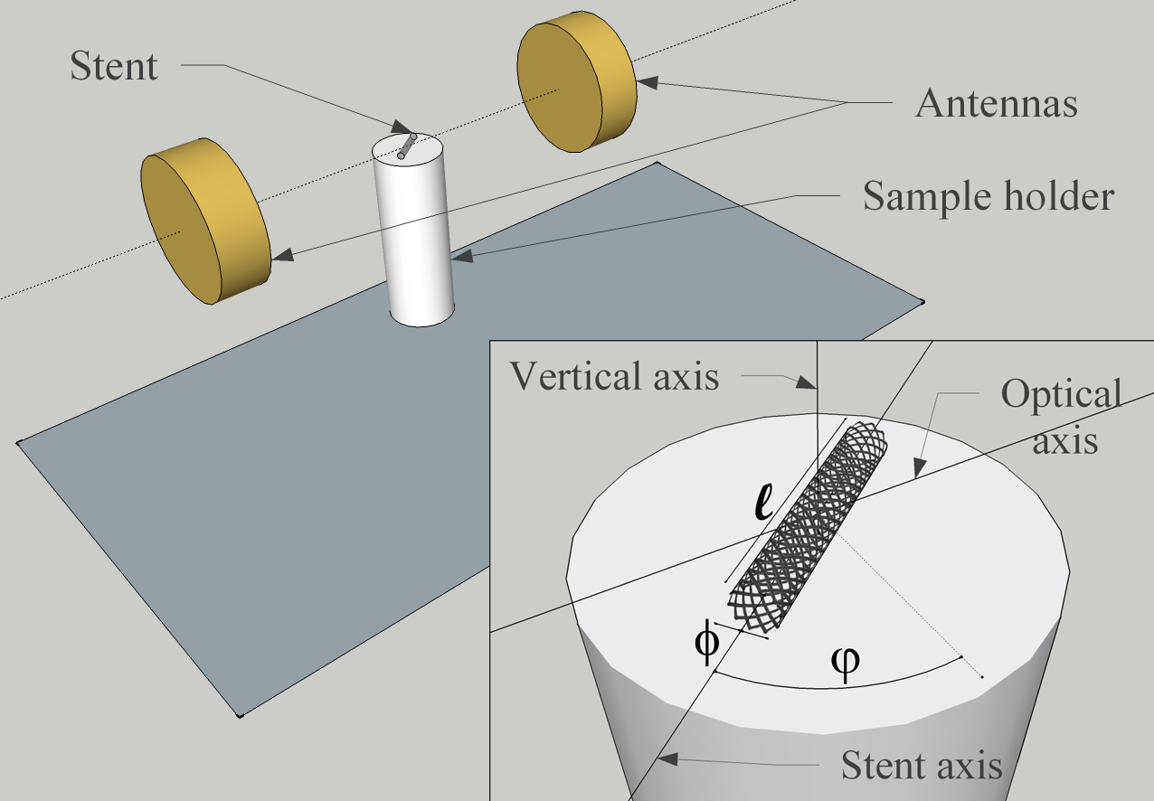
Figure 1 shows a sketch of the experimental set-up used in the present work. In vitro microwave absorbance spectra were measured in open air for thirty commercial drug-eluting stents Medtronic of different nominal length and diameter . To obtain the spectra a pair of right-handed circularly-polarized cavity-backed spiral antennas A-info was connected to a 2-port vector network analyzer HP via coaxial feed lines. The antennas have a - GHz spectral range. This set-up provides the transmission coefficient of port 1 to port 2 as a function of frequency, , or vice versa , from which we can calculate the absorbance spectrum as
| (1) |
where and respectively denote the transmission coefficients measured as a function of frequency with and without a stent, and subindices have been dropped for simplicity. Results were obtained from the average of 100 acquisitions of both magnitudes. Measurements were performed in a symmetrical configuration in which the center of the stent was placed at the midpoint of the line joining the two antennas (optical axis), which are set at a constant separation of 16 cm. The stents were positioned with the aid of an expanded polystyrene sample holder which allows to rotate the stent longitudinal axis (stent axis) at an azimuth angle around a vertical axis that is perpendicular to the optical axis (see the inset in Figure 1). spectra were obtained at 4∘ steps over a complete rotation of each stent around the vertical axis.
III RESULTS AND DISCUSSION
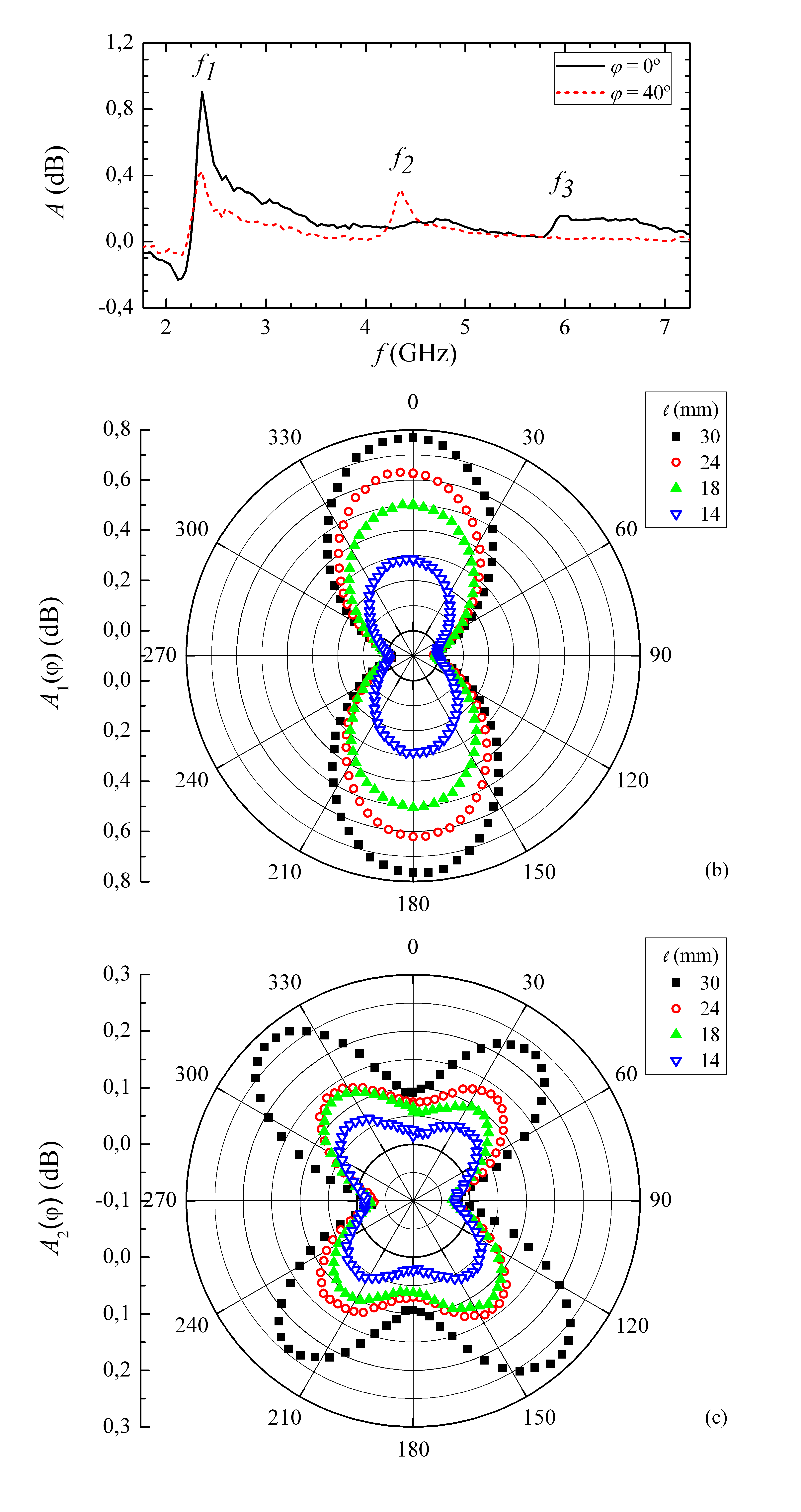
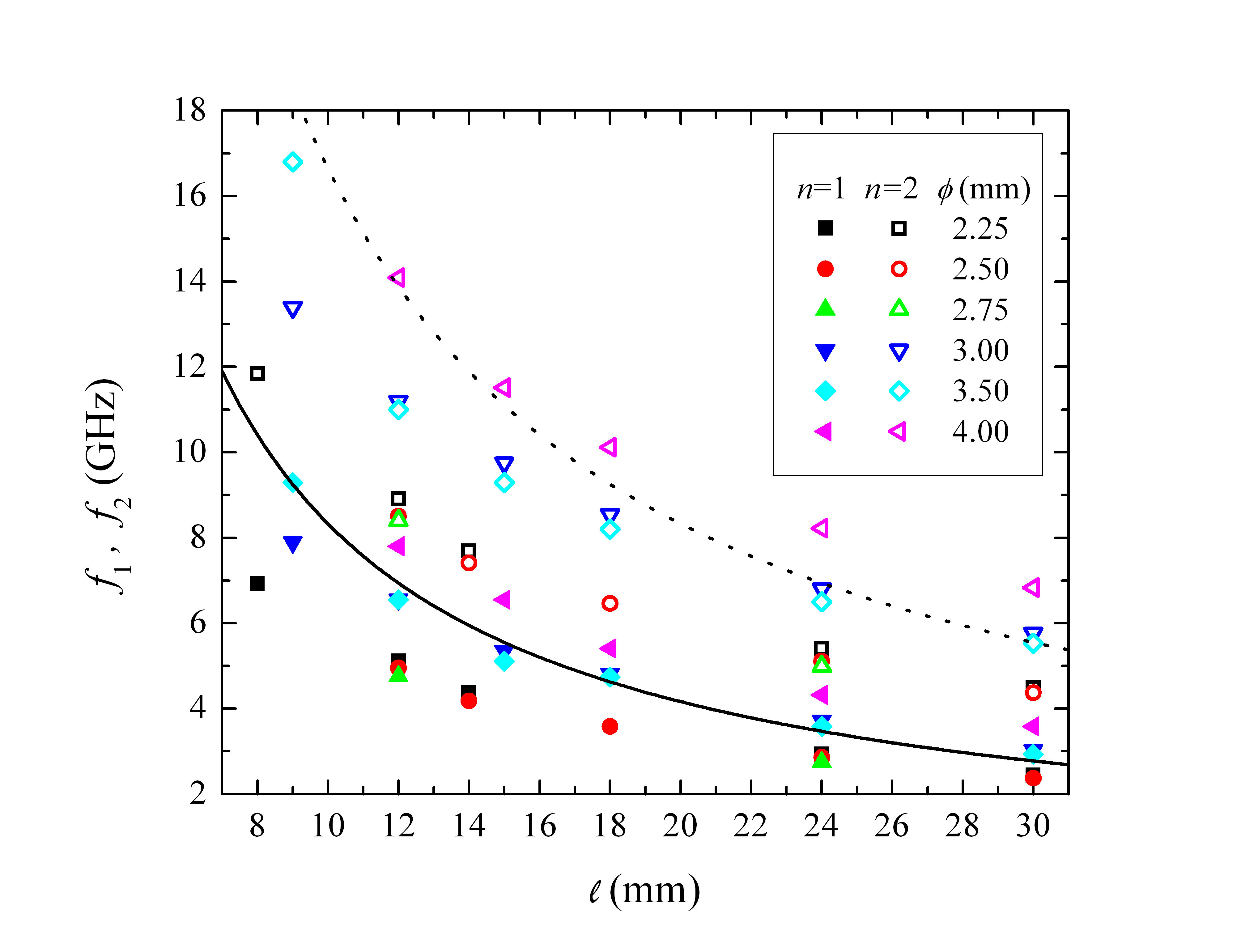
Figure 2(a) shows typical spectra obtained at two values of for a stent of well-defined dimensions. Similar spectra were obtained at any value for the thirty stents investigated. Discrete resonant frequencies with quality factors close to 20 are exhibited by both spectra. Although up to five orders of resonance were detected, we focus here on the characterization of the first two, and . All first orders on the one hand, and all second orders on the other, shared the same features for each studied stent. Figures 2(b) and 2(c) present respectively the azimuthal dependence of the amplitude of the first two resonances, and , for four stents with the same diameter and different lengths. shows a bilobular pattern, with peaks occurring when the stent axis is perpendicular to the optical axis, while shows a tetralobular pattern, with peaks occurring when is in the range 32∘ - 40∘. Higher resonances show more complex dependences, like hexalobular patterns for , for example. The ability to discern these resonances by means of the inspection of their patterns allows to create a database with their values for each stent. Figure 3 shows and as a function of for several values of the thirty stents investigated. Notice how both frequencies are inversely proportional to and roughly proportional to .
The shapes of the experimental curves in Figures 2(b) and 2(c) closely resemble gain patterns of a center-feed half-wave dipole antenna Orfanidis08 . This fact, along with the dependence shown in Figure 3, suggests that the microwave scattering of stents is remarkably similar to that produced by such device PreNoteChow09 . The microwave electromagnetic field couples here to the antenna conductive structure and induces a standing electric current along it. Consequently the scattering is enhanced at resonant frequencies given by Balanis97
| (2) |
where and are respectively the permittivity and permeability of the stent surrounding medium, is the dipole antenna length, and is the resonance mode. We found out that the resonant frequencies in a stent of length are significantly lower than those corresponding to a dipole antenna with . This means that, in terms of microwave scattering, metallic stents of length behave akin to dipole antennas of length , where is a scaling factor. As an example, in Figure 3 we have plotted Equation 2 for . There is an acceptable agreement between experimental resonant frequencies and theoretical estimations. The presence of a scaling factor greater than one should be considered reasonable due to the characteristic folded structure of coronary stents. The precise value of this scaling factor would actually give a hint about the folding degree of a particular stent architecture.
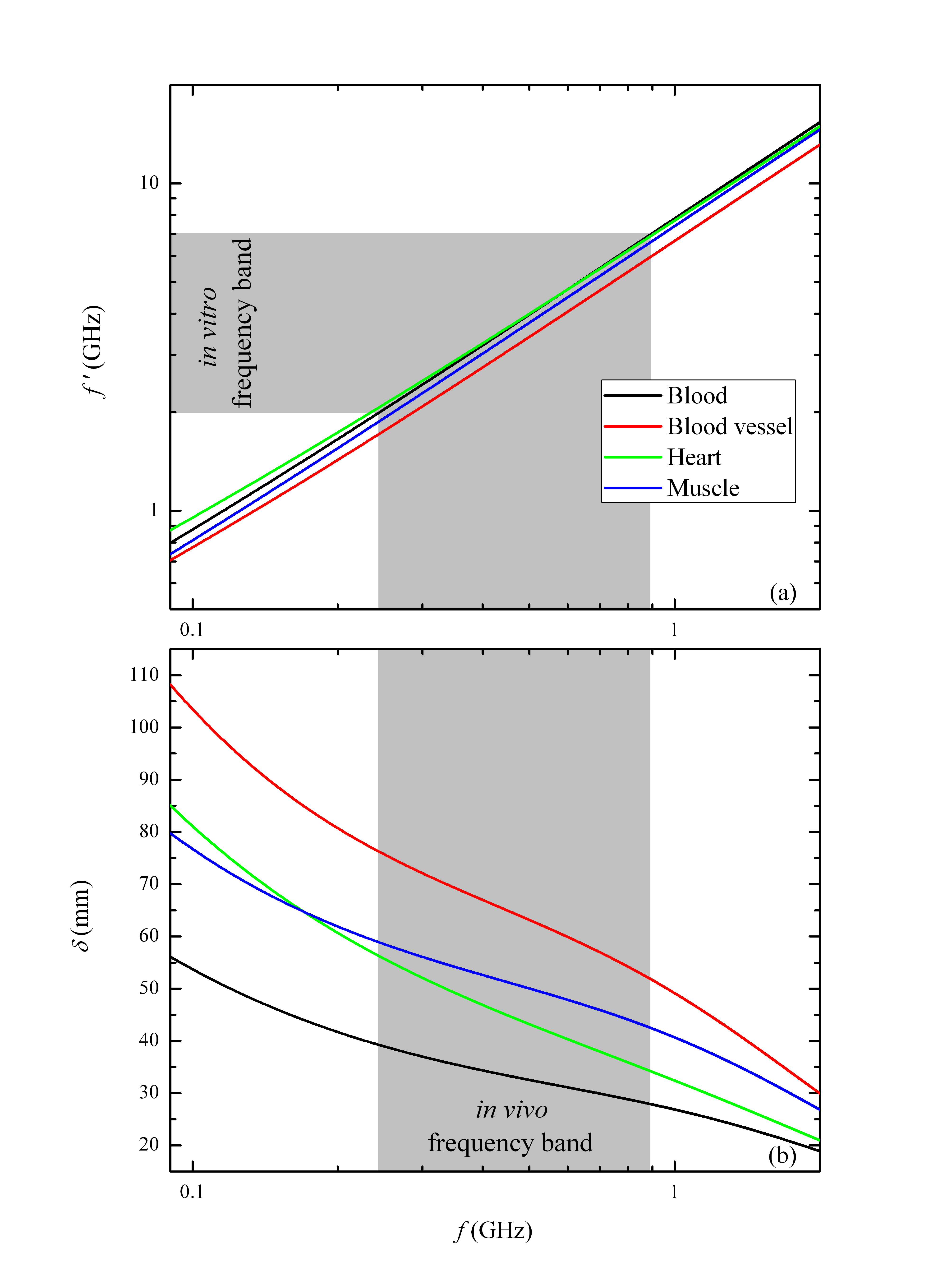
These results show that a method for the detection of structural distortions of stents by measuring their resonant frequencies is possible, at least under in vitro conditions. We will then discuss the in vivo applicability of this method in terms of the skin depth of microwaves in the human body. Due to the factor of Equation 2, it is expected that the in vitro resonant frequencies of stents will shift down with respect to the in vivo frequencies, since the relative permittivity of biological tissues in the microwave range is higher than one. We can thus obtain the relation between the in vitro and in vivo values, and respectively, using Equation 2 and assuming air as the surrounding in vitro medium of the stent,
| (3) |
Introducing the dependences of and given by parametric models Gabriel96III in Equation 3, may be determined numerically. Figure 4(a) shows such dependence for several representative tissues and highlights that the shift between and is of about one order of magnitude.
Conduction and displacement currents induced by electromagnetic waves are of the same order for most biological tissues VanderVorst06 , so in this case the skin depth for media limited by plane boundaries is generally expressed as Jordan50
| (4) |
where is the conductivity of the medium. Using again parametric models Gabriel96III , we can determine numerically . Figure 4(b) shows that is found to be of several tens of millimeters for in vivo operating frequencies. This fact demonstrates that microwaves travel through the thoracic cavity and reach the heart, thereby enabling the medical applicability of microwave spectrometry for the evaluation of the dimensions of implanted coronary stents.
IV CONCLUSIONS
We have proved experimentally that metallic stents of a given nominal length and diameter exhibit characteristic resonant frequencies in their open air microwave scattering spectra. We have also proposed a simple theoretical model based on a dipole antenna that allows to estimate the values of such frequencies. It is expected that some structural distortions experienced by implanted stents due to aging should be reflected in a variation of the resonant frequencies. Finally, we have estimated the in vivo frequency band corresponding to the in vitro frequencies of the investigated stents and its associated skin depth range for several representative tissues, demonstrating the medical applicability of the method here discussed. Our work will allow the achievement of techniques able to prevent alterations, such as stent recoil or fracture, that nowadays have strong medical impact. Characterization of stents with induced geometrical distortions within biological tissue mimics are under development. Further experiments will include ex vivo and in vivo trials. We hope that our work will stimulate the development of non-invasive and non-ionizing intensive monitoring medical techniques of patients with coronary stents.
V ACKNOWLEDGMENTS
G. A.-G. thanks L. Humbert-Vidan for her backing, and ICREA Acadèmia and Universitat de Barcelona for the financial support. V. L.-D. and J. T. appreciate financial support from ICREA Acadèmia and Universitat de Barcelona. A. G.-S. thanks Universitat de Barcelona for backing his research. O. R.-L. and A. B.-G. thanks Medtronic, Inc. for providing the stents studied in the present work. J. O’C. thanks the Spanish Government project MAT2011-29269-C03-02.
References
- (1) S. Garg and P. W. Serruys, “Coronary stents: Looking forward,” J. Am. Coll. Cardiol., vol. 56, no. 10, Supplement, pp. S43–S78, august 2010.
- (2) S. Garg and P. W. Serruys, “Coronary stents: Current status,” J. Am. Coll. Cardiol., vol. 56, no. 10, Supplement, pp. S1–S42, august 2010.
- (3) R. Schatz, J. Palmaz, F. Tio, F. Garcia, O. Garcia, and S. Reuter, “Balloon-expandable intracoronary stents in the adult dog,” Circulation, vol. 76, no. 2, pp. 450–457, august 1987.
- (4) S. P. Karas, M. B. Gravanis, E. C. Santoian, K. A. Robinson, K. A. Anderberg, and S. B. King III, “Coronary intimal proliferation after balloon injury and stenting in swing: An animal model of restenosis,” Journal of the American College of Cardiology, vol. 20, no. 2, pp. 467–474, august 1992.
- (5) P. C. Gordon, C. Gibson, D. J. Cohen, J. P. Carrozza, R. E. Kuntz, and D. S. Baim, “Mechanisms of restenosis and redilation within coronary stents - quantitative angiographic assessment,” Journal of the American College of Cardiology, vol. 21, no. 5, pp. 1166–1174, april 1993.
- (6) R. Hoffmann, G. S. Mintz, G. R. Dussaillant, J. J. Popma, A. D. Pichard, L. F. Satler, K. M. Kent, J. Griffin, and M. B. Leon, “Patterns and mechanisms of in-stent restenosis,” Circulation, vol. 94, no. 6, pp. 1247–1254, may 1996.
- (7) D. J. Moliterno, “Healing achilles - sirolimus versus paclitaxel,” New England Journal of Medicine, vol. 353, no. 7, pp. 724–727, august 2005.
- (8) C. Stettler, S. Wandel, S. Allemann, A. Kastrati, M. C. Morice, A. Schömig, M. E. Pfisterer, G. W. Stone, M. B. Leon, J. S. de Lezo, J.-J. Goy, S.-J. Park, M. Sabaté, M. J. Suttorp, H. Kelbaek, C. Spaulding, M. Menichelli, P. Vermeersch, M. T. Dirksen, P. Cervinka, A. S. Petronio, A. J. Nordmann, P. Diem, B. Meier, M. Zwahlen, S. Reichenbach, S. Trelle, S. Windecker, and P. Jüni, “Outcomes associated with drug-eluting and bare-metal stents: a collaborative network meta-analysis,” The Lancet, vol. 370, no. 9591, pp. 937–948, september 2007.
- (9) C. Spaulding, J. Daemen, E. Boersma, D. E. Cutlip, and P. W. Serruys, “A pooled analysis of data comparing sirolimus-eluting stents with bare-metal stents,” New England Journal of Medicine, vol. 356, no. 10, pp. 989–997, march 2007.
- (10) G. W. Stone, J. W. Moses, S. G. Ellis, J. Schofer, K. D. Dawkins, M.-C. Morice, A. Colombo, E. Schampaert, E. Grube, A. J. Kirtane, D. E. Cutlip, M. Fahy, S. J. Pocock, R. Mehran, and M. B. Leon, “Safety and efficacy of sirolimus- and paclitaxel-eluting coronary stents,” New England Journal of Medicine, vol. 356, no. 10, pp. 998–1008, march 2007.
- (11) L. Mauri, W.-h. Hsieh, J. M. Massaro, K. K. Ho, R. D’Agostino, and D. E. Cutlip, “Stent thrombosis in randomized clinical trials of drug-eluting stents,” New England Journal of Medicine, vol. 356, no. 10, pp. 1020–1029, march 2007.
- (12) A. Kastrati, J. Mehilli, J. Pache, C. Kaiser, M. Valgimigli, H. Kelbaek, M. Menichelli, M. Sabaté, M. J. Suttorp, D. Baumgart, M. Seyfarth, M. E. Pfisterer, and A. Schömig, “Analysis of 14 trials comparing sirolimus-eluting stents with bare-metal stents,” New England Journal of Medicine, vol. 356, no. 10, pp. 1030–1039, march 2007.
- (13) J. A. Ormiston and P. W. Serruys, “Bioabsorbable coronary stents,” Circ. Cardiovasc. Interv., vol. 2, no. 3, pp. 255–260, june 2009.
- (14) E. Chow, A. Chlebowski, S. Chakraborty, W. Chappell, and P. Irazoqui, “Fully wireless implantable cardiovascular pressure monitor integrated with a medical stent,” IEEE Trans. Biomed. Eng., vol. 57, no. 6, pp. 1487–1496, june 2010.
- (15) K. Takahata, Y. Gianchandani, and K. Wise, “Micromachined antenna stents and cuffs for monitoring intraluminal pressure and flow,” J. Microelectromech. Syst., vol. 15, no. 5, pp. 1289–1298, october 2006.
- (16) K. Takahata and Y. Gianchandani, “A planar approach for manufacturing cardiac stents: design, fabrication, and mechanical evaluation,” J. Microelectromech. Syst., vol. 13, no. 6, pp. 933–939, december 2004.
- (17) H. Eggebrecht, H. Kühl, G. M. Kaiser, S. Aker, M. O. Zenge, F. Stock, F. Breuckmann, F. Grabellus, M. E. Ladd, R. H. Mehta, R. Erbel, and H. H. Quick, “Feasibility of real-time magnetic resonance-guided stent-graft placement in a swine model of descending aortic dissection,” Eur. Heart J., vol. 27, no. 5, pp. 613–620, january 2006.
- (18) S. M. Wetzner, L. C. Kiser, and J. S. Bezreh, “Duplex ultrasound imaging: vascular applications.” Radiology, vol. 150, no. 2, pp. 507–514, february 1984.
- (19) B. Perrenot, R. Vaillant, R. Prost, G. Finet, P. Douek, and F. Peyrin, “Motion correction for coronary stent reconstruction from rotational x-ray projection sequences,” IEEE Trans. Med. Imaging, vol. 26, no. 10, pp. 1412–1423, october 2007.
- (20) S. E. Nissen and P. Yock, “Intravascular ultrasound,” Circulation, vol. 103, no. 4, pp. 604–616, january 2001.
- (21) B. Wang, J. Su, A. Karpiouk, K. Sokolov, R. Smalling, and S. Emelianov, “Intravascular photoacoustic imaging,” IEEE J. Sel. Top. Quantum Electron., vol. 16, no. 3, pp. 588–599, may-june 2010.
- (22) C. Kauffmann, P. Motreff, and L. Sarry, “In vivo supervised analysis of stent reendothelialization from optical coherence tomography,” IEEE Trans. Med. Imaging, vol. 29, no. 3, pp. 807–818, march 2010.
- (23) T. Tsunoda, M. Nakamura, M. Wada, N. Ito, Y. Kitagawa, M. Shiba, S. Yajima, R. Iijima, R. Nakajima, M. Yamamoto, T. Takagi, T. Yoshitama, H. Anzai, T. Nishida, and T. Yamaguchi, “Chronic stent recoil plays an important role in restenosis of the right coronary ostium,” Coronary Artery Dis., vol. 15, no. 1, pp. 39–44, february 2004.
- (24) T. Chakravarty, A. J. White, M. Buch, H. Naik, N. Doctor, J. Schapira, S. Kar, J. S. Forrester, R. E. Weiss, and R. Makkar, “Meta-analysis of incidence, clinical characteristics and implications of stent fracture,” Am. J. Cardiol., vol. 106, no. 8, pp. 1075–1080, june 2010.
- (25) T. Canan and M. S. Lee, “Drug-eluting stent fracture: Incidence, contributing factors, and clinical implications,” Catheter. Cardiovasc. Interv., vol. 75, no. 2, pp. 237–245, december 2010.
- (26) Endeavor Sprint Zotarolimus-Eluting Coronary Stent System model, Medtronic, Inc., Minneapolis, USA.
- (27) JXTXLX-20180 model, Chengdu A-info Inc., Chengdu, China.
- (28) HP 8510C model, Agilent Technologies, Santa Clara, CA, USA.
- (29) S. J. Orfanidis, Electromagnetic Waves and Antennas. Piscataway, New Jersey: Rutgers University, 2002, chapter 16, pp. 637–660, electronic book. Revision date august 31, 2010.
- (30) E. Chow and coauthors have recently evaluated the use of stents as antennas for implantable wireless medical applications, which reinforces our hypothesis. See for instance E. Chow, Y. Ouyang, B. Beier, W. Chappell, and P. Irazoqui, ”Evaluation of cardiovascular stents as antennas for implantable wireless applications, ”IEEE Trans. Microw. Theory Tech., vol. 57, no. 10, pp. 2523–2532, october 2009.
- (31) C. A. Balanis, Antenna Theory: Analysis and Design, 2nd ed. New York: Wiley, 1997, chapter 4, pp. 133–202.
- (32) S. Gabriel, R. W. Lau, and C. Gabriel, “The dielectric properties of biological tissues: III. parametric models for the dielectric spectrum of tissues,” Phys. Med. Biol., vol. 41, no. 11, pp. 2271–2293, april 1996.
- (33) A. Vander Vorst, A. Rosen, and Y. Kotsuka, RF/Microwave Interaction with Biological Tissues, 1st ed. New Jersey: John Wiley & Sons, Inc., 2006, chapter 1, pp. 39–44.
- (34) E. C. Jordan and K. G. Balmain, Electromagnetic waves and radiating systems, 2nd ed. New Jersey: Prentice-Hall, Inc., 1950, chapter 5, pp. 130–133.