Unraveling siRNA Unzipping Kinetics with Graphene
Abstract
Using all atom molecular dynamics simulations, we report spontaneous unzipping and strong binding of small interfering RNA (siRNA) on graphene. Our dispersion corrected density functional theory based calculations suggest that nucleosides of RNA have stronger attractive interactions with graphene as compared to DNA residues. These stronger interactions force the double stranded siRNA to spontaneously unzip and bind to the graphene surface. Unzipping always nucleates at one end of the siRNA and propagates to the other end after few base-pairs get unzipped. While both the ends get unzipped, the middle part remains in double stranded form because of torsional constraint. Unzipping probability distributions fitted to single exponential function give unzipping time () of the order of few nanoseconds which decrease exponentially with temperature. From the temperature variation of unzipping time we estimate the energy barrier to unzipping.
I Introduction
The interaction of nucleic acids with carbon nanotubes (CNTs)
and graphene have attracted much attention due to the potential
applications in nanotube separation zheng1 ; zheng2 ,
sensing biosensor ; mohanty , sequencing
garaj ; schneider ; christopher and nanomedicine
liu1 ; liu2 ; liu3 ; lu2010 . Basic understanding of
the interaction mechanism of nucleic acids with CNT or
graphene anindyacpl ; varghese_cpc ; ralph2007 ; santoshjcp ; santoshjbs ; narahari is
essential for such applications.
Small interfering ribonucleic acid (siRNA)
molecules are a class of double stranded non-coding RNA
which are typically 21 to 23 nucleotides
in length. The properties of siRNA are
actively being studied due to their potential
influence on cell functionality and applications
in medicine to achieve RNA interference
(RNAi) napoli ; fire ; couzin .
The mechanism of RNAi involves RNA-induced
silencing complex (RISC) comprising
of Dicer, Argonaute2 and siRNA binding
protein that induces unzipping of siRNA
into two single strand RNAs zamore2000 ; hutvagner2002 ; tomari2004 ; zamore2005 ; Ghildiyal2009 .
One of these two strands acts as a guiding
strand to form specific base-pairs with mRNA
and silences the gene.
For using RNAi technology in the treatment
of diseases such as cancer, HIV, viral infections and eye
diseases kurreck , efforts are being made for the efficient
and safe siRNA delivery systems to achieve
the desired RNAi effect. Dendrimers tsubouchi ; huang ; vasumati and carbon nanotubes liu1 ; liu2 ; liu3 ; santoshjcp
are good carriers of siRNA into disease infected cell.
The discovery of graphene has
led to its possible use in efficient delivery
of siRNA as well as various oligonucleotides.
Several attempts have been made to study the
properties of nucleic acid interaction with graphene
mohanty ; varghese_cpc ; ralph2007 ; zhao2011 ; narahari ; lu2010 ; lv2010 .
However the interaction between graphene and siRNA/DNA
has not been understood.
In this paper, we show the unzipping
of siRNA on graphene and subsequent binding
between the two. Graphene is a
free-standing two dimensional monolayer of
carbon atoms arranged into a honeycomb
lattice novoselov2004 ; meyer2007 .
Graphene is a partially hydrophobic molecule:
its faces are highly hydrophobic while edges,
depending on functionalization, can be
hydrophilic in nature swatijpcc2011 .
Thus it has the ability to pass through
hydrophobic lipid bilayer and can also interact with
the hydrophilic head groups. Dispersion
interaction including stacking serves as
the major attractive interactions
between the non-polar molecules paton .
The importance of dispersion interactions has been
verified recently in analyzing the structure and
energetics of the graphene-nucleobase complexes
swatijpcc2012 and in studying the unzipping of
siRNA with single walled CNT santoshjcp .
Using state of the art all atom molecular
dynamics simulation along with the ab-initio
quantum mechanical calculations, we give a
comprehensive understanding of the structure
and thermodynamics of the siRNA-graphene complex.
In the supplementary material supplementary , we discuss the
effect of force fields (FF) on the unzipping
and adsorption of siRNA/dsDNA on graphene.
II Computational Details
II.1 Classical simulations
We have used AMBER11 suite of programs amber11
for simulating the systems with Amber 2003
(along with ff99) force fields duan and the TIP3P
model jorgensen for water. Latest
improvements of torsion angle parameters
are reported for DNA/RNA in a new force field called
parmbsc0 and ff10 parmbsc0 ; banas ; yildirim .
The comparison of force fields ff99, parmbsc0 and ff10
are discussed in the supplementary material.
Similar conformational changes are observed with ff99,
parmbsc0 and ff10.
Use of ff99 also helps us to compare
the present results with our previous
simulations on siRNA-dendrimer complex
that used ff99 vasumati .
The initial structure of
the small interfering ribonucleic
acid (siRNA) was taken from the protein
data bank (PDB code: 2F8S)yuan .
The sequence of the siRNA used is
r(UU AGA CAG CAU AUA UGC UGU CU)2
with sticky ends of sequence UU on the two
ends of the strands. We have built
graphene sheet of Å2 wider
dimensions to ensure sufficient sliding
area for the siRNA before optimum binding on graphene.
For comparison, we have also simulated dsDNA of the
same length on graphene. The dsDNA has same sequence
as the siRNA where the nucleobase Uracil (U) is
replaced by Thymine (T) nucleobase, i.e., d(TT AGA
CAG CAT ATA TGC TGT CT)2. Structure of the dsDNA
in B-form was generated using NUCGEN module of AMBER amber11 .
The siRNA/dsDNA-graphene
complex structure is then solvated with TIP3P
model water box using the LEaP module in AMBER 11 amber11 .
The box dimensions were chosen such that there
is at least 20 Å solvation shell from the surface of
siRNA/dsDNA-graphene complex to the edge of the water box.
In addition, some water residues were replaced
by 44 Na+ counterions for siRNA and 42 Na+
counterions for dsDNA to neutralize the negative
charge on the phosphate backbone groups of the
siRNA/dsDNA structure. This has resulted in box
dimensions of 18818672 Å3
with total system size of 214602 atoms for
siRNA-graphene complex and 18818669 Å3
with total system size of 206865 atoms for dsDNA-graphene
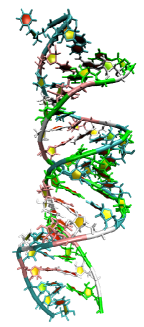
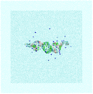
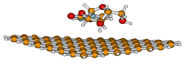
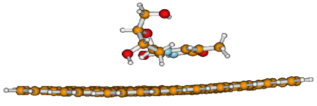
complex. The crystal structure of
siRNA and the initial system containing
siRNA and graphene with added water plus neutralizing
counterions are shown in Figures 1
and 1, respectively.
We have modeled the carbon atoms in graphene as
uncharged Lennard-Jones particles with LJ
parameters listed in Table 1.
The bonded interactions viz., stretching, torsion and
dihedral terms were also included. To keep the
graphene fixed during simulations, all the
carbon atom positions in graphene were
restrained with harmonic potential of spring
constant of 1000 kcal/mol-Å2. The translational
center of mass motions were removed every 1000 steps.
The system is subjected to standard simulation protocol
described in Refs. maiti2004 ; maiti2006 .
The long range electrostatic interactions were calculated
with the Particle Mesh Ewald (PME) method darden
using a cubic B-spline interpolation of order 4 and
a tolerance is set for the direct space sum cutoff.
A real space cutoff of 9 Å was used both for the long range
electrostatic and short range van der Waals interactions with
a non-bond list update frequency of 10. The trajectory was
saved at a frequency of 2 ps for the entire simulation
scale of 55-85 ns for each system.
We have used periodic boundary
conditions in all three directions during the simulation.
Bond lengths involving bonds to hydrogen
atoms were constrained using SHAKE algorithm ryckaert .
This constraint enabled us to use a time step of 2 fs for
obtaining long trajectories of each 50 ns. During
the minimization, the siRNA/dsDNA-graphene complex
structures were fixed in their starting conformations
using harmonic constraints with a force constant
of 500 kcal/mol-Å2. This allowed the water
molecules to reorganize which eliminates bad
contacts with the siRNA/dsDNA and the graphene.
The minimized structures were then subjected to 40 ps of MD,
using 1 fs time step for integration. During the MD, the
system was gradually heated from 0 to 300 K using
weak 20 kcal/mol-Å harmonic constraints on the
solute to its starting structure. This allows slow
relaxation of the siRNA/dsDNA-graphene complex structure.
Subsequently, simulations were performed under constant
pressure-constant temperature conditions (NPT), with
temperature regulation achieved
using the Berendsen weak coupling method berendsen
(0.5 ps time constant for heat bath coupling and 0.5 ps
pressure relaxation time). Constant temperature-pressure
MD was used to get the
correct solvent density corresponding to experimental
condition. Finally, for analysis of structures and
properties, we have carried out 50 ns of NVT production
MD with 2 fs integration time step using a heat bath
coupling time constant of 1 ps.
The binding free energy for the non-covalent association of two molecules in solution can be written as . For any species on the right hand side , accordingly,
| (1) |
where is the change in enthalpy and is calculated by summing the gas-phase energies and solvation free energies ; , where is the electrostatic energy calculated from the Coulomb potential, is the non-bond van der Waals energy and is the internal energy contribution from bonds, angles and torsions. where is the electrostatic energy calculated from a Generalized Born (GB) method and is the non-electrostatic energy calculated as ; where is the surface tension parameter ( = 0.0072 kcal/mol-Å), is the solvent-accessible surface area of the molecule and is the solvation free energy for a point solute ( = 0). For the entropy calculation we have used two-phase thermodynamic (2PT) model lin2003 ; lin2010 ; hemant , based on density of states (DoS) function. The DoS function can be calculated from the Fourier transform of the velocity auto-correlation function which provides information on the normal mode distribution of the system. The method has found successful application in several related problems lin2003 ; maitinl ; lin2010 ; hemant ; nandy2011 .
II.2 Quantum simulations
We have carried out quantum chemical calculations to
understand the interaction of the planar
nanographene with the nucleosides of RNA and DNA.
For the present investigation, we have taken a graphene sheet of
dimension with alternate armchair and zig-zag
edges with C-C bond lengths of 1.42 Å,
C-H bond length 1.09 Å and all the angles kept at .
The edges are terminated with hydrogen atoms
to avoid any unwanted terminal effects koskinen .
We have modeled adenine, guanine, cytosine, thymine
and uracil with furanose sugar connected to the respective
nucleobases by the -glycosidic bond.
As structural features that distinguish siRNA
from DNA are the presence of uracil nucleobase and
hydroxyl-OH group in the constituted ribose sugar
in siRNA, we have modeled thymine with the deoxyribose
sugar and all other bases are modeled with ribose sugar to
be consistent with siRNA structure.
These structures act as miniature models
of siRNA and DNA to understand the interaction with
the planar graphene molecule. Initially the nucleosides
are placed vertically above, around 4 Å from the center
of the planar hexagonal ring of the graphene and
the nucleobases lie parallel to the graphene sheet.
All the model building was done with the help of Discovery
studio 2.0 studio and Molden molden
software.
We have optimized all the graphene-nucleosides
complex systems to get the energy minimized configuration.
For all the quantum chemical calculations, we have
employed dispersion corrected density functional
approach using B97XD/6-31G** basis setchai
using Gaussian 09 gaussian . The total interaction
energy of each system has been calculated
using BSSE, where BSSE
represents basis set superposition error, arising
from the overlapping of the atomic orbitals. The
BSSE corrections has been calculated using Boys
and Bernardi function counterpoise
method boysbernadi . We have also carried out
frequency calculation on the optimized
geometry using the same method and basis set.
To analyze the degree of binding, we have also
carried out the charge transfer analysis of
the nucleosides and graphene sheet using the
Natural Bond Orbital (NBO) approach reed1985 ; reed1988 ; carpenter1988 .
III Results and discussion
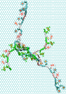
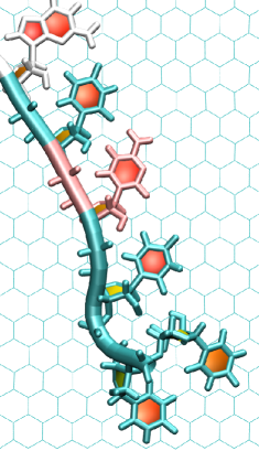
III.1 siRNA Unzipping
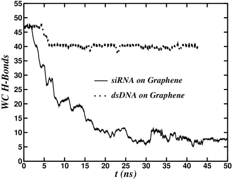
Figure 1 shows the instantaneous snapshots of siRNA as it binds to graphene. The unzipping of base-pairs starts within 3 ns and by 22 ns, 41 of the initial 48 Watson-Crick (WC) H-bonds are broken resulting in almost complete unzipping of siRNA into two single strand RNAs. Figure 2 shows the number of intact H-bonds of siRNA and dsDNA with time when bound to graphene. Breaking of WC H-bonds in siRNA/dsDNA manifest the deformation mechanism of nucleic acid molecule voet ; santoshjpcm ; santoshbj . It can be seen that a large decrease of H-bonds occurs in the first few nanoseconds when siRNA starts unzipping and within 22 ns, 41 H-bonds are broken leaving only 7 intact H-bonds. The unzipped bases are then free to interact with graphene surface via van der Waals forces. Figure 2 shows that H-bonds are saturated at 7. At this stage, siRNA optimally binds to graphene and the complex is very stable after 22 ns.In the optimum bound configuration where H-bonds are constant with time, there are few transient H-bonds due to room temperature thermal fluctuations. Because of these transient H-bonds, the curve has fluctuations about its mean value with standard deviations ranging from 1 to 3 H-bonds. The different nucleosides interact with different strength with graphene making the binding possible. Interestingly, dsDNA of same sequence and length show much less unzipping and binding on graphene compared to siRNA. Within 6 ns, only 8 of total 48 H-bonds in dsDNA get unzipped and remain constant at 40 H-bonds throughout the rest of simulation time of 44 ns. This difference has its origin in the extra hydroxyl group of uridine that is present in siRNA. To have a molecular level understanding of this binding affinity we calculate the binding free energy of different nucleosides with graphene both from the classical MD simulations (using MM-GBSA method) as well as from our dispersion corrected DFT calculations.
III.2 Binding free energy
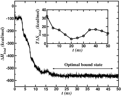
Figure 3 shows the enthalpy
contribution to the total
binding free energy as a function of time as
siRNA binds to graphene. In the plot, we have marked
the time interval at which optimal binding
happens as reflected by the binding energy.
After this binding the complex is stable for
the entire duration of the simulation
with fluctuations ranging only 1.5 % of its average
value in the optimum bound state. From the stable
trajectory of siRNA-graphene complex,
the enthalpy contribution ()
to the total binding free
energy was calculated for 250 snapshots
separated each by 2 ps.
The enthalpy contribution ()
to the total binding free
energy is -562.6 6.2 kcal/mol.
Entropy is calculated
at every 5 ns along the trajectory. For this,
we simulate the system for
40 ps with velocities and coordinates
saved at a frequency of 4 fs.
The velocity auto-correlation function
converges within 10 ps. When siRNA is
binding with graphene during initial stage,
siRNA entropy decreases since
the graphene substrate suppress the fluctuations
of unzipped bases (inset of Figure 3).
After siRNA binds optimally
to graphene, the entropy starts increasing
again due to inherent kinetics of unzipped
bases. However the entropy contribution () to
the total binding free energy is small
compared to the enthalpy contribution ()
arising due to dispersive interaction between the
graphene and the aromatic nucleobases.
The enthalpy and entropy contributions
in Eqn. 1 give the total binding
free energy () of siRNA
when binding to graphene. The value of
is -573.0 8 kcal/mol. However, the binding
energy of the dsDNA bound to graphene is calculated
to be -190 9 kcal/mol which is very low
compared to siRNA bound to graphene.
To get further molecular level picture of the binding
mechanism we also compute the histograms of the
closest approach of nucleoside to graphene.
This will allow us to understand the relative
binding affinity as well as how different
nucleosides are oriented on the graphene substrate.
We track the center of mass position of different nucleobases
with respect to graphene as a function of simulation time.
We calculate the closest
approach of nucleoside to graphene as the perpendicular
distance (because graphene is in plane) where and
are the centers of mass position of nucleoside and graphene,
respectively. is calculated for 50 ns trajectory
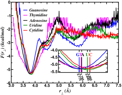
and the histogram of nucleosides is shown in
Figure 4. From this plot, we can understand
the probability () of finding the
nucleoside at a given position
from the graphene. Using histogram, the free energy of nucleoside is calculated
as
(where is the Boltzmann constant) and
plotted as a function of distance in Figure 4.
The minimum in the free energy indicates the most
optimum bound configuration for nucleoside-graphene
complex. In the inset we show the zoomed part of
the occurrence of free energy minima.
has minima at 3.775 Å , 3.790 Å , 3.795 Å ,
3.840 Å and 3.860 Å for guanosine, Thymidine, adenosine,
uridine and cytidine, respectively. Guanine can
form strongest H-bonding interaction
and stacking interaction with graphene.The distance of closest approach in the increasing
order for guanosine, thymidine, adenosine, uridine and
cytidine is .
Hence guanine has most interaction
strength with graphene and cytidine has least
interaction strength as G T A U C.
The binding energy order of nucleosides
is consistent with experimental and
theoretical calculations anindyacpl ; varghese_cpc ; ralph2007 .
The stable complex structure with most of the
base-pairs already unzipped in siRNA can be
delivered to the target
virus infected cell for RNAi applications.
Since siRNA has to undergo unwinding process with the
effect of RISC, our proposed
delivery mechanism by graphene possesses potential
advantages in achieving RNAi. Toxic effects of
graphene inside cell may be suppressed with proper
surface functionalization lam ; jia ; cui .
III.3 Structural deformation
Snapshots shown in Figure 1 indicate that the siRNA
molecule exhibit large structural deformation on
binding to graphene. This structural
deformation is characterized by the number
of siRNA atoms that come close to the graphene
in a specified cutoff distance and the root mean
square deviation (RMSD) of siRNA with respect
to its crystal structure. We calculate close
contacts when any siRNA atoms are
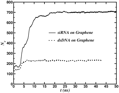
within 5 Å of the graphene sheet. The number
of close contacts between siRNA
and graphene is plotted in Figure 5
as a function of time. Since siRNA is getting
unzipped within 21 ns, is increasing
rapidly within 21 ns and reaches a constant
value of 710 siRNA atoms. The fluctuations are
very less in after the complete binding
of siRNA to the graphene. Interestingly, for
dsDNA is much less than that of siRNA.
The dsDNA has only 240 atoms within 5 Å from
graphene sheet in the stable configuration.
This also demonstrates very low binding affinity
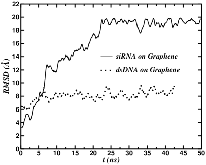
of dsDNA compared to siRNA with graphene. In Figure 5 we
plot the RMSD of siRNA/dsDNA as a function of time
for both the siRNA/dsDNA-graphene complex.
For the calculation of RMSD, the reference
structure of siRNA is taken to be the crystal
structure of siRNA after initial minimization.
Note that the plot shows RMSD for only
production NVT simulation time scale.
In the most optimum bound
configuration, the average RMSD
of siRNA is 19.4 0.4 Å on graphene
whereas
of dsDNA is 8.5 0.6 Å.
As the binding of siRNA is more on graphene, the siRNA
structure deforms leading to a large
value of .
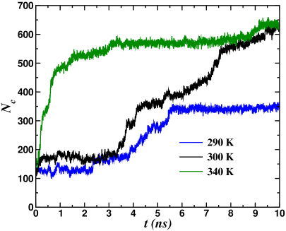
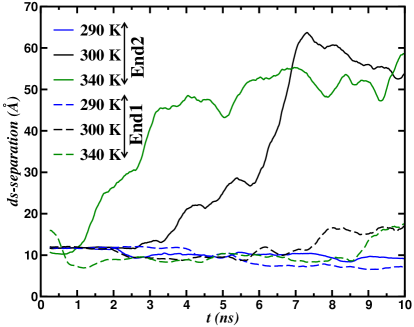
III.4 Unzipping kinetics
In Figures 6 and 6,
we plot the number of contacts
for siRNA as well the distance between the two strands
of the double stranded siRNA (ds-separation) at three
different temperatures. It takes few ns to nucleate the
unzipping. Once critical numbers of contacts are created
between the siRNA and graphene, unzipping starts from
one end (end2 for the current situation). Unzipped bases
at one end help to make more contacts with graphene and
thereby enhancing the interaction between siRNA and graphene.
This facilitates unzipping at the other
end. Figure 6 shows that it takes almost 3 ns at
300 K and less than 1 ns at 340 K for the critical
number of contacts to be created. Once this is done,
strong interaction between graphene and siRNA force
the rapid unzipping of siRNA as is evident from the
rapid increase of ds-separation in Figure 6.
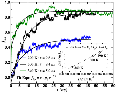
To get an estimate of the unzipping time (), we plot the
unzipping probability distributions () at three different
temperatures in Figure 7 and fit them to single
exponential functions as done in ref. mathe2004 .
This gives rise to = 9.8 ns, 8.4 ns and 5.0 ns
at temperatures 290 K, 300 K and 340 K, respectively.
By fitting the unzipping time as a function of temperature
to , we get to
be 100.8 ps and the activation energy, to be
2.637 kcal/mol or 0.114 . Plot of versus
and the fit was shown in the inset
of Figure 7.
III.5 Insights from Quantum simulations
To understand the difference in binding affinity
of siRNA and dsDNA with graphene we
have calculated the binding energy of the
graphene/siRNA and graphene/dsDNA miniature
complexes using dispersion corrected DFT method (DFT-D).
The sequence of the siRNA studied has A:U base pairs
at both the ends, which is also the case
of most of the confirmed siRNA sequences chalk2005 ,
which are known to open up quite easily as compared to
G:C base pairs. It may be noted that most of the DNA/RNA
oligonucleotide sequences whose three-dimensional structures
in double helical forms are available have G:C base pairs
at both the termini dhananjay2009 . The siRNA
sequences need to unzip soon for their functionality and
probably that drives design of sequences with terminal
loosely bound A:U base pairs. However, it appeared that
the dsDNA molecule, containing terminal A:T base pairs
does not unzip at physiological condition. In order to
check whether this is due to stronger attraction in A:T
as compared to A:U base pairs, we have optimized both
these base pairs using wB97XD/6-31G** method and found
their interaction energies to be -15.80 and -15.91 kcal/mol,
respectively. This clearly indicates that the terminal
A:U base pair is not weaker one as compared to its DNA
counterpart. Thus, the other component of differential
interaction, i.e. interactions between Uracil residues
and Thymine residues with graphene might be driving the
RNA molecules to unzip.
We are mainly interested in
thymidine-graphene and uridine-graphene
complex systems since these are the
principal nucleosides, which can
differentiate between DNA and RNA.
Moreover these nucleosides remain
unpaired in the siRNA as well as dsDNA.
On analyzing the optimized geometry
of the complex systems as shown in
Figure 8 (graphene-uridine and
graphene-thymidine complex), we find
that in both the cases
O3′-H3′ of the
constituent sugar points towards the
planar graphene sheet with close approach
forming O-H… contacts. In case of
thymidine nucleoside, the closest
O3′-H3′…ring center is found
to be around 2.54 Å , and the angle
O3′-H3ring center is
obtained as . These distances
and angles are sufficient to form O-H…
types of H-bonds. While in case of uridine
nucleoside, the O3′-H3′…ring center
bond distances are found to be 2.34 Å , and the angle
O3′-H3′…C is obtained
as , forming significantly stronger H-bond
between the graphene and uridine sugar,
as compared to that
of thymidine sugar complex swatijmsd .
The O2 and O4 groups of the thymine and uracil
also interact with the graphene sheet, but the
magnitudes of interaction seems to be very
low as compared to that of O-H…
types of contact. Nevertheless these
carbon oxygen atoms can form lone
pair type of contacts giving
extra stabilization jain2009 .
In addition to these contacts,
the O2′ may also form H-bond
with the graphene, which however was not found in
the energy minimized structures, but can not
be ignored at physiological temperature
and between the oligonucleotides.
Furthermore several of 2′-OH groups, which are
equivalent to 3′-OH, are present in the
siRNA strands and absent in the dsDNA strands.
Thus the siRNA strands tend to dissociate
from their double helical forms and bind
strongly to the graphene sheet.
The BSSE corrected interaction energy has been
calculated for all the complex systems and
they follows the trends G A U T C.
Interaction energy of the graphene-uridine
nucleoside is found to be -22.26 kcal/mol,
while that of graphene-thymidine nucleoside
is found to be -20.30 kcal/mol. So we can
infer that uridine nucleoside interacts with
the graphene more strongly than that of the
thymidine nucleoside. These interaction energy
values also well correlate the H-bond
lengths and angles values obtained in both
the cases. Our previous studies swatijpcc2012
on interactions of the graphene with the
nucleobases gives the interaction energy
strengths as G A C T U. So it
proves that inclusion of sugar in the
nucleobases alters the interaction energy
strengths, therefore plays significant
role in stabilizing the systems due to
availability of more hydrogen bond donor
sites.
Frequency calculation of the entire complex as well
as the isolated systems give no imaginary frequency
indicating the structures are at their
local minima. Frequency calculation enables us to carry out
thermochemical analysis of the system. All the
calculations are carried out at 298.15 K and 1 atm.
pressure. We have calculated the change in enthalpy
and free energy of the systems. The of
graphene-uridine is found to be -26.39
kcal/mol, whereas that of of
graphene-thymidine is calculated to be
-23.80 kcal/mol. Similarly the of
graphene-uridine and graphene-thymidine
are found to be -11.95 and -10.22 kcal/mol,
respectively. As it is well known that the system is
more favorable with increase in the negative value
of , graphene-uridine complex is more stable.
The formation of stable graphene-uridine nucleoside
complex may initiate and enhance the unzipping of
the siRNA structure as observed by our counterpart
MD simulation studies.
We have also calculated the NBO charges of the
thymidine and uridine nucleosides of the graphene-nucleosides
systems and compared with the isolated nucleosides.
The difference in NBO
charges of the major hydrogen bond donor atoms
of the nucleosides which interacts with the
graphene sheet are given in Table 2.
We observed that charge transfer is more
significant for the O3′-H3′
and O4 atoms for uridine molecule with graphene,
since they interacts strongly with the graphene
sheet. The O2 of thymidine shows negligible amount
of charge transfer with the graphene sheet.
It may also be noted that due to
the methyl group of thymine, close to the O4 atom,
the O4 atom of thymine may not be allowed to come close to the
graphene plane, thereby reducing its interaction strength.
We can conclude that
uridine interacts with the graphene sheet more
strongly than thymidine, which is
well correlated with the hydrogen bond strengths,
interaction energy, and thermochemical analysis
of the systems.
IV Conclusion
We demonstrated very unusual phenomena of complete
siRNA unzipping and binding on graphene substrate.
One of the major goals of the current study is to understand the binding mechanism of
siRNA/dsDNA on graphene. Our study also shows that siRNA unzips and binds to graphene
forming graphene-siRNA hybrid. This allows us to study the siRNA unzipping kinetics.
siRNA unzipping is very important in the context of RNAi therapeutics where a short siRNA
enters into cell and gets unzipped for its further action. Studying unzipping kinetics through
graphene also offers an alternate route to nanopore assisted unzipping where an electric field
is applied to translocate dsRNA/dsDNA. The stable graphene-siRNA hybrid may also be used
for efficient delivery of siRNA. The complex may penetrate the hydrophobic regions of the
bilayer due to hydrophobicity of graphene. Inside the cell, the graphene-siRNA may remain
as complex between graphene and two single stranded RNA chains. One of the chains might
easily dissociate from the complex whenever a competitive messenger RNA chain approaches
the complex. This can silence the required gene. Another interesting observation is that
dsDNA of same sequence as siRNA except thymine in place of uracil has less unzipping and
less binding on graphene. This interesting property could be used to detect or separate siRNA
and dsDNA molecules.
We support these
findings through long classical MD
simulation as well as calculating the
relative binding affinity of nucleosides with
graphene through dispersion corrected DFT methods.
It is shown that the unpaired uracil residues make
strongest contacts with the graphene molecule
through van der Waals and specific H-bonding
interaction involving -OH group of the
ribose sugar. These interactions can be responsible
for the double helical siRNA to unzip, which are
stabilized subsequently by several such O-H…
interaction. The equivalent double stranded DNA does
not have the -OH group and hence remains stable throughout
the simulation time. The spontaneous unzipping
helps us to study the siRNA unzipping kinetics
for the first time. Unzipping time is of the
order of 5-10 ns and decreases with increasing
temperature. Unzipping time follows Arrhenius
behavior and allows us to get an estimate of
the energy barriers for the siRNA unzipping.
In contrast to the unzipping kinetics study
through nanopore unzipping which requires
application of voltage and takes longer
time branton2003 , unzipping in
graphene is very fast and happen
spontaneously.
V Acknowledgements
We acknowledge computational resource supported
by the DST Centre for Mathematical Biology at IISc.
We thank DBT, India for the financial support.
SM thank UGC, India for senior research fellowship.
| Atom | (K) | (Å) |
|---|---|---|
| C | 43.7 | 3.40 |
| Na+ | 1.41 | 3.328 |
| Atom No. | Graphene + Thymidine | Graphene + Uridine |
|---|---|---|
| difference in NBO charge | difference in NBO charge | |
| O3′-H3′ | 0.005 | 0.007 |
| O2 | 0.002 | 0.000 |
| O4 | 0.007 | 0.014 |

References
- (1) M. Zheng, A. Jagota, E. D. Semke, B. A. Diner, R. S. Mclean, S. R. Lustig, R. E. Richardson, and N. G. Tassi. Nature Materials, 2(5):338–342, (2003).
- (2) M. Zheng, A. Jagota, M. Strano, A. Santos, P. Barone, S. G. Chou, B. A. Diner, M. S. Dresselhaus, R. S. McLean, G. B. Onoa, G. G. Samsonidze, E. D. Semke, M. Usrey, and D. J. Walls. Science, 302(5650):1545–1548, (2003).
- (3) C.-H. Lu, H.-H. Yang, C.-L. Zhu, X. Chen, and G.-N. Chen. Angew. Chem. Int. Ed., 48(26):4785–4787, (2009).
- (4) N. Mohanty and V. Berry. Nano Lett., 8(12):4469–4476, (2008).
- (5) S. Garaj, W. Hubbard, A. Reina, J. Kong, D. Branton, and J. A. Golovchenko. Nature, 467(7312):190–193, (2010).
- (6) G. F. Schneider, S. W. Kowalczyk, V. E. Calado, G. Pandraud, H. W. Zandbergen, L. M. K. Vandersypen, and C. Dekker. Nano Lett., 10(8):3163–3167, (2010).
- (7) C. A. Merchant, K. Healy, M. Wanunu, V. Ray, N. Peterman, J. Bartel, M. D. Fischbein, K. Venta, Z. Luo, A. T. C. Johnson, and M. Drndic. Nano Lett., 10(8):2915–2921, (2010).
- (8) Z. Liu, M. Winters, M. Holodniy, and H. Dai. Angew. Chem. Int. Ed., 46(12):2023–2027, (2007).
- (9) Z. Liu, K. Chen, C. Davis, S. Sherlock, Q. Cao, X. Chen, and H. Dai. Cancer Res., 68(16):6652–6660,(2008).
- (10) Z. Liu, S. Tabakman, K. Welsher, and H. Dai. Nano Research, 2(2):85–120, (2009).
- (11) C.-H. Lu, C.-L. Zhu, J. Li, J.-J. Liu, X. Chen, and H.-H. Yang. Chem. Commun., 46(18):3116–3118, (2010).
- (12) A. Das, A. K. Sood, P. K. Maiti, M. Das, R. Varadarajan, and C. N. R. Rao. Chem. Phys. Lett., 453(4-6):266–273, (2008).
- (13) N. Varghese, U. Mogera, A. Govindaraj, A. Das, P. K. Maiti, A. K. Sood, and C. N. R. Rao. ChemPhysChem, 10(1):206–210, (2009).
- (14) S. Gowtham, R. H. Scheicher, R. Ahuja, R. Pandey, P. Karna, and P. Shashi. Phys. Rev. B., 76(3):033401, (2007).
- (15) M. Santosh, S. Panigrahi, D. Bhattacharyya, A. K. Sood, and P. K. Maiti. J. Chem. Phys., 136(24):065106, (2012).
- (16) B. Nandy, M. Santosh, and P. K. Maiti. J. Biosci., 37(3):457–474, (2012).
- (17) D. Umadevi and G. N. Sastry. J. Phys. Chem. Lett., 2(13):1572–1576, (2011).
- (18) C. Napoli, C. Lemieux, and R. Jorgensen. Plant Cell, 2(4):279–289, (1990).
- (19) A. Fire, S. Q. Xu, M. K. Montgomery, S. A. Kostas, S. E. Driver, and C. C. Mello. Nature, 391(6669):806–811, (1998).
- (20) J. Couzin. Science, 298(5602):2296–2297,(2002).
- (21) P. D. Zamore, T. Tuschl, P. A. Sharp, and D. P. Bartel. Cell, 101(1):25 – 33, (2000).
- (22) G. Hutvagner and P. D. Zamore. Current Opinion in Genetics & amp; Development, 12(2):225 – 232, (2002).
- (23) Y. Tomari, C. Matranga, B. Haley, N. Martinez, and P. D. Zamore. Science, 306(5700):1377–1380, (2004).
- (24) P. D. Zamore and B. Haley. Science, 309(5740):1519–1524, (2005).
- (25) M. Ghildiyal and P. D. Zamore. Nature Rev. Genet., 10(2):94–108, (2009).
- (26) J. Kurreck. Angew. Chem. Int. Ed., 48(8):1378–1398, (2009).
- (27) A. Tsubouchi, J. Sakakura, R. Yagi, Y. Mazaki, E. Schaefer, H. Yano, and H. Sabe. J. Cell Biol., 159(4):673–683, (2002).
- (28) Y. Z. Huang, M. W. Zang, W. C. Xiong, Z. J. Luo, and L. Mei. J. Biol. Chem., 278(2):1108–1114, (2003).
- (29) V. Vasumathi and P. K. Maiti. Macromolecules, 43(19):8264–8274, (2010).
- (30) X. Zhao. J. Phys. Chem. C, 115(14):6181–6189, (2011).
- (31) W. Lv, M. Guo, M.-H. Liang, F.-M. Jin, L. Cui, L. Zhi, and Q.-H. Yang. J. Mater. Chem., 20(32):6668–6673, (2010).
- (32) K. Novoselov, A. Geim, S. Morozov, D. Jiang, Y. Zhang, S. Dubonos, I. Grigorieva, and A. Firsov. Science, 306(5696):666–669, (2004).
- (33) J. C. Meyer, A. K. Geim, M. I. Katsnelson, K. S. Novoselov, T. J. Booth, and S. Roth. Nature, 446(7131):60–63, (2007).
- (34) S. Panigrahi, A. Bhattacharya, D. Bandyopadhyay, S. J. Grabowski, D. Bhattacharyya, and S. Banerjee. J. Phys. Chem. C, 115(30):14819–14826, (2011).
- (35) R. S. Paton and J. M. Goodman. J. Chem. inf. mod., 49(4):944–955, (2009).
- (36) S. Panigrahi, A. Bhattacharya, S. Banerjee, and D. Bhattacharyya. J. Phys. Chem. C, 116(7):4374–4379, (2012).
- (37) See Supplementary Material Document No. — for figures, plots and discussion on the effect of force fields for ff99, parmbsc0 and ff10.
- (38) D. A. Case, T. A. Darden, T. E. Cheatham, III., C. L. Simmerling, J. Wang, R. E. Duke, R. Luo, R. C. Walker, W. Zhang, K. M. Merz, B. Roberts, B. Wang, S. Hayik, A. Roitzberg, G. Seabra, I. Kolossvai, K. F. Wong, F. Paesani, J. Vanicek, J. Liu, X. Wu, S. R. Brozell, T. Steinbrecher, H. Gohlke, Q. Cai, X. Ye, J. Wang, M.-J. Hsieh, G. Cui, D. R. Roe, D. H. Mathews, M. G. Seetin, C. Sagui, V. Babin, T. Luchko, S. Gusarov, A. Kovalenko, and P. A. Kollman. Amber 11, (2010).
- (39) Y. Duan, C. Wu, S. Chowdhury, M. C. Lee, G. M. Xiong, W. Zhang, R. Yang, P. Cieplak, R. Luo, T. Lee, J. Caldwell, J. M. Wang, and P. Kollman. J. Comput. Chem., 24(16):1999–2012, (2003).
- (40) Jorgensen, W. L. and Chandrasekhar, J and Madura, J. D. and Impey, R. W. and Klein, M. L. J. Chem. Phys., 79(2):926–935, (1983).
- (41) A. Perez, I. Marchan, D. Svozil, J. Sponer, T. E. Cheatham, III, C. A. Laughton, and M. Orozco. Biophys. J., 92(11):3817–3829, (2007).
- (42) P. Banas, D. Hollas, M. Zgarbova, P. Jurecka, M. Orozco, T. E. Cheatham, III, J. Sponer, and M. Otyepka. J. Chem. Theory Comput., 6(12):3836–3849, (2010).
- (43) I. Yildirim, H. A. Stern, S. D. Kennedy, J. D. Tubbs, and D. H. Turner. Journal of Chemical Theory and Computation, 6(5):1520–1531, (2010).
- (44) Y.-R. Yuan, Y. Pei, H.-Y. Chen, T. Tuschl, and D. J. Patel. Structure, 14(10):1557–1565, (2006).
- (45) P. K. Maiti, T. A. Pascal, N. Vaidehi, and W. A. Goddard. Nuc. Acids Res., 32(20):6047–6056, (2004).
- (46) P. K. Maiti, T. A. Pascal, N. Vaidehi, J. Heo, and W. A. Goddard. Biophys. J., 90(5):1463–1479, (2006).
- (47) T. Darden, D. York, and L. Pedersen. J. Chem. Phys., 98(12):10089–10092,(1993).
- (48) J. P. Ryckaert, G. Ciccotti, and H. J. C. Berendsen. J. Comput. Phys., 23(3):327–341, (1977).
- (49) H. J. C. Berendsen, J. P. M. Postma, W. F. Vangunsteren, A. Dinola, and J. R. Haak. J. Chem. Phys., 81(8):3684–3690, (1984).
- (50) S. T. Lin, M. Blanco, and W. A. Goddard. J. Chem. Phys., 119(22):11792–11805, (2003).
- (51) P. K. Maiti and B. Bagchi Nano Lett., 6:2478–2485, (2006).
- (52) S.-T. Lin, P. K. Maiti, and W. A. Goddard, III. J. Phys. Chem. B, 114(24):8191–8198,(2010).
- (53) H. Kumar, B. Mukherjee, S.-T. Lin, C. Dasgupta, A. K. Sood, and P. K. Maiti. J. Chem. Phys., 134(12):124105, (2011).
- (54) B. Nandy and P. K. Maiti. J. Phys. Chem. B, 115:217–230, (2011).
- (55) P. Koskinen, S. Malola, and H. Hakkinen. Phys. Rev. Lett., 101(11),115502, (2008).
- (56) Discovery Studio 2.0, Accelerys Software Inc. San Diego, CA, USA, (2007).
- (57) G. Schaftenaar and J. Noordik. J. Comput.-Aided Mol. Des., 14(2):123–134, (2000).
- (58) J.-D. Chai and M. Head-Gordon. Phys. Chem. Chem. Phys., 10(44):6615–6620, (2008).
- (59) M. J. Frisch, G. W. Trucks, H. B. Schlegel, G. E. Scuseria, M. A. Robb, J. R. Cheeseman, G. Scalmani, V. Barone, B. Mennucci, G. A. Petersson, H. Nakatsuji, M. Caricato, X. Li, H. P. Hratchian, A. F. Izmaylov, J. Bloino, G. Zheng, J. L. Sonnenberg, M. Hada, M. Ehara, K. Toyota, R. Fukuda, J. Hasegawa, M. Ishida, T. Nakajima, Y. Honda, O. Kitao, H. Nakai, T. Vreven, J. A. Montgomery, Jr., J. E. Peralta, F. Ogliaro, M. Bearpark, J. J. Heyd, E. Brothers, K. N. Kudin, V. N. Staroverov, R. Kobayashi, J. Normand, K. Raghavachari, A. Rendell, J. C. Burant, S. S. Iyengar, J. Tomasi, M. Cossi, N. Rega, J. M. Millam, M. Klene, J. E. Knox, J. B. Cross, V. Bakken, C. Adamo, J. Jaramillo, R. Gomperts, R. E. Stratmann, O. Yazyev, A. J. Austin, R. Cammi, C. Pomelli, J. W. Ochterski, R. L. Martin, K. Morokuma, V. G. Zakrzewski, G. A. Voth, P. Salvador, J. J. Dannenberg, S. Dapprich, A. D. Daniels, . Farkas, J. B. Foresman, J. V. Ortiz, J. Cioslowski, and D. J. Fox. Gaussian 09 Revision A.1. Gaussian Inc. Wallingford CT (2009).
- (60) S. Boys and F. Bernardi. Molecular Physics, 19(4):553–566, (1970).
- (61) A. E. Reed, L. A. Curtiss, and F. Weinhold. Chem. Rev., 88(6):899–926, (1988).
- (62) A. E. Reed, R. B. Weinstock, and F. Weinhold. J. Chem. Phys., 83(2):735–746, (1985).
- (63) J. E. Carpenter and F. Weinhold. J. Mol. Struc.-Theochem, 46:41–62, (1988).
- (64) D. Voet and J. G. Voet. Biochemistry. John Wiley & Sons. Inc.,, 3 edition, (2005).
- (65) M. Santosh and P. K. Maiti. J. Phys.: Condens. Matter, 21(3):034113, (2009).
- (66) M. Santosh and P. K. Maiti. Biophys. J., 101(6):1393–1402,(2011).
- (67) C.-W. Lam, J. T. James, R. McCluskey, and R. L. Hunter. Toxicological Sciences, 77(1):126–134, (2004).
- (68) G. Jia, H. Wang, L. Yan, X. Wang, R. Pei, T. Yan, Y. Zhao, and X. Guo. Environmental Science & Technology, 39(5):1378–1383, (2005).
- (69) D. Cui, F. Tian, C. S. Ozkan, M. Wang, and H. Gao. Toxicology Lett., 155(1):73 – 85, (2005).
- (70) J. Mathe, H. Visram, V. Viasnoff, Y. Rabin, and A. Meller. Biophys. J., 87(5):3205–3212, (2004).
- (71) A. M. Chalk, R. E. Warfinge, P. Georgii-Hemming, and E. E. L. Sonnhammer. Nuc. Acids Res., 33:D131–D134, (2005).
- (72) S. Samanta, S. Mukherjee, J. Chakrabarti, and D. Bhattacharyya. Nuc. Acids Res., 130:115103, (2009).
- (73) S. Panigrahi, R. Pal, and D. Bhattacharyya. J. Biomol. Struct. Dyn., 29(3):541–556, (2011).
- (74) A. Jain, V. Ramanathan, and R. Sankararamakrishnan. Protein Sci., 18(3):595–605, (2009).
- (75) A. Sauer-Budge, J. Nyamwanda, D. Lubensky, and D. Branton. Phys. Rev. Lett., 90(23):238101,(2003).
- (76) W. Humphrey, A. Dalke, and K. Schulten. VMD: Visual molecular dynamics. J. Mol. Graph., 14(1):33–&, (1996).