Investigation of tracer diffusion in crowded cylindrical channel
Abstract
Based on a coarse-grained model, we carry out molecular dynamics simulations to analyze the diffusion of a small tracer particle inside a cylindrical channel whose inner wall is covered with randomly grafted short polymeric chains. We observe an interesting transient subdiffusive behavior along the cylindrical axis at high attraction between the tracer and the chains, however, the long time diffusion is always normal. This process is found to be enhanced for the case that we immobilize the grafted chains, i.e. the sub-diffusive behavior sets in at an earlier time and spans over a longer time period before becoming diffusive. Even if the grafted chains are replaced with a frozen sea of repulsive, non-connected particles in the background, the transient subdiffusion is observed. The intermediate subdiffusive behavior only disappears when the grafted chains are replaced with a mobile background sea of mutually repulsive particles. Overall, the long time diffusion coefficient of the tracer along the cylinder axis decreases with the increase in system volume fraction, strength of attraction between the tracer and the background and also on freezing the background. We believe that the simple model presented here could be useful for a qualitative understanding of the process of macromolecular diffusion inside the nuclear pore complex.
I Introduction
Diffusion in crowded environment has been an active area of experimental and theoretical research Sokolov (2012); Fakhri et al. (2010); Raccis et al. (2011); Fillippidi et al. (2007); Höfling et al. (2011); Wong et al. (2004); Jeon et al. (2011); Frey et al. (2006); Gorman et al. (2007); van den Broek et al. (2008); Roosen-Runge et al. (2011). Especially in biology it is not uncommon to find aqueous crowded environments with agents that are important for biological functions. Many examples where crowding and/or sticky interaction between the diffusing species and the surroundings changes the rate and even the nature of the diffusion process have been found in biological or synthetic contexts:
DNA-binding proteins search for specific binding sites on a DNA molecule by combining one dimensional diffusion along the DNA strand and three dimensional diffusion in the bulk Gorman et al. (2007); van den Broek et al. (2008). The self diffusivity of proteins in crowded solutions has been shown to be slowed down to one fifth of the dilute limiting value Roosen-Runge et al. (2011). Also diffusion of proteins across the nuclear pore complex (NPC) Bayliss et al. (1999); Alberts et al. (2002); Cohen et al. (2011); Isgro and Schulten (2007); Bird and Baker (2011); Zilman et al. (2007); Kowalczyk et al. (2011) is an example of diffusion in a crowded environment where the crowding is due to the presence of proteins called nucleoporins that are rich in hydrophobic amino acids and form a brush Frey and Gölich (2007); Elbaum (2006) or a reversible hydrogel Lim et al. (2006); Elbaum (2006). Proteins diffusing through NPC bind to these nucleoporins and experience a slowdown. A thorough explanation of this effect would lead to a better understanding of the biological functioning of the NPC Frey et al. (2006); Lim et al. (2006).
Another issue is the emergence of non-Fickian, anomalous diffusion in presence of crowding Sokolov (2012): Evidence of anomalous diffusion was found for colloidal tracer particles in entangled actin filament networks Wong et al. (2004) and for soluble proteins in highly viscoelastic cytoplasm and nucleoplasm Guigas et al. (2007). Very recently it has been shown that diffusion inside the NPC could also be anomalous Lowe et al. (2010). On the other hand sticky interaction can also lead to subdiffusive behavior even in absence of crowding. It has been shown experimentally that functionalized colloidal particles with DNA sticky ends diffuse anomalously on a complementarily coated surface Xu et al. (2011). Diffusion in hydrogels and gel-like media Tabatabaei et al. (2011); Amsden (1998); Cherdhirankorn et al. (2009); Raccis et al. (2011); Viehman and Schweizer (2008) are also examples of diffusion in crowded environment and subdiffusive behavior in an intermediate time scale has been observed in computer simulation Tabatabaei et al. (2011) and signatures of subdiffusive behavior has been observed experimentally Raccis et al. (2011).
These observations suggest that similar physical principles govern the diffusion processes in under very different conditions. We investigate these principles in a simple and generic model and shed light the role of crowding and sticky interaction with means of molecular dynamics simulations.
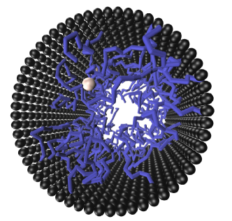
We study the diffusion of tracer particles inside a cylindrical channel grafted with polymeric chains from the inside. The polymeric chains consists of mutually repulsive monomers connected with springs. In the field of polymer science, this is a typical model for a polymer in a good solvent Grest and Kremer (1989). Tracers are added to the system and the impact of their interaction with the polymer chain on their diffusional property is studied. The thermal effects of the surrounding water is replaced by a standard LAngevin thermostat. In this sense it is a solvent–implicit model where hydrodynamic effects are neglected.
From a biological perspective this model, although being very simple, is inspired by the brush model of the central plug of NPC Patel et al. (2007). Nucleoporins rich in hydrophobic units form a brush–like structure Lim et al. (2006) and it is believed that proteins being transported through the central plug bind to these nucleoporins Frey and Gölich (2007); Herrmann et al. (2009). These bindings are believed to be hydrophobic in nature, and each binding is in the range of Moussavi-Baygi et al. (2011a, b).
To mimic the aspect of crowding we chose a repulsive interaction between all chain monomers and the tracer particles. To investigate the role of interaction we incorporate a Lennard–Jones potential between the tracer and the monomers of which we vary the interaction strength in a range from corresponding to the interaction strength of hydrophobic contacts between proteins and nucleoporins in the NPC. The resulting diffusive behaviour is characterized by investigating the mean–square–displacement for the tracer on different timescales.
The paper is structured as follows. In section II, the model, the simulation methods and the classification of observed diffusion processes are discussed. In Section IV the results and interpretations for the different models are given. The paper ends with the conclusions in section IV.
II Model and Method
The model described in the following section is evaluated using molecular dynamics. We use a generic unit system in which the Lennard–Jones parameter is chosen as the unit of length, the temperature multiplied with Boltzmann’s constant as the unit of energy and mass is measured in units of the mass of the monomers in the chain molecules. All quantities in the following are expressed in this unit system.
A cylinder with height and radius is created from particles with a diameter and a mutual distance of 1 so that a closed cylindrical surface is formed on which each particle has four next neighbors. These particles, we call them wall particles, are fixed in space, i. e. their equations of motions are not integrated, independent of the force acting on them. The cylinder axis is chosen to be the axis. Each of the polymeric chains is made of monomers, connected through a finite extensible nonlinear elastic (FENE) potential:
| (1) |
Here is the force constant and is the maximum displacement of the bond. In the simulation the parameters are chosen to be , , . The cylindrical surface is grafted randomly with polymeric chains by fixing the end monomer of each of the chains to one of the wall particles.
The repulsive interaction between the monomers themselves and with the wall is modeled via the purely repulsive variant of the Lennard-Jones potential also known as the Weeks-Chandler-Andersen (WCA) potential Weeks et al. (1971):
| (2) |
with and .
The tracer particles are of the same size and of the same mass as the monomers. They interact with the wall particles by means of the WCA potential, and with the grafted polymers either via the WCA potential (for simple crowding), or by an attractive (=“usual”) Lennard-Jones potential:
| (3) |
where the cut off radius is fixed at and the attraction strength is varied.
The degree of crowding is characterized by the grafting density , i. e. the number of chains per unit are of the cylinder. A typical value of chains corresponds to .
A typical representation of the investigated system is shown in Fig. 1 by using VMD Humphrey et al. (1996) .
We performed five independent simulations for each data point with 2 million timesteps each in order to allow to estimate the statistical errors indicated in all figures. All simulations are carried out with the Langevin thermostat, thus solving the full phase space stochastic equation of motion known as the Langevin equation of motion. Periodic boundary conditions along the cylindrical axis are used throughout the simulation and the velocity Verlet algorithm is used with a time step of .
For a particle with mass and the position co-ordinate (where stands for the the particle), interacting with all other particles () through a general potential the full phase space (underdamped) Langevin equation of motion reads as
| (4) |
where
| (5) |
is the frictional drag acting on the particle with friction coefficient , is the random force acting on the particle. The first moment of the random force is chosen to be zero:
| (6) |
The magnitude of the random forces obeys
| (7) |
where is the Boltzmann constant and is the temperature of the Langevin thermostat.
The delta function in time between the random forces in the above equation ensures the spectrum of the random force to be white, corresponding to a Markovian process in phase space with no memory. What is that: This also means dissipation is Ohmic. Throughout the simulation hydrodynamic interactions are neglected. With the above choice of random forces a fluctuation–dissipation theorems, that connects the diffusion coefficient and the friction coefficient holds: The diffusion coefficient of a single particle, up to a factor is the inverse of the friction coefficient: . In all simulations we apply a friction coefficient of and the diffusion coefficients reported in the paper are given in units of , being also 1 in our unit system.
The molecular dynamics simulation have been carried out with the help of the simulation package ESPResSo Limbach et al. (2006) in version 3.1 Arnold et al. (2012).
To describe the diffusion of tracer particles it is a common practice to use the mean square displacement (MSD), as a measure, where is the displacement of the particle at time from its initial position at . The angular bracket denotes an average over all possible realizations of the process. Under the time translational invariance this quantity is independent of and this index can be dropped, thus can also be interpreted as the lag time. For ergodic systems, the average over realizations can be replaced by an ensemble average. In case of normal diffusion, MSD increases linearly with time and in dimension reads as , where is the diffusion coefficient of the tracer particle. On the other hand, a process where the MSD is not linear in time but proportional to with is referred to as anomalous or non-Fickian diffusion Sokolov (2012). Here is the diffusion exponent and if , the process is called subdiffusive and if , the process is called superdiffusive. For anomalous diffusion there is no well defined diffusion coefficient. While the definition given above is very helpful for isotropic environments, in anisotropic environments the diffusion constant needs to be replaced by a diffusion tensor. As we are only interested in the diffusive behaviour along the cylinder axis (=–axis), we measure the MSD in direction:
| (8) |
corresponding to the component of the diffusion tensor. Throughout the paper always represents the tracer diffusion coefficient along the cylinder axis and MSD is the mean square displacement of the tracer along the cylinder axis.
III Results
In this section, we present the results of the MD simulations. We compute the MSD of the tracer and analyze the resulting transient diffusive behaviour with respect to the question if anomalous diffusion appears and try to elucidate the reasons for this by comparison to reference systems.
To explore the role of chain connectivity we compare all our results to a system when the chain monomers are not connected via the FENE potential, but free to move thus forming a gas or a ”sea“ of independent particles. The other reference is a system in which the grafted chains are moving much slower than the tracer particle, we call this a frozen background. For brevity we characterize the four possible combinations by the keywords pairs (chains/particles) and (mobile/frozen). We will show that both properties, the chain connectivity and the background motion, have a distinct impact on the diffusion properties of the tracer particles.
First we discuss the structure of the polymer coating: The radius of gyration of the chains is under all reported conditions and the mutual distance between grafting points is smaller also for the smallest grafting densities. Thus we are in the regime where the grafted chains overlap considerably. In Fig. LABEL:fig:rhomandgamma we report the local volume fractions of chain monomers, i.e. the fraction of the volume locally occupied by monomers, assuming they are spheres of diamter . The local volume fractions of up to 10% and more also indicate that locally the crowding is significant: Under these conditions mean free paths are to be expected.
III.1 Repulsive Tracer–Chain Interaction
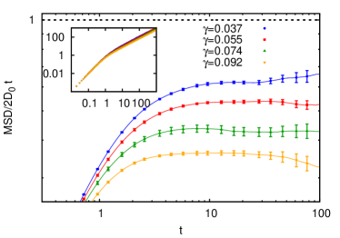
We now discuss the situation where the interaction between tracer particles and grafted chains is purely repulsive. We perform simulations with different numbers of grafted chains () hence different grafting densities () and calculate the mean square displacement (MSD). We investigate four grafting densities seen in Fig. 3. In the double-logarithmic plot of the MSD, all lines nearly collapse (seen in the inset of Fig. 3). We thus plot the MSD of the tracer along the cylinder axis divided by , where is the free diffusion coefficient along the cylinder axis. Plotting this reduced MSD allows to see the different characteristics of diffusion. On the timescale a ballistic regime where the MSD is proportional to the square of is observed, corresponding to a linear increase of the reduced MSD, and on longer timescales the diffusion becomes normal. As depicted in Fig. 3 the mean square displacement of the tracer particles shows no sign of anomalous diffusion for all investigated grafting densities.
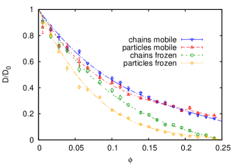
The long time diffusion coefficient is reduced compared to a freely diffusing particle by collisions with the polymer chains. For low volume fractions of polymer chains we thus expect the diffusion coefficient to be reduced by a factor proportional to the (average) volume fraction, thus the probability to collide with a monomer per unit time. Neither the chain connectivity nor the question if the background moves on the same velocity scale as the tracer particles significantly influences this effect. In Fig. 4 we report the dependence of the diffusion coefficient on the monomer volume fraction, . In all cases the tracer diffusion coefficient decreases similarly with increasing monomer volume fraction. If the monomers are connected and form chains, the impact on the diffusion coefficient is lower, as the volume from which the tracer is excluded is smaller. Also notice that the diffusion becomes faster with a mobile background than with a frozen background. This is because in the frozen background the tracer practically collides with an infinite mass and is more likely to get bounced back to its initial position. At higher volume fractions the topology of the background becomes less important. As can be seen from the Fig. 4, the two data sets corresponding to frozen particles and chains coincide at higher volume fractions as do the data sets for mobile particles and chains.
III.2 Attractive Tracer-Chain Interaction
The dynamics of a tracer particle which interacts with the chains through an attractive Lennard-Jones potential as defined in (Eq.(3)) is more complex and shows interesting features that are missing in the case of a purely repulsive interaction. Very different modes of motion are important in case of attractive polymer–tracer interaction: An attractive tracer can get attached to one of the chains, follow the chains movement, move along the chain, hop from one chain to the other or hop back to the same chain. It can also move back to the bulk where it executes normal diffusion. The emerging process is a combination of all these modes of transport and should strongly depend on the strength of attraction between the tracer and the chain and also on the density of the grafted chains. We do not expect anomalous diffusion on long time scales, but see this rather as a transient phenomenon. This is what one would expect as long as the noise is white and the background is translationally invariant. In the simulations the strength of attraction between the tracer and the polymer () is varied from to . The choice of this range of attraction is inspired by the fact that during the transport through NPC proteins bind to the nucleoporins through hydrophobic contacts which are in the range of . Moussavi-Baygi et al. (2011b).
First we investigate the influence of the attractive interaction on the position where the tracer can be found. In Fig. LABEL:fig:rhomandrhot the probability density to find the tracer at a given distance from the cylinder axis for different attraction strength at a fixed grafting density 0.055 is reported. Notice that the probability of finding the tracer along the radial direction () changes profoundly on making the tracer–chain interaction attractive. In case of purely repulsive tracers, is high at the center of the cylinder () and gradually decreases towards the chains and vanishes at the cylinder wall (). On the other hand in case of attractive tracers the density profile is qualitatively different. The tracer has a low density around the center of the cylinder () and has a high density close to the chains. As expected the attractive interaction significantly pushes the tracers from the center of the cylinder towards the area of the grafted chains. For comparison we also insert the density profile of chain monomers.
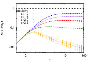
Increasing the attraction strenght between the tracer particle and the chains in general slows down the diffusion. In table 1 we report the long time diffusion coefficients obtained for different attraction strengths () at the grafting density . When the attraction strength is increased, a subdiffusive regime for an intermediate time period is observed in the plot of the reduced MSD against (Fig. 6) for .
| 1.0 | 0.38 0.02 |
|---|---|
| 1.5 | 0.23 0.01 |
| 2.0 | 0.125 0.005 |
| 2.5 | 0.074 0.003 |
We give the following interpretation: The energy landscape seen by a tracer particle for a fixed configuration of the polymer chain has minima in which the particle gets trapped on these intermediate timescales. These minima are especially deep in vicinity of kinks in the polymer chain, where the attractive regions of several monomers overlap. This also occurs in vicinity of points, where two chains closely pass by each other. When a particle gets trapped, it moves together with the monomers responsible for the trapping. Since the chains are connected the corresponding displacement will only be small. The structure of the polymer chains itself is subject to Brownian motion, and this motion eventually helps the tracer to escape the traps.
To justify this interpretation we perform independent control experiments with a system where the chain monomers are not connected (a sea of particles) and investigate the impact of strongly slowing down the motion of the background, hence look the motion of the tracer particle on a frozen background. We report here only the results for attraction strength , but qualitatively the results that were obtained for the other investigated attraction strengths of 1, 1.5 and 2.5 are similar.
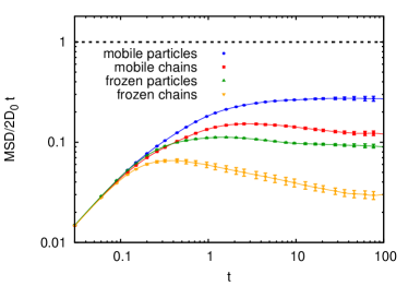
The observed reduced MSDs in all four cases are shown in Fig. 7. By giving up the chain structure of the environment we expect to facilitate diffusion as the deep traps where the attractive regions of several monomers overlap are less likely as entropy drives them away from each other. The long time diffusion coefficient is increased by a factor of and the reduced MSD exhibits no subdiffusive behaviour any more. Fixing the position of the disconnected monomers slows down diffusion again, as this suppresses the joint diffusion of complexes of tracers and monomers, but merely stops the tracers until they can escape from their traps. For this stopping effect is so pronounced that a transient subdiffusive behaviour occurs (corresponding to the negative slope of the green curve in Fig. 7), but for weaker attraction it disappears.
Freezing the structure of the chains does not change the distribution of traps, but their lifetimes. Trapped particles have to escape by their own Brownian motion and the random motion of the chains does no more facilitate their escape due to random displacements of the traps. It is seen that a substantially more pronounced subdiffusive regime occcurs. The long time diffusion coefficient is decreased by by freezing the background. This observation qualitatively supports the proposition of Bickel and Bruinsma Bickel and Bruinsma (2002). They showed with a very simple model for the transport across NPC that the fluctuating network of chains in the background acts as an extra noise and actually enhances the diffusion of a protein immersed in it as compared to in a frozen network.
IV Conclusions
In this work, we have investigated the process of diffusion of tracer particles inside a crowded cylindrical channel. Our coarse grained simulations show that if the interaction between the environment and the tracer particle is purely repulsive, the diffusion is slowed down significantly: Up to 80% if the background moves, up to 95% if the background is frozen. No subdiffusive behaviour is observed on any timescale. If the tracer interaction, however, is only weakly attractive (1-2.5 ) we observe a stronger slowdown than in the repulsive case, and also a pronounced subdiffusive regime. The subdiffusive regime is enlarged by freezing the background, corresponding to a much slower motion of the background particles. These findings are summarized in table 2.
Our findings suggest that the subdiffusive behaviour observed by Lowe et. al.Lowe et al. (2010) for the diffusion of tracers in the NPC is not caused by crowding from a polymer brush alone. Our results clearly demonstrate that simple attractive interactions between the crowding polymer-brush and the transported particles are sufficient to observe transient subdiffusive behaviour. The presence of attractive interactions were recently revealed in the work of Lim et al.Lim et al. (2007) who could show that proteins that help transporting cargo through the NPC, do have an attractive interaction with the nucleoporins polymers in the NPC. It, however, remained unanswered if the nucleoporins form a gel rather than a brush phase and what consequences this has for the cargo transport through the NPC. In the future we want to refine and extend our presented model into this direction.
| Chains | Transient subdiffusion at intermediate attraction | Transient subdiffusion at intermediate attraction |
|---|---|---|
| Particles | No subdiffusion | Transient subdiffusion at high attraction |
V Acknowledgments
Illuminating discussion with Dr. Olaf Lenz and Dr. Felix Höfling are gratefully acknowledged . We also thank Volkswagenstiftung and the Collaborative Research Centres 716 (SFB 716) for providing the necessary financial funds.
References
- Sokolov (2012) I. M. Sokolov, Soft Matter DOI: 10.1039/C2SM25701G, Advanced Article (2012).
- Fakhri et al. (2010) N. Fakhri, F. C. Mackintosh, B. Lounis, L. Cognet, and M. Pasquali, Science 330, 1804 (2010).
- Raccis et al. (2011) R. Raccis, R. Roskamp, I. Hopp, B. Menges, K. Koynor, U. Jonas, W.Knoll, H. J. Butt, and G. Fytas, Soft Matter 7, 7042 (2011).
- Fillippidi et al. (2007) E. Fillippidi, V. Michalidou, B. Loppinet, J. Rühe, and G. Fytas, Langmuir 23, 5139 (2007).
- Höfling et al. (2011) F. Höfling, K.-U. Bamberg, and T. Franosch, Soft Matter 7, 1358 (2011).
- Wong et al. (2004) I. Wong, M. L. Gardel, D. R. Reichman, E. R. Weeks, M. T. Valentine, A. R. Bausch, and D. A. Weitz, Phys. Rev. Lett. 92, 178101 (2004).
- Jeon et al. (2011) J.-H. Jeon, V. Tejedor, S. Burov, E. Barkai, C. Selhuber-Unkel, K. Berg-Sørensen, L. Oddershede, and R. Metzler, Phys. Rev. Lett. 106, 048103 (2011).
- Frey et al. (2006) S. Frey, R. P. Richter, and D. Görlich, Science 314, 815 (2006).
- Gorman et al. (2007) J. Gorman, A. Chowdhury, J. A. Surtees, J. Shimada, D. R. Reichman, E. Alani, and E. C. Greene, Molecular Cell 28, 359 (2007).
- van den Broek et al. (2008) B. van den Broek, M. A. Lomholt, S.-M. J. Kalisch, R. Metzler, and G. J. L. Wuite, Proc. Nalt. Acad. Sci. 105, 15738 (2008).
- Roosen-Runge et al. (2011) F. Roosen-Runge, M. Hennig, F. Zhang, R. M. J. Jacobs, M. Sztucki, H. Schober, T. Seydel, and F. Schreiber, Proc. Natl. Acad. Sci. 108, 11815 (2011).
- Bayliss et al. (1999) R. Bayliss, K. Ribbeck, D. Akin, H. kent, C. M. Feldherr, D. Görlich, and M. Stewart, J. Mol. Biol. 293, 579 (1999).
- Alberts et al. (2002) B. Alberts, A. Johnson, J. Lewis, M. Raff, K. Roberts, and P. Walter, Molecular Biology of the Cell, fourth edition (Gerland Publishing Inc. New York & London, 2002).
- Cohen et al. (2011) J. A. Cohen, A. Chaudhury, and R. Golestanian, Phys. Rev. Lett. 107, 238102 (2011).
- Isgro and Schulten (2007) T. A. Isgro and K. Schulten, J. Mol. Biol 366, 330 (2007).
- Bird and Baker (2011) S. P. Bird and L. A. Baker, Biomacromolecules 12, 3119 (2011).
- Zilman et al. (2007) A. Zilman, S. Talia, B. Chait, M. Rout, and M. Magnasco, PLoS Computational Biology 3, 1281 (2007).
- Kowalczyk et al. (2011) S. W. Kowalczyk, L. Kapinos, T. R. Blosser, T. Magalhaes, P. van Nies, R. Y. H. Lim, and C. Dekker, Nature Nanotechnology 6, 433 (2011).
- Frey and Gölich (2007) S. Frey and D. Gölich, Cell 130, 512 (2007).
- Elbaum (2006) M. Elbaum, Science 314, 766 (2006).
- Lim et al. (2006) R. Lim, N.-P. Huang, J. Köser, J. Deng, K. H. Aaron-Lau, K. Schwarz-Herion, B. Fahrenkrog, and U. Aebi, Proc. Natl. Acad. Sci. USA. 103, 9512 (2006).
- Guigas et al. (2007) G. Guigas, C. Kalla, and M. Weiss, FEBS Lett 581, 5094 (2007).
- Lowe et al. (2010) A. R. Lowe, J. J. Siegel, P. Kalab, M. Siu, and J. T. Liphardt, Nature 467, 600 (2010).
- Xu et al. (2011) Q. Xu, L. Feng, R. Sha, N. C. Seeman, and P. M. Chaikin, Phys. Rev. Lett. 106, 228102 (2011).
- Tabatabaei et al. (2011) F. Tabatabaei, O. Lenz, and C. Holm, Colloid Polym. Sci 289, 523 (2011).
- Amsden (1998) B. Amsden, Macromolecules 31, 8382 (1998).
- Cherdhirankorn et al. (2009) T. Cherdhirankorn, A. Best, K. Koynov, K. Peneva, K. Müllen, and G. Fytas, J. Phys. Chem. B 113, 3355 (2009).
- Viehman and Schweizer (2008) D. C. Viehman and K. S. Schweizer, J. Phys. Chem. B 112, 16110 (2008).
- Grest and Kremer (1989) G. S. Grest and K. Kremer, Phys. Rev. A 33, 3628 (1989).
- Patel et al. (2007) S. S. Patel, B. J. Belmont, J. M. Sante, and M. F. Rexach, Cell 129, 83 (2007).
- Herrmann et al. (2009) M. Herrmann, N. Neuberth, J. Wissler, J. Pérez, D. Gradl, and A. Naber, Nano Letters 9, 3330 (2009).
- Moussavi-Baygi et al. (2011a) R. Moussavi-Baygi, Y. Jamali, R. Karimi, and M. R. K. Mofrad, Biophys J. 100, 1410 (2011a).
- Moussavi-Baygi et al. (2011b) R. Moussavi-Baygi, Y. Jamali, R. Karimi, and M. R. K. Mofrad, PLoS Computational Biology 7, 1 (2011b).
- Weeks et al. (1971) J. D. Weeks, D. Chandler, and H. C. Andersen, J. Chem. Phys. 54, 5237 (1971).
- Humphrey et al. (1996) W. Humphrey, A. Dalke, and K. Schulten, J. Molec. Graphics 14, 33 (1996).
- Limbach et al. (2006) H. J. Limbach, A. Arnold, B. A. Mann, and C. Holm, Comput. Phys. Commun. 174, 704 (2006).
- Arnold et al. (2012) A. Arnold, O. Lenz, S. Kesselheim, R. Weeber, F. Fahrenberger, D. Roehm, P. Košovan, and C. Holm, in Proceedings of the Sixth International Workshop on Meshfree Methods for Partial Differential Equations, edited by M. Griebel, C. Rieger, and M. A. Schweitzer (Springer, Berlin, Germany, 2012), Lecture Notes in Computational Science and Engineering.
- Bickel and Bruinsma (2002) T. Bickel and R. Bruinsma, BioPhys. J. 83, 3079 (2002).
- Lim et al. (2007) R. Y. H. Lim, B. Fahrenkrog, J. Köser, K. Schwarz-Herion, J. Deng, and U. Aebi, Science 318, 640 (2007).