Amino-acid-dependent main-chain torsion-energy terms for protein systems
Abstract
Many commonly used force fields for protein systems such as AMBER, CHARMM, GROMACS, OPLS, and ECEPP have amino-acid-independent force-field parameters of main-chain torsion-energy terms. Here, we propose a new type of amino-acid-dependent torsion-energy terms in the force fields. As an example, we applied this approach to AMBER ff03 force field and determined new amino-acid-dependent parameters for and angles for each amino acid by using our optimization method, which is one of the knowledge-based approach. In order to test the validity of the new force-field parameters, we then performed folding simulations of -helical and -hairpin peptides, using the optimized force field. The results showed that the new force-field parameters gave structures more consistent with the experimental implications than the original AMBER ff03 force field.
I Introduction
Computer simulations of protein folding into native structures can be achieved when both of the following two requirements are met: (1) potential energy functions (or, force fields) for the protein systems are sufficiently accurate and (2) sufficiently powerful conformational sampling methods are available. Professor Harold A. Scheraga has been one of the most important pioneers in studies of both of the above requirements Liwo et al. (2008); Scheraga (2011). By the developments of the generalized-ensemble algorithms (for reviews, see, e.g., Refs. Hansmann and Okamoto (1999); Mitsutake, Sugita, and Okamoto (2001)) and related methods, Requirement (2) seems to be almost fulfilled. In this article, we therefore concentrate our attention on Requirement (1).
There are several well-known all-atom (or united-atom) force fields, such as AMBER Cornell et al. (1995); Kollman et al. (1997a); Wang, Cieplak, and Kollman (2000); Hornak et al. (2006); Duan et al. (2003a), CHARMM MacKerell Jr et al. (1998); MacKerell Jr, Feig, and Brooks III (2004), OPLS Jorgensen, Maxwell, and Tirado-Rives (1996); Kaminski et al. (2001), GROMOS van Gunsteren et al. (1996); Oostenbrink et al. (2004), GROMACS Berendsen, van der Spoel, and van Drunen (1995); Lindahl, Hess, and van der Spoel , and ECEPP Némethy et al. (1992); Arnautova, Jagielska, and Scheraga (2006). Generally, the force-field parameters are determined based on experimental results for small molecules and theoretical results using quantum chemistry calculations of small peptides such as alanine dipeptide.
In a force field, the potential energy is usually composed of the bond-stretching term, the bond-bending term, the torsion-energy term, and the nonbonded energy term. In these energy terms, it is known that the torsion-energy term is the most problematic. For instance, the ff94 Cornell et al. (1995) and ff96 Kollman et al. (1997b) versions of AMBER differ only in the main-chain torsion-energy parameters. Nevertheless, the secondary-structure-forming tendencies of the two force fields are quite different Yoda, Sugita, and Okamoto (2004a, b); Sakae and Okamoto (2003, 2004a, 2004b). Therefore, many researchers have studied this main-chain torsion-energy terms and their force-field parameters. For instance, newer force-field parameters of the main-chain torsion-energy terms about and angles have been developed, which are, e.g., AMBER ff99SB Hornak et al. (2006), AMBER ff03 Duan et al. (2003a), CHARMM22/CMAP MacKerell Jr, Feig, and Brooks III (2004) and OPLS-AA/L Kaminski et al. (2001). The methods of the force-field refinement thus mainly concentrate on the torsion-energy terms. These modifications of the torsion energy are usually based on quantum chemistry calculations Simmerling, Strockbine, and Roitberg (2002); Duan et al. (2003b); Iwaoka and Tomoda (2003); MacKerell Jr, Feig, and Brooks III ; Kamiya et al. (2005) or NMR experimental results Best and Hummer (2009); Mittal and Best (2010).
We have also proposed a new main-chain torsion-energy term, which is represented by a double Fourier series in two variables, the main-chain dihedral angles and Sakae and Okamoto (2006, 2010a). This expression gives a natural representation of the torsion energy in the Ramachandran space Ramachandran and Sasisekharan (1968) in the sense that any two-dimensional energy surface periodic in both and can be expanded by the double Fourier series. We can then easily control secondary-structure-forming tendencies by modifying the main-chain torsion-energy surface. We have presented preliminary results for AMBER ff94 and AMBER ff96 Sakae and Okamoto (2006, 2010a). Moreover, we have introduced several optimization methods of force-field parameters Sakae and Okamoto (2003, 2004a, 2004b, 2010b, 2010c). These methods are based on the minimization of some score functions by simulations in the force-field parameter space, where the score functions are derived from the protein coordinate data in the Protein Data Bank (PDB). One of the score functions consists of the sum of the square of the force acting on each atom in the proteins with the structures from the PDB Sakae and Okamoto (2003, 2004a, 2004b). Other score functions are taken from the root-mean-square deviations between the original PDB structures and the corresponding minimized structures Sakae and Okamoto (2010b, c).
In this article, we propose a new type of the main-chain torsion-energy terms for protein systems, which can have amino-acid-dependent force-field parameters. As an example of this formulation, we applied this approach to the AMBER ff03 force field and determined new amino-acid-dependent main-chain torsion-energy parameters for (N-Cα-C-N) and (Cβ-Cα-C-N) by using our optimization method in Refs Sakae and Okamoto (2003, 2004a, 2004b).
In section 2 the details of the new main-chain torsion-energy terms are given. In section 3 the results of applications of the method to AMBER ff03 force field and those of folding simulations of two peptides are presented. Section 4 is devoted to conclusions.
II Methods
II.1 Amino-acid-dependent force-field parameters
The existing force fields for protein systems such as AMBER Cornell et al. (1995); Kollman et al. (1997a); Wang, Cieplak, and Kollman (2000); Hornak et al. (2006); Duan et al. (2003a), CHARMM MacKerell Jr et al. (1998); MacKerell Jr, Feig, and Brooks III (2004), and OPLS Jorgensen, Maxwell, and Tirado-Rives (1996); Kaminski et al. (2001), etc. use essentially the same functional forms for the potential energy except for minor differences. The conformational potential energy can be written as, for instance,
| (1) |
Here, , , , and represent the bond-stretching term, the bond-bending term, the torsion-energy term, and the nonbonded energy term, respectively. Each force field has similar but slightly different parameter values. For example, the torsion-energy term is usually given by
| (2) |
where the first summation is taken over all dihedral angles (both in the main chain and in the side chains), is the number of waves, is the phase, and is the Fourier coefficient. Namely, the energy term has and as force-field parameters.
We can further write the torsion-energy term as
| (3) |
where and are the torsion-energy terms for dihedral angles around main-chain bonds and around side-chain bonds, respectively. Examples of the dihedral angles in are (C-N-Cα-C), (N-Cα-C-N), (Cβ-Cα-N-C), (Cβ-Cα-C-N), and (Cα-C-N-Cα). The force-field parameters in can readily depend on amino-acid residues. However, those in are usually taken to be independent of amino-acid residues and the common parameter values are used for all the amino-acid residues (except for proline). This is because the amino-acid dependence of the force field is believed to be taken care of by the very existence of side chains. In Table I, we list examples of the parameter values for (N-Cα-C-N) and (Cβ-Cα-C-N) in general AMBER force fields.
However, this amino-acid independence of the main-chain torsion-energy terms is not an absolute requirement, because we are representing the entire force field by rather a small number of classical-mechanical terms. In order to reproduce the exact quantum-mechanical contributions, one can introduce amino-acid dependence on any force-field term including the main-chain torsion-energy terms. Hence, we can generalize in Eq. (3) from the expression in Eq. (2) to the following amino-acid-dependent form:
| (4) |
where () is the label for the 20 kinds of amino-acid residues and are dihedral angles around the main-chain bonds in the -th amino-acid residue.
II.2 Optimization method for force-field parameters
In the previous subsection, we have generalized the main-chain torsion-energy term so that its parameters are amino-acid dependent. The question is then how to obtain optimal parameter values for this new main-chain torsion-energy term.
One method is to use the parameter optimization method that was introduced in Refs. Sakae and Okamoto (2003, 2004a, 2004b). We first retrieve native structures (one structure per protein) from PDB. We try to choose proteins from different amino-acid sequence homology as much as possible. If the force-field parameters are of ideal values, then all the chosen native structures are stable without any force acting on each atom in the molecules on the average. Hence, we expect
| (5) |
where
| (6) |
and
| (7) |
Here, is the total number of atoms in molecule , is the total potential energy for molecule , and is the force acting on atom . In reality, , and because , we can optimize the force-field parameters by minimizing with respect to these parameters in the main-chain torsion-energy term in Eq. (4). In practice, we perform a minimization simulation in the main-chain torsion-energy force-field parameter space for this minimization Sakae and Okamoto (2003, 2004a, 2004b).
III Results and Discussion
III.1 An example of the amino-acid-dependent force-field parameter optimizations
We present the results of our optimizations of the force-field parameters for the main-chain angles (N-Cα-C-N) and (Cβ-Cα-C-N) in Eq. (4). We did this for the case of AMBER ff03 force field. We determined these values for the 19 amino-acid residues except for proline.
At first, we chose 100 PDB files with resolution 2.0 Å or better, with sequence similarity of amino acid 30.0 % or lower, and with less than 200 residues (the average number of residues is 117.0) from PDB-REPRDB Noguchi et al. (1997) (see Table 2). We then refined these selected 100 structures. Generally, data from X-ray experiments do not have coordinates for hydrogen atoms. Therefore, we have to add hydrogen coordinates. Many protein simulation software packages provide with routines that add hydrogen atoms to the PDB coordinates. We used the AMBER11 program package Case et al. (2005). We thus minimized the total potential energy with respect to the coordinates for each proten conformation, where is the harmonic constraint energy term (), and is the solvation energy term. Here, is the force constant of the restriction and are the original coordinate vectors of heavy atoms in PDB. As one can see from , the coordinates of hydrogen atoms will be mainly adjusted, but unnatural heavy-atom coordinates will also be modified. We performed this minimization for all the 100 protein structures separately and obtained 100 refined structures by using (kcal/mol). As for the solvation energy term , we used the GB/SA solvent included in the AMBER program package ( and ) Onufriev, Bashford, and Case (2004); Weiser, Shenkin, and Still (1999).
For these refined protein structures, we performed the optimization of force-field parameters of and angles for AMBER ff03 force field by using the fucntion in Eq. (6) as the total potential energy function () for the Monte Carlo simulations in the parameter space. Here, we used AMBER11 Case et al. (2005) for the force calculations in Eq. (7). We have to optimize the 38 () parameters simultaneously by the simulations in 38 parameters. However, here, for simplicity, we just optimized two parameters, and , for each amino-acid residue separately, keeping the other values as the original values. In order to obtain the optimal parameters, we performed Monte Carlo simulations of two parameters ( of and ) for the 19 amino-acid residues except for proline. In Table 3, the optimized parameters are listed.
III.2 Test simulations with two peptides
In order to check the force-field parameters obtained by our optimization method, we performed the folding simulations using two peptides, namely, C-peptide of ribonuclease A and the C-terminal fragment of the B1 domain of streptococcal protein G, which is sometimes referred to as G-peptide Honda, Kobayashi, and Munekata (2000). The C-peptide has 13 residues and its amino-acid sequence is Lys-Glu--Thr-Ala-Ala-Ala-Lys+-Phe-Glu-Arg+-Gln-His+-Met. This peptide has been extensively studied by experiments and is known to form an -helix structure Shoemaker et al. (1985); Osterhout Jr. et al. (1989). Because the charges at peptide termini are known to affect helix stability Shoemaker et al. (1985); Osterhout Jr. et al. (1989), the N and C termini of the peptide was blocked with acetyl and N-methyl groups, respectively. The G-peptide has 16 residues and its amino-acid sequence is Gly-Glu--Trp-Thr-Tyr-Asp--Asp--Ala-Thr-Lys+-Thr-Phe-Thr-Val-Thr-Glu-. The termini were kept as the usual zwitter ionic states, following the experimental conditions Honda, Kobayashi, and Munekata (2000); Blanco, Rivas, and Serrano (1994); Kobayashi et al. (1995). This peptide is known to form a -hairpin structure by experiments Honda, Kobayashi, and Munekata (2000); Blanco, Rivas, and Serrano (1994); Kobayashi et al. (1995).
For the folding simulations, we used replica-exchange molecular dynamics (REMD) Sugita and Okamoto (1999). REMD is one of the generalized-ensemble algorithms, and has high conformational sampling efficiency by allowing configurations to heat up and cool down while maintaining proper Boltzmann distributions. We used the AMBER11 program package Case et al. (2005). The unit time step was set to 2.0 fs, and the bonds involving hydrogen atoms were constrained by SHAKE algorithm Ryckaert, Ciccotti, and Berendsen (1977). Each simulation was carried out for 30.0 ns (hence, it consisted of 15,000,000 MD steps) with 16 replicas by using Langevin dynamics. The exchange procedure for each replica were performed every 3,000 MD steps. The temperature was distributed exponentially: 650, 612, 577, 544, 512, 483, 455, 428, 404, 380, 358, 338, 318, 300, 282, and 266 K. As for solvent effects, we used the GB/SA model in the AMBER program package ( and ) Onufriev, Bashford, and Case (2004); Weiser, Shenkin, and Still (1999). The initial conformations for each peptide were fully extended ones for all the replicas. The REMD simulations were performed with different sets of randomly generated initial velocities for each replica.
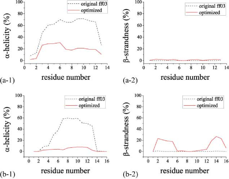
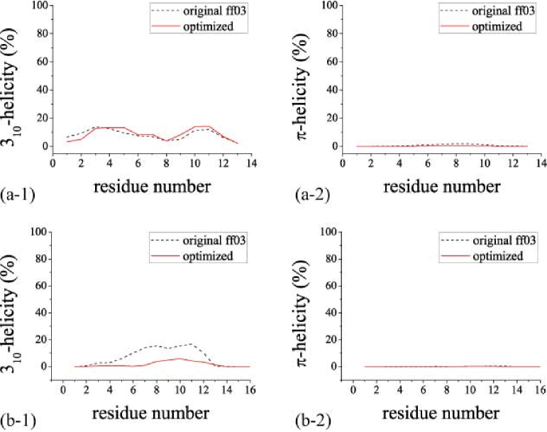
In Fig. 1, -helicity and -strandness of two peptides obtained from the REMD simulations are shown. We checked the secondary-structure formations by using the DSSP program Kabsch and Sander (1983), which is based on the formations of the intra-main-chain hydrogen bonds. As is shown in Fig. 1, for the original AMBER ff03 force field, the -helicity is clearly higher than the -strandness not only in C-peptide but also in G-peptide. Namely, the original AMBER ff03 force field clearly favors -helix and does not favor -structure. On the other hand, for the optimized force field, in the case of C-peptide, the -helicity is higher than the -strandness, and in the case of G-peptide, the -strandness is higher than the -helicity. We conclude that these results obtained from the optimized force field are in better agreement with the experimental results in comparison with the original force field. In Fig. 2, 310-helicity and -helicity of two peptides obtained from the REMD simulations are shown. For 310 helicity, there is no large difference for both force fields in C-peptide, and in the case of G-peptide, the value of the optimized force field slightly decreases in comparison with the original force field. -helicity has almost no value in the both cases of the original and optimized force fields in two peptides.
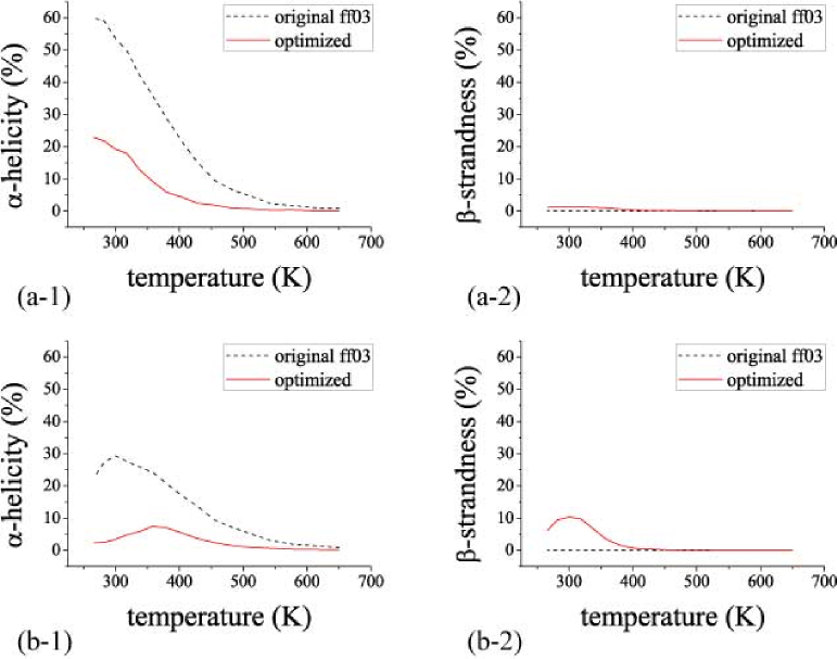
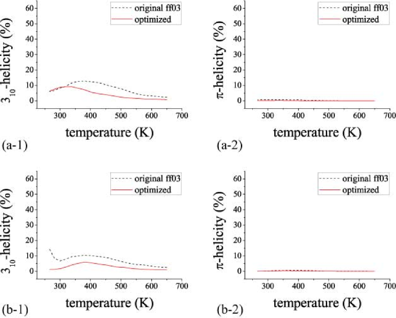
In Fig. 3, -helicity and -strandness as functions of temperature for the two peptides obtained from the REMD simulations are shown. For -helicity, the values of both force fields decrease gradually from low temperature to high temperature in the case of C-peptide. On the other hand, in the case of G-peptide, there are small peaks at around 300 K and 358 K for the original and optimized force fields, respectively. For -strandness, in the case of C-peptide, it is almost zero for both force fields. In the case of G-peptide, for the optimized force field, there is clearly a peak around 300 K. In Fig. 4, 310-helicity and -helicity of the two peptides as functions of temperature are shown. For 310-helicity, in the case of both peptides, the values of the optimized force field are lower than the original force field as a whole except around low temperature in C-peptide. For -helicity, it is almost zero for both force fields in the two peptides.
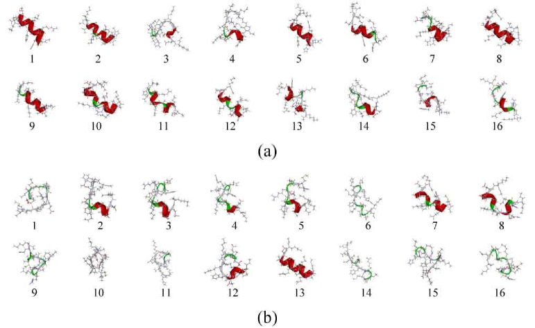
In Fig. 5, the lowest-energy conformations of C-peptide obtained from the REMD simulations in the case of the original and the optimized force fields are shown. In the case of the original force field, all the conformations have helices. No. 3 has only 310-helix, No. 13 has both -helix and 310-helix, and the rest of the conformations have only -helix. In the case of the optimized force field, seven conformations (Nos. 2, 4, 5, 7, 8, 12, and 13) have helices. Nos. 2, 5, 7, 13 have only -helix, Nos. 4, 12 have only 310-helix, and No. 8 has both -helix and 310-helix. Additionally, there is one -bridge structure in No. 10. In Fig. 6, the lowest-energy conformations of G-peptide are shown. In the case of the original force field, all the conformations except for No. 4 have helices. No. 6 has both -helix and 310-helix, Nos. 5, 7, 11 have only 310-helix, and the rest have only -helix. In the case of the optimized force field, Nos. 11 and 12 have -helix, No. 8 has 310-helix, No. 5 has -bridge, and Nos. 7, 9, 10 have -strand. These results clearly show that the optimized force field favors helix structure much less than the original force field, and, additionally, in the case of G-peptide, slightly favors -structure.
IV Conclusions
The main-chain torsion-energy terms are the most problematic terms in the force field for protein systems. We therefore concentrate our attention on these terms in order to obtain optimal protein force field. In this article, we proposed amino-acid-dependent main-chain torsion-energy terms in the force field for protein systems. This generalization gives more freedom to the force-field optimization problem. In principle, we can introduce amino-acid dependence on any force-field term. The present work introduced this dependence on even the main-chain torsion-energy terms, which previously had been treated independent of the amino-acid residue type.
As an example of the present general formalism, we modified the AMBER ff03 force field so that the parameters of the main-chain and angles may be amino-acid dependent except for proline (hence, 38 parameters were optimized). Although preliminary because we did not optimize the 38 parameters simulataneously, our optimized parameters already gave structures more consistent with the experimental implications than the original AMBER force field in the folding simulations of two small peptides.
We can easily apply the present formulations to other popular force fields such as AMBER ff99SB, CHARMM22/CMAP, etc. This will be our future work.
Acknowledgements.
This article is dedicated to the 90-th birthday of Professor Harold A. Scheraga. The computations were performed on the computers at the Research Center for Computational Science, Institute for Molecular Science, Information Technology Center, Nagoya University, and Center for Computational Sciences, University of Tsukuba. This work was supported, in part, by the Grants-in-Aid for the Academic Frontier Project, “Intelligent Information Science”, for Scientific Research on Innovative Areas (“Fluctuations and Biological Functions” ), and for the Next Generation Super Computing Project, Nanoscience Program and Computational Materials Science Initiative from the Ministry of Education, Culture, Sports, Science and Technology (MEXT), Japan.REFERENCES
References
- Liwo et al. (2008) A. Liwo, C. Czaplewski, O. Stanislaw, and H. A. Scheraga, “Computational techniques for efficient conformational sampling of proteins,” Curr. Opin. Struct. Biol. 18, 134–139 (2008)
- Scheraga (2011) H. A. Scheraga, “Respice, adspice, and prospice,” Ann. Rev. Biophys. 40, 1–39 (2011)
- Hansmann and Okamoto (1999) U. H. E. Hansmann and Y. Okamoto, “New monte carlo algorithms for protein folding,” Curr. Opin. Struct. Biol. 9, 177–183 (1999)
- Mitsutake, Sugita, and Okamoto (2001) A. Mitsutake, Y. Sugita, and Y. Okamoto, “Generalized-ensemble algorithms for molecular simulations of biopolymers,” Biopolymers 60, 96–123 (2001)
- Cornell et al. (1995) W. D. Cornell, P. Cieplak, C. I. Bayly, I. R. Gould, J. Kenneth M. Merz, D. M. Ferguson, D. C. Spellmeyer, T. Fox, J. W. Caldwell, and P. A. Kollman, “A second generation force field for the simulation of proteins, nucleic acids, and organic molecules,” J. Am. Chem. Soc. 117, 5179–5197 (1995)
- Kollman et al. (1997a) P. A. Kollman, R. Dixon, W. Cornell, T. Fox, C. Chipot, and A. Pohorille, Computer simulations of biological systems, edited by W. F. van Gunsteren and P. K. Weiner, Vol. 3 (ESCOM, Dordrecht, 1997) pp. 83–96
- Wang, Cieplak, and Kollman (2000) J. Wang, P. Cieplak, and P. A. Kollman, “How well does a restrained electrostatic potential (resp) model perform in calculating conformational energies of organic and biological molecules?” J. Comput. Chem. 21, 1049–1074 (2000)
- Hornak et al. (2006) V. Hornak, A. Abel, R. Okur, B. Strockbine, A. Roitberg, and C. Simmerling, “Comparison of multiple amber force fields and development of improved protein backbone parameters,” Proteins 65, 712–725 (2006)
- Duan et al. (2003a) Y. Duan, C. Wu, S. Chowdhury, M. C. Lee, G. Xiong, W. Zhang, R. Yang, P. Cieplak, R. Luo, and T. Lee, “A point-charge force field for molecular mechanics simulations of proteins based on condensed-phase quantum mechanical calculations,” J. Comput. Chem. 24, 1999–2012 (2003a)
- MacKerell Jr et al. (1998) A. D. MacKerell Jr, D. Bashford, M. Bellott, J. Dunbrack, R. L., J. D. Evanseck, M. J. Field, S. Fischer, J. Gao, H. Guo, S. Ha, D. Joseph-McCarthy, L. Kuchnir, K. Kuczera, F. T. K. Lau, C. Mattos, S. Michnick, T. Ngo, D. T. Nguyen, B. Prodhom, I. Reiher, W. E., B. Roux, M. Schlenkrich, J. C. Smith, J. Stote, R.; Straub, M. Watanabe, J. Wiorkiewicz-Kuczera, D. Yin, and M. Karplus, “All-atom empirical potential for molecular modeling and dynamics studies of proteins,” J Phys Chem B 102, 3586–3616 (1998)
- MacKerell Jr, Feig, and Brooks III (2004) A. D. MacKerell Jr, M. Feig, and C. Brooks III, “Extending the treatment of backbone energetics in protein force fields: limitations of gas-phase quantum mechanics in reproducing protein conformational distributions in molecular dynamics simulations,” J. Comput. Chem. 25, 1400–1415 (2004)
- Jorgensen, Maxwell, and Tirado-Rives (1996) W. L. Jorgensen, D. S. Maxwell, and J. Tirado-Rives, “Development and testing of the opls all-atom force field on conformational energetics and properties of organic liquids,” J. Am. Chem. Soc. 118, 11225–11236 (1996)
- Kaminski et al. (2001) G. A. Kaminski, R. A. Friesner, J. Tirado-Rives, and W. L. Jorgensen, “Evaluation and reparametrization of the opls-aa force field for proteins via comparison with accurate quantum chemical calculations on peptides,” J. Phys. Chem. B 105, 6474–6487 (2001)
- van Gunsteren et al. (1996) W. F. van Gunsteren, S. R. Billeter, A. A. Eising, P. H. Hünenberger, P. Krüger, A. E. Mark, W. R. P. Scott, and I. G. Tironi, Biomolecular Simulation: The GROMOS96 Manual and User Guide (Vdf Hochschulverlag AG an der ETH Zürich, Zürich, 1996)
- Oostenbrink et al. (2004) C. Oostenbrink, A. Villa, A. E. Mark, and W. F. van Gunsteren, “A biomolecular force field based on the free enthalpy of hydration and solvation: the gromos force-field parameter sets 53a5 and 53a6,” J. Comput. Chem. 25, 1656–1676 (2004)
- Berendsen, van der Spoel, and van Drunen (1995) H. J. C. Berendsen, D. van der Spoel, and R. van Drunen, “Gromacs: a message-passing parallel molecular dynamics implementation,” Comput. Phys. Commun. 91, 43–56 (1995)
- (17) E. Lindahl, B. Hess, and D. van der Spoel, “Gromacs 3.0: a package for molecular simulation and trajectory anaylysis,”
- Némethy et al. (1992) G. Némethy, K. D. Gibson, K. A. Palmer, C. N. Yoon, G. Paterlini, A. Zagari, S. Rumsey, and H. A. Scheraga, “Energy parameters in polypeptides. 10. improved geometrical parameters and nonbonded interactions for use in the ecepp/3 algorithm, with application to proline-containing peptides,” J. Phys. Chem. 96, 6472–6484 (1992)
- Arnautova, Jagielska, and Scheraga (2006) Y. A. Arnautova, A. Jagielska, and H. A. Scheraga, “A new force field (ecepp-05) for peptides, proteins, and organic molecules,” J. Phys. Chem. B 110, 5025–5044 (2006)
- Kollman et al. (1997b) P. A. Kollman, R. Dixon, W. Cornell, T. Fox, C. Chipot, and A. Pohorille, “Computer simulations of biological systems,” (Escom, Netherlands, 1997) Chap. The development/application of a ‘minimalist’ organic/biochemical molecular mechanic force field using a combination of ab initio calculations and experimental data, pp. 83–96
- Yoda, Sugita, and Okamoto (2004a) T. Yoda, Y. Sugita, and Y. Okamoto, “Comparisons of force fields for proteins by generalized-ensemble simulations,” Chem. Phys. Lett. 386, 460–467 (2004a)
- Yoda, Sugita, and Okamoto (2004b) T. Yoda, Y. Sugita, and Y. Okamoto, “Secondary-structure preferences of force fields for proteins evaluated by generalized-ensemble simulations,” Chem. Phys. 307, 269–283 (2004b)
- Sakae and Okamoto (2003) Y. Sakae and Y. Okamoto, “Optimization of protein force-field parameters with the protein data bank,” Chem. Phys. Lett. 382, 626–636 (2003)
- Sakae and Okamoto (2004a) Y. Sakae and Y. Okamoto, “Protein force-field parameters optimized with the protein data bank. i. force-field optimizations,” J. Theo. Comput. Chem. 3, 339–358 (2004a)
- Sakae and Okamoto (2004b) Y. Sakae and Y. Okamoto, “Protein force-field parameters optimized with the protein data bank. ii. comparisons of force fields by folding simulations of short peptides,” J. Theo. Comput. Chem. 3, 359–378 (2004b)
- Simmerling, Strockbine, and Roitberg (2002) C. Simmerling, B. Strockbine, and A. E. Roitberg, “All-atom structure prediction and folding simulations of a stable protein,” J. Am. Chem. Soc. 124, 11258–11259 (2002)
- Duan et al. (2003b) Y. Duan, C. Wu, S. Chowdhury, M. C. Lee, G. Xiong, W. Zhang, R. Yang, P. Cieplak, R. Luo, T. Lee, J. Caldwell, J. Wang, and P. Kollman, “A point-charge force field for molecular mechanics simulations of proteins based on condensed-phase quantum mechanical calculations,” J. Comput. Chem. 24, 1999–2012 (2003b)
- Iwaoka and Tomoda (2003) M. Iwaoka and S. Tomoda, “The saap force field. a simple approach to a new all-atom protein force field by using single amino acid potential (saap) functions in various solvents,” J. Comput. Chem. 24, 1192–1200 (2003)
- (29) A. D. MacKerell Jr, M. Feig, and C. L. Brooks III, “Extending the treatment of backbone energetics in protein force fields: Limitations of gas-phase quantum mechanics in reproducing protein conformational distributions in molecular dynamics simulations,” J Comput. Chem.
- Kamiya et al. (2005) N. Kamiya, Y. Watanabe, S. Ono, and J. Higo, “Amber-based hybrid force field for conformational sampling of polypeptides,” Chem. Phys. Lett. 401, 312–317 (2005)
- Best and Hummer (2009) R. B. Best and G. Hummer, “Optimized molecular dynamics force field applied to the helix-coil transition of polypeptides,” J. Phys. Chem. B 113, 9004–9015 (2009)
- Mittal and Best (2010) J. Mittal and R. B. Best, “Tackling force-field bias in protein folding simulations: Folding of villin hp35 and pin ww domains in explicit water,” Biophys. J. 99, L26–L28 (2010)
- Sakae and Okamoto (2006) Y. Sakae and Y. Okamoto, “Secondary-structure design of proteins by a backbone torsion energy,” J. Phys. Soc. Jpn. 75 (2006), 054802 (9 pages)
- Sakae and Okamoto (2010a) Y. Sakae and Y. Okamoto, “Controlling the secondary-structure-forming tendencies of proteins by a backbone torsion-energy term,” Mol. Sim. 36, 138–158 (2010a)
- Ramachandran and Sasisekharan (1968) G. N. Ramachandran and V. Sasisekharan, “Conformation of polypeptides and proteins,” Adv. Protein Chem. 23, 283–438 (1968)
- Sakae and Okamoto (2010b) Y. Sakae and Y. Okamoto, “Determination method of the balance of the secondary-structure-forming tendencies of force fields,” Mol. Sim. 36, 159–165 (2010b)
- Sakae and Okamoto (2010c) Y. Sakae and Y. Okamoto, “Optimisation of opls-ua force-field parameters for protein systems using protein data bank,” Mol. Sim. 36, 1148–1156 (2010c)
- Noguchi et al. (1997) T. Noguchi, K. Onizuka, Y. Akiyama, and M. Saito, “Pdb-reprdb: A database of representative protein chains in pdb (protein data bank),” in Proc. of the Fifth International Conference on Intelligent Systems for Molecular Biology (AAAI press, Menlo Park, CA, 1997)
- Case et al. (2005) D. A. Case, T. Cheatham, T. Darden, H. Gohlke, R. Luo, K. M. Merz, Jr., A. Onufriev, C. Simmerling, B. Wang, and R. Woods, “The amber biomolecular simulation programs,” J. Computat. Chem. 26, 1668–1688 (2005)
- Onufriev, Bashford, and Case (2004) A. Onufriev, D. Bashford, and D. A. Case, “Exploring protein native states and large-scale conformational changes with a modified generalized born model,” Proteins 55, 383–394 (2004)
- Weiser, Shenkin, and Still (1999) J. Weiser, P. S. Shenkin, and W. C. Still, “Approximate atomic surfaces from linear combinations of pairwise overlaps (lcpo),” J. Comput. Chem. 20, 217–230 (1999)
- Honda, Kobayashi, and Munekata (2000) S. Honda, N. Kobayashi, and E. Munekata, “Thermodynamics of a β-hairpin structure: evidence for cooperative formation of folding nucleus,” J. Mol. Biol. 295, 269–278 (2000)
- Shoemaker et al. (1985) K. R. Shoemaker, P. S. Kim, D. N. Brems, S. Marqusee, E. J. York, I. M. Chaiken, J. M. Stewart, and R. L. Baldwin, “Nature of the charged-group effect on the stability of the c-peptide helix,” Proc. Natl. Acad. Sci. U.S.A. 82, 2349–2353 (1985)
- Osterhout Jr. et al. (1989) J. J. Osterhout Jr., R. L. Baldwin, E. J. York, J. M. Stewart, H. J. Dyson, and P. E. Wright, “1h nmr studies of the solution conformations of an analogue of the c-peptide of ribonuclease a,” Biochemistry 28, 7059–7064 (1989)
- Blanco, Rivas, and Serrano (1994) F. J. Blanco, G. Rivas, and L. Serrano, “A short linear peptide that folds into a native stable bold beta-hairpin in aqueous solution,” Nature Struct. Biol. 1, 584–590 (1994)
- Kobayashi et al. (1995) N. Kobayashi, S. Honda, H. Yoshii, H. Uedaira, and E. Munekata, “Complement assembly of two fragments of the streptococcal protein g b1 domain in aqueous solution,” FEBS Lett. 366, 99–103 (1995)
- Sugita and Okamoto (1999) Y. Sugita and Y. Okamoto, “Replica-exchange molecular dynamics method for protein folding,” Chem. Phys. Lett. 314, 141–151 (1999)
- Ryckaert, Ciccotti, and Berendsen (1977) J.-P. Ryckaert, G. Ciccotti, and H. J. C. Berendsen, “Numerical integration of the cartesian equations of motion of a system with constraints: Moecular dynamics of n-alkanes,” J. Comput. Phys. 23, 327–341 (1977)
- Kabsch and Sander (1983) W. Kabsch and C. Sander, “Dictionary of protein secondary structure: Pattern recognition of hydrogen-bonded and geometrical features,” Biopolymers 22, 2577–2637 (1983)
| force field | (N-Cα-C-N) | (Cβ-Cα-C-N) | ||||
|---|---|---|---|---|---|---|
| ff94 | 1 | 0.75 | 2 | 0.07 | 0 | |
| 2 | 1.35 | 4 | 0.10 | 0 | ||
| 4 | 0.40 | |||||
| ff96 | 1 | 0.85 | 0 | 2 | 0.07 | 0 |
| 2 | 0.30 | 4 | 0.10 | 0 | ||
| ff99 | 1 | 1.70 | 2 | 0.07 | 0 | |
| 2 | 2.00 | 4 | 0.10 | 0 | ||
| ff99SB | 1 | 0.45 | 1 | 0.20 | 0 | |
| 2 | 1.58 | 2 | 0.20 | 0 | ||
| 3 | 0.55 | 3 | 0.40 | 0 | ||
| ff03 | 1 | 0.6839 | 1 | 0.7784 | ||
| 2 | 1.4537 | 2 | 0.0657 | |||
| 3 | 0.4615 | 3 | 0.0560 | 0 |
| fold | PDB ID | chain | PDB ID | chain | PDB ID | chain | PDB ID | chain |
|---|---|---|---|---|---|---|---|---|
| all | 1DLW | A | 1N1J | B | 1U84 | A | 1HBK | A |
| 1TX4 | A | 1V54 | E | 1SK7 | A | 1TQG | A | |
| 1V74 | B | 1DVO | A | 1HFE | S | 1J0P | A | |
| 1Y02 | A71-114 | 1IJY | A | 1I2T | A | 1G8E | A | |
| 1VKE | C | 1FS1 | A109-149 | 1D9C | A | 1AIL | A | |
| 1Q5Z | A | 1T8K | A | 1OR7 | C | 1NG6 | A | |
| 1C75 | A | 2LIS | A | 1NH2 | B | 1Q2H | A | |
| 1NKP | A | |||||||
| all | 1XAK | A | 1T2W | A | 1GMU | C1-70 | 1AYO | A |
| 1PK6 | A | 1OFS | B | 1BEH | A | 1JO8 | A | |
| 1UXZ | A | 1UB4 | C | 1LGP | A | 1CQY | A | |
| 1PM4 | A | 1OU8 | A | 1V76 | A | 1R6J | A | |
| 1OA8 | D | 1IFG | A | |||||
| 1IO0 | A | 1U7P | A | 1JKE | C | 1MXI | A | |
| 1LY1 | A | 1NRZ | A | 1IM5 | A | 1VC1 | A | |
| 1OGD | A | 1IIB | A | 1PYO | D | 1MUG | A | |
| 1H75 | A | 1K66 | A | 1COZ | A | 1D4O | A | |
| 1VCC | A | 1PP0 | B | 1PZ4 | A | 1TU1 | A | |
| 1Q2Y | A | 1M4J | A | 1N9L | A | 1LQV | B | |
| 1A3A | A | 1K2E | A | 1TT8 | A | 1HUF | A | |
| 1SXR | A | 1CYO | A | 1ID0 | A | 1UCD | A | |
| 1F46 | B | 1KPF | A | 1BYR | A | 1Y60 | D | |
| 1SEI | A | 1RL6 | A | 1WM3 | A | 1FTH | A | |
| 1APY | B | 1N13 | E | 1LTS | C | 1UGI | A | |
| 1MWP | A | 1PCF | A | 1IHR | B | 1H6H | A |
| (N-Cα-C-N) | (Cβ-Cα-C-N) | ||
| original ff03 | 0.6839 | 0.7784 | |
| Ala | 0.122 | 0.150 | |
| Arg | 0.409 | 0.200 | |
| Asn | |||
| Asp | 0.182 | ||
| Cys | 0.361 | 0.089 | |
| Gln | 0.144 | ||
| Glu | 0.180 | 0.152 | |
| Gly | 0.258 | ||
| His | 0.020 | 0.237 | |
| Ile | 0.643 | 0.194 | |
| Leu | 0.382 | 0.257 | |
| Lys | 0.222 | 0.042 | |
| Met | 0.141 | 0.346 | |
| Phe | 0.553 | ||
| Ser | 0.475 | ||
| Thr | 0.512 | 0.328 | |
| Trp | 0.027 | 0.477 | |
| Tyr | 0.082 | 0.652 | |
| Val | 0.142 | 0.590 |