Coherent storage and phase modulation of single hard x-ray photons using nuclear excitons
Abstract
Coherent storage and phase modulation of x-ray single-photon wave packets in resonant scattering of light off nuclei is investigated theoretically. We show that by switching off and on again the magnetic field in the nuclear sample, phase-sensitive storage of photons in the keV regime can be achieved. Corresponding phase modulation of the stored photon can be accomplished if the retrieving magnetic field is rotated by . The development of such x-ray single-photon control techniques is a first step towards forwarding quantum optics and quantum information to shorter wavelengths and more compact photonic devices.
pacs:
78.70.Ck, 42.50.Md, 42.50.Nn, 76.80.+ySeeking for versatile solutions for quantum and classical computing on the most compact scale is one of the crucial objectives in both fundamental physics and information technology. The photon as flying qubit is anticipated to be the fastest information carrier and to provide the most efficient computing implementation. However, extending Moore’s law Moore1998 to the future quantum photonic circuits must meet the bottleneck of the diffraction limit, i.e., few hundred nm for the optical region. Forwarding optics and quantum information to shorter wavelengths in the x-ray region has the potential of shrinking computing elements in future photonic devices such as the quantum photonic circuit Politi2008 . This is strongly related to the development and availability of compact x-ray sources based on table-top plasma wigglers kneip2010bright and magnet undulators fuchs2009laser or x-ray high-harmonic generation with optical coherent light sources chen2010bright . The realization of a short wavelength quantum photonic circuit requires mastery of x-ray optics and powerful control tools of single-photon wave packet amplitude, frequency, polarization and phase Specht2009 . The development of x-ray optics elements has made already significant progress with the realization of x-ray diamond mirrors Shvydko2004 ; Shvydko2010 ; Shvydko2011 and cavities Ishikawa2008 , hard x-ray waveguides PfeifferWGuide ; JarreWGuide and the Fabry-Pérot resonator Liss2000 ; Shvydko2003 ; Ishikawa2005 . Efficient coherent photon storage for photon delay lines and x-ray phase modulation, preferably even for single-photon wave packets, are next milestones yet to be reached.
Moving towards the interactions in the x-ray regime Buth2007 ; Zepf2009 ; Schafer2009 ; Shwartz2011 ; Young2011 ; Rohringer2012 , also new physical systems come into play, e.g., nuclei with low-lying collective states naturally arise as candidates for x-ray quantum optics studies. Nuclear quantum optics Kocharovskaya1999 ; Coussement2002 ; Buervenich2006 and nuclear coherent population transfer Liao2011 are rendered experimentally possible by the advent and commissioning of x-ray free electron lasers (XFEL) slac ; Sacla ; xfel . Coherent control tools based on nuclear cooperative effects Van1999 ; shvydko2000 ; Roehlsberger2004 ; Roehlsberger2010 ; Roehlsberger2012 are known also from nuclear forward scattering (NFS) experiments with third-generation synchrotron light sources. The underlying physics here relies on the delocalized nature of the nuclear excitation produced by coherent XFEL or synchrotron radiation (SR) light, i.e., the formation of so-called nuclear excitons. Key examples in this direction is how manipulation of the hyperfine magnetic field in NFS systems provides means to store nuclear excitation energy Shvydko1996 and in turn to generate keV single-photon entanglement Palffy2009 .
In this Letter, we present two important control tools for single hard x-ray photons using resonant scattering of light off nuclei in a NFS setup. The formation of a nuclear exciton consisting of a single delocalized excitation opens the possibility to control the coherent decay and therefore emission of the scattered photon. Making use of this feature, we first put forward how to coherently store a single hard x-ray photon for time intervals of 10-100 ns by turning off the hyperfine magnetic field in a NFS system. The stored single photon can be released by turning on the magnetic field. We emphasize that our scheme conserves not only the excitation energy, as already pioneeringly demonstrated in Ref. Shvydko1996 , but also the photonic polarization and phase beyond the ps time range. Next, we show how to modulate the stored photon with a phase shift of by using a releasing hyperfine magnetic field oriented in the opposite direction to the initial one. For the measurement of this -phase shift of the retrieved photon, we refer to the echo technique using two nuclear targets Smirnov1996NE ; jex1997 ; Smirnov2005 and demonstrate for the first time a magnetically induced nuclear exciton echo without any mechanical vibration of the targets. This feasible echo two-sample setup can also be used for phase-sensitive photon storage involving a mere rotation of the hyperfine magnetic field by 180∘.
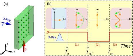
The typical NFS setup involves a solid-state target containing 57Fe. A x-ray pulse with meV bandwidth (either SR or coherent XFEL light) tuned on the 14.413 keV nuclear transition from the ground state to the first excited state shines perpendicular to the nuclear sample, as shown in Fig. 1(a). SR typically produces at most one excited nucleus per pulse, thus providing a reliable single-excitation and single released photon scenario. The disadvantage here is that the initial photonic phase is undefined. Coherent x-ray light from seeded or oscillator XFEL sxfel.Feldhaus ; sxfel.Saldin ; XFELO with a well-defined photonic phase can be used at low intensities such as to keep the excitation rate below one nucleus per pulse in the sample and guarantee single photons. Control over the number of excited nuclei per pulse can be achieved either by using x-ray partial reflection or partial transmission on silicon mirrors Shvydko2004 in order to limit the laser beam intensity or by varying the concentration of 57Fe nuclei in the target. An externally applied magnetic field B parallel to the axis induces the nuclear hyperfine splitting of the ground and excited 57Fe nuclear states of spins =1/2 and =3/2, respectively. Depending on the pulse polarization, different hyperfine transitions will be driven. In the following we consider the x-ray field linearly polarized parallel to the axis driving the two magnetic dipole transitions, where and denote the projections of the excited and ground state nuclear spins on the quantization axis, respectively.
The dynamics of the density matrix is governed by the Maxwell-Bloch equations Crisp1970 ; Shvydko1999N ; Scully2006 ; Palffy2008 :
| (1) |
with the interaction Hamiltonian
In the equations above is the x-ray detuning to the 14.4 keV transition assumed to be zero and denotes the Zeeman energy splitting of the nuclear ground (excited) state proportional to the magnetic field B. In Eq. (1), for and are the density matrix elements of for the nuclear wave function . The ket vectors are the eigenvectors of the two ground and two excited states hyperfine levels with , , and , respectively. Furthermore, are the corresponding Clebsch-Gordan coefficients Shvydko1998 ; Palffy2008 for the transitions and describes the spontaneous decay Scully2006 . The parameter is defined as , where GHz is the spontaneous decay rate of excited states, represents the effective resonant thickness Crisp1970 ; Shvydko1998 ; Shvydko1999N and m the thickness of the target, respectively. Further notations are for the Rabi frequency which is propotional to the electric field of the x-ray pulse Scully2006 ; Palffy2008 and the speed of light.
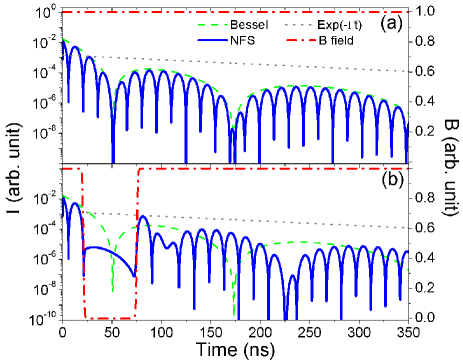
Fig. 1(b) illustrates the time evolution of our photon storage scheme. The external magnetic field B, depicted by the red line, is present before the x-ray pulse impinges on the target at . At the B field is turned off and later turned back on at . The orange arrows depict the time evolution of the nuclear transition current matrix elements as defined in Ref. Shvydko1996 . In our treatment, this is equivalent with investigating the coherence terms and Crisp1970 ; Shvydko1999N .
Initially, the ensemble of 57Fe nuclei is excited by the x-ray pulse at . Subsequently, the purely real currents are abruptly built. In the time interval (1), the two currents start to rotate in opposite directions on the complex plane with the factor of caused by the magnetic field until when B is turned off. The corresponding phase gain is . Here and in the following we have used for simplicity the notations and . Within the time interval (2), the quantum beat (arising from the interference between the two transitions) is frozen with the factor of since the hyperfine field has vanished, and only the dynamical beat Crisp1970 ; Shvydko1998 ; Van1999 due to interference between multiple scattering processes in the sample persists. During the time interval (3), the presence of the magnetic field makes the quantum beat emerge again.
We numerically solve Eq. (1) with and , and present our results in Figs. 2 and 3. The NFS signal intensities are compared with the spontaneous decay curves and the pure dynamical beat (for the case of no hyperfine splitting) Crisp1970 ; Shvydko1999H , where is the Bessel function of first kind. Fig. 2(a) shows the unperturbed NFS time spectrum where both quantum beat and dynamical beat are observed. In Fig. 2(b) we demonstrate photon storage by turning off the magnetic field at ns (corresponding to a quantum beat minimum, with odd). Both nuclear currents corresponding to the transitions are frozen on the imaginary axis (see Fig. 1(b)) and undergo destructive interference. In this case the intensity of the emitted radiation is suppressed by three orders of magnitude. Later on, by turning the hyperfine magnetic field on again at ns, the unsuppressed photon signal is observed again within the time interval (3). Fig. 2 also shows that the stored nuclear excitation energy experiences spontaneous decay during the storage time Shvydko1996 .
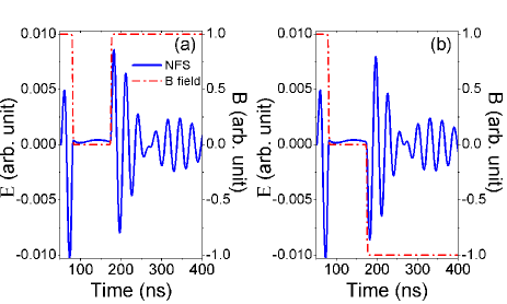
The electric field envelopes of the scattered photon are presented in Fig. 3. In Fig. 3(a), the magnetic field before ns and that after ns are the same and the phase before storage and after retrieving is continuous. If, however, the retrieving magnetic field is applied in opposite direction as shown in Fig. 3(b), the phase of the released photonic wave packet will be modulated with a shift of . This is caused by the effect of reversed time related with the change of sign of the hyperfine magnetic field Shvydko1994P ; Shvydko1995 , i.e., all the nuclear currents evolve backwards in time. Our density matrix calculations have been double-checked by the comparison with results from the iterative solution of the wave equations developed in Ref. Shvydko1996 . The agreement is complete for both electric field envelope and scattered light intensity, proving the equivalence of the two methods.
The most significant advantage of our scheme is the conservation of the photonic polarization and phase. Storage of nuclear excitation energy by magnetic field rotations in NFS experiments with SR was presented in Ref. Shvydko1996 . This pioneering work has opened the avenue of coherent control applications with nuclei using magnetic switching. However, the scheme in Ref. Shvydko1996 is not phase-sensitive. Since the magnetic Hamiltonian is not zero during the storage, both the polarization Palffy2010 and the phase of the particular polarization components cannot be stored and the properties of the released photon depend on the switching instants. With the advent of coherent XFEL sources and x-ray quantum optics and quantum information experiments, phase storage and modulation become crucial for many applications. So far, coherent trapping of hard x-rays in crystal cavities provides photon storage for time intervals in the ps range Ishikawa2008 . Our scheme provides robust phase and polarization storage of the x-ray photon on the 10-100 ns scale determined by the nuclear lifetime.
In order to implement our phase-sensitive storage scheme experimentally, a material with no intrinsic nuclear Zeeman splitting like stainless steel Fe55Cr25Ni20 Smirnov1996NE ; jex1997 is required. The remaining challenge is to turn off and on the external magnetic fields of few Tesla on the ns time scale. According to our calculations for the case of Fig. 2, the raising time of the B field should be shorter than 50 ns (the raising time was considered 4 ns for all presented cases). This could be achieved by using small single- or few-turn coils and a moderate pulse current of approx. 15 kA from low-inductive high-voltage “snapper” capacitors Miura2003 . Another mechanical solution, e.g., the lighthouse setup Roehlsberger2000 could be used to move the excited target out of and into a region with confined static B field. The sample is first excited while located in a first confined static magnetic field region. A fast rotation moves the sample out of this magnetic field region, and later on brings it under the action of a second static magnetic field. Simple geometrical considerations show that a displacement of the size of the sample thickness (about 3.5 m) corresponds to a time interval of 10 ns at a rotation frequency of 70 kHz and rotor radius of 5 mm Roehlsberger2000 . The sample can be thus rotated out of the confined magnetic field region fast enough to provide switching times on the order of 10 ns.
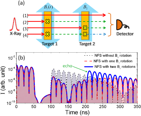
Let us now turn to the measurement of the phase shift. A typical x-ray optics setup would require to let the -modulated photon interfere with a part of the original pulse on a triple Laue interferometer Laue1 ; Laue2 . We adopt here another approach, namely, the simple and elegant photon echo solution used in NFS experiments with SR helistoe1982 ; helistoe1991 ; Smirnov1996NE ; jex1997 ; Smirnov2005 to allow the scattered photon to interfere with itself in the two-target setup presented in Fig. 4(a). A dynamical magnetic field is applied to target 1, and a static is applied to target 2. The target response is determined by and helistoe1991 ; Roehlsberger2004 , and the forward-scattered x-ray field is then given by Smirnov2005 . Using as x-ray input, the resulting electric field is the real part of
| (2) | |||||
This depicts the interference of four possible coherent scattering channels Roehlsberger2004 ; Smirnov2005 : (1) , no scattering; (2), the photon is scattered by target 1 only; (3) , the photon is scattered by target 2 only; (4) the mutual integral, the photon is first scattered by target 1 and then by target 2. Channel (2) and (3) cancel each other out when the effective thicknesses of the two targets are equal and , i.e, is reversed at . Hence a significant suppression of the NFS signal can serve as signature for the effective phase shift magnetically modulated in target 1.
In order to obtain the total scattered field intensity, we solve Eqs. (1) for both targets using the scattered field of target 1 as incoming field for target 2. Our numerical results are illustrated in Fig. 4(b). The presence of two targets results in the faster coherent decay that proceeds with effective resonant depth of , i.e., double the thickness of each target Smirnov1996NE . The magnetic field in target 1 is switched off at ns and back on at ns. For continuous phase, the intensity of the scattered field does not change. If, however, the phase of the retrieved field is -modulated by turning on the opposite magnetic field , the detected signal is significantly suppressed due to destructive interference between the two scattering channels. In turn, a second magnetic field rotation back at a node value produces an echo due to constructive interference as it can be seen in Fig. 4(b) for the rotation of back at ns.
This magnetically induced nuclear exciton echo itself provides another convenient solution for photon storage. A sequence of two rotations of the magnetic field direction in target 1 at the quantum beat minima can lead to storage and retrieval of the x-ray photon phase-modulated. This can be experimentally achieved in antiferromagnets as 57FeBO3 with strong intrinsic hyperfine magnetic fields that can be rotated with the help of a weak 10 G external field Shvydko1996 . Fast magnetic field rotations in such materials have been demonstrated Shvydko1994P . This specific case of magnetic switching in a two-target setup preserves the photon polarization and can modulate the photonic phase but is less robust compared to our scheme since both efficiency of the storage and the phase of the released photon depend on the rotation moment. Nevertheless, the magnetically induced nuclear exciton echo might provide an additional experimentally accessible setup to investigate mechanical-free x-ray storage and phase modulation of a single-photon wave packet.
In conclusion, we have put forward the possibilities of phase-sensitive storage and phase modulation for single hard x-ray photons in a NFS setup. These x-ray coherent control tools are important milestones for optics and quantum information applications at shorter wavelengths aiming towards more compact future photonic devices.
We would like to thank R. Röhlsberger for fruitful discussions and T. Herrmannsdörfer for his advice on the generation of strong magnetic fields.
References
- [1] G. E. Moore. Proc. IEEE, 86, 82 (1998).
- [2] A. Politi, M. J. Martin J. Cryan, J. G. Rarity, S. Yu, and J. L. O’Brien. Science, 320, 646 (2008).
- [3] S. Kneip, C. McGuffey, JL Martins, SF Martins, C. Bellei, V. Chvykov, F. Dollar, R. Fonseca, C. Huntington, G. Kalintchenko, et al. Nature Phys., 6, 980 (2010).
- [4] M. Fuchs, R. Weingartner, A. Popp, Z. Major, S. Becker, J. Osterhoff, I. Cortrie, B. Zeitler, R. Hörlein, G.D. Tsakiris, et al. Nature Phys., 5, 826 (2009).
- [5] M.C. Chen, P. Arpin, T. Popmintchev, M. Gerrity, B. Zhang, M. Seaberg, D. Popmintchev, MM Murnane, and HC Kapteyn. Phys. Rev. Lett., 105, 173901 (2010).
- [6] H. P. Specht, J. Bochmann, M. Mücke, B. Weber, E. Figueroa, D. L. Moehring, and G. Rempe. Nature Photon., 3, 469 (2009).
- [7] Yu. Shvyd’ko. X-Ray Optics: High-Energy-Resolution Applications. Springer-Verlag, 2004.
- [8] Yu. Shvyd’ko, S. Stoupin, A Cunsolo, A. H. Said, and X. Huang. Nature Phys., 6, 196 (2010).
- [9] Yu. Shvyd’ko, S. Stoupin, V. Blank, and S. Terentyev. Nature Photon., 5, 539 (2011).
- [10] S.-Y. Chen, H.-H. Wu, Y.-Y. Chang, Y.-R. Lee, W.-H. Sun, S.-L. Chang, Yu. P. Stetsko, M.-T. Tang, M. Yabashi, and T. Ishikawa. Appl. Phys. Lett., 93, 141105 (2008).
- [11] F. Pfeiffer, C. David, M. Burghammer, C. Riekel, and T. Salditt. Science, 297, 230 (2002).
- [12] A. Jarre, C. Fuhse, C. Ollinger, J. Seeger, R. Tucoulou, and T. Salditt. Phys. Rev. Lett., 94, 074801 (2005).
- [13] K.-D. Liss, R. Hock, M. Gomm, B. Waibel, A. Magerl, M. Krisch, and R. Tucoulou. Nature, 404, 371 (2000).
- [14] Yu. V. Shvyd’ko, M. Lerche, H.-C. Wille, E. Gerdau, M. Lucht, H. D. Rüter, E. E. Alp, and R. Khachatryan. Phys. Rev. Lett., 90, 013904 (2003).
- [15] S.-L. Chang, Yu. P. Stetsko, M.-T. Tang, Y.-R. Lee, W.-H. Sun, M. Yabashi, and T. Ishikawa. Phys. Rev. Lett., 94, 174801 (2005).
- [16] C. Buth, R. Santra, and L. Young. Phys. Rev. Lett., 98, 253001 (2007).
- [17] B. Dromey et al. Nature Phys., 5, 146 (2009).
- [18] D. C. Yost, T. R. Schibli, J. Ye, J. L. Tate, J. Hostetter, M. B. Gaarde, and Kenneth J. Schafer. Nature Phys., 5, 815 (2009).
- [19] S. Shwartz and S. E. Harris. Phys. Rev. Lett., 106, 080501 (2011).
- [20] E. P. Kanter et al. Phys. Rev. Lett., 107, 233001 (2011).
- [21] N. Rohringer et al. Nature, 481, 488 (2012).
- [22] O. Kocharovskaya, R. Kolesov, and Y. Rostovtsev. Phys. Rev. Lett., 82, 3593 (1999).
- [23] R. Coussement et al. Phys. Rev. Lett., 89, 107601 (2002).
- [24] T. J. Bürvenich, J. Evers, and C. H. Keitel. Phys. Rev. Lett., 96, 142501 (2006).
- [25] W.-T. Liao, A. Pálffy, and C. H. Keitel. Phys. Lett. B, 705, 134 (2011).
- [26] J. Arthur et al. Linac Coherent Light Source (LCLS). Conceptual Design Report. SLAC, Stanford, 2002.
- [27] XFEL @ SACLA. Official Website, 2012. http://xfel.riken.jp/eng/sacla/.
- [28] Massimo Altarelli et al. XFEL: The European X-Ray Free-Electron Laser. Technical Design Report. DESY, Hamburg, 2006.
- [29] U. Van Bürck. Hyperfine Interact., 123/124, 483 (1999).
- [30] Yu. V. Shvyd’ko. Hyperfine Interact., 125, 173 (2000).
- [31] R. Röhlsberger. Nuclear Condensed Matter Physics With Synchrotron Radiation: Basic Principles, Methodology and Applications. Springer-Verlag, 2004.
- [32] R. Röhlsberger, K. Schlage, B. Sahoo, S. Couet, and R. Rüffer. Science, 328, 1248 (2010).
- [33] R. Röhlsberger, H. C. Wille, K. Schlage, and B. Sahoo. Nature, 482, 199 (2012).
- [34] Yu. V. Shvyd’ko et al. Phys. Rev. Lett., 77, 3232 (1996).
- [35] A. Pálffy, C. H. Keitel, and J. Evers. Phys. Rev. Lett., 103, 017401 (2009).
- [36] G. V. Smirnov, U. van Bürck, J. Arthur, A. Q. R. Baron, A. I. Chumakov, S. L. Ruby, W. Potzel, and Brown G. S. Phys. Rev. Lett., 77, 183 (1996).
- [37] H. Jex, A. Ludwig, F. J. Hartmann, E. Gerdau, and O. Leupold. Europhys. Lett., 40, 317 (1997).
- [38] G. V. Smirnov, U. van Bürck, W. Potzel, P. Schindelmann, S. L. Popov, E. Gerdau, Yu. V. Shvydo’ko, H. D. Rüter, and O. Leupold. Phys. Rev. A, 71, 023804 (2005).
- [39] J. Feldhaus, E. L. Saldin, J. R. Schneider, E. A. Schneidmiller, and M. V. Yurkov. Opt. Commun., 140, 341 (1997).
- [40] E. L. Saldin, E. A. Schneidmiller, Yu. V. Shvyd’ko, and M. V. Yurkov. Nucl. Instrum. Methods A, 475, 357 (2001).
- [41] Kwang-Je Kim, Yuri Shvyd’ko, and Sven Reiche. Phys. Rev. Lett., 100, 244802 (2008).
- [42] M. D. Crisp. Phys. Rev. A, 1, 1604 (1970).
- [43] Yu. V. Shvyd’ko. Phys. Rev. B, 59, 9132 (1999).
- [44] M. O. Scully and M. S. Zubairy. Quantum Optics. Cambridge University Press, 2006.
- [45] A. Pálffy, J. Evers, and C. H. Keitel. Phys. Rev. C, 77, 044602 (2008).
- [46] Yu. V. Shvyd’ko, U. van Bürck, W. Potzel, P. Schindelmann, E. Gerdau, O. Leupold, J. Metge, H. D. Rüter, and G. V. Smirnov. Phys. Rev. B, 57, 3552 (1998).
- [47] Yu. V. Shvyd’ko and U. Van Bürck. Hyperfine Interact., 123/124, 511 (1999).
- [48] Yu. V. Shvyd’ko. Hyperfine Interact., 90, 287 (1994).
- [49] Yu. V. Shvyd’ko, T. Hertrich, J. Metge, O. Leupold, E. Gerdau, and H.D. Rüter. Phys. Rev. B, 52, R711 (1995).
- [50] A. Pálffy and J. Evers. J. Mod. Opt., 57, 1993 (2010).
- [51] N. Miura, T. Osada, and S. Takeyama. J. Low Temp. Phys., 133, 139 (2003).
- [52] R. Röhlsberger, T. S. Toellner, W. Sturhahn, K. W. Quast, E. E. Alp, A. Bernhard, E. Burkel, O. Leupold, and E. Gerdau. Phys. Rev. Lett., 84, 1007 (2000).
- [53] Y. Hasegawa, Y. Yoda, K. Izumi, T. Ishikawa, S. Kikuta, X. W. Zhang, and M. Ando. Phys. Rev. Lett., 75, 2216 (1995).
- [54] Y. Hasegawa and S. Kikuta. Hyperfine Interact., 123/124, 721 (1999).
- [55] P. Helistö, E. Ikonen, T. Katila, and K. Riski. Phys. Rev. Lett., 49, 1209 (1982).
- [56] P. Helistö, I. Tittonen, M. Lippmaa, and T. Katila. Phys. Rev. Lett., 66, 2037 (1991).