Functional quantum biology in photosynthesis and magnetoreception
Abstract
Is there a functional role for quantum mechanics or coherent quantum effects in biological processes? While this question is as old as quantum theory, only recently have measurements on biological systems on ultra-fast time-scales shed light on a possible answer. In this review we give an overview of the two main candidates for biological systems which may harness such functional quantum effects: photosynthesis and magnetoreception. We discuss some of the latest evidence both for and against room temperature quantum coherence, and consider whether there is truly a functional role for coherence in these biological mechanisms. Finally, we give a brief overview of some more speculative examples of functional quantum biology including the sense of smell, long-range quantum tunneling in proteins, biological photoreceptors, and the flow of ions across a cell membrane.
keywords:
1 Introduction
The role of quantum mechanics in biological processes has a long history [1, 2, 3]. After all, biological molecules are constructed of atoms, and bound together by forces of a ‘quantum origin’. However the role of quantum coherence (and more recently entanglement) on time or energy scales which play a direct role in biological function has remained controversial. Recent experiments [4, 5, 6, 7, 8] on photosynthetic ‘Light Harvesting Complexes’ (LHC) and their constituents (e.g., the Fenna–Matthews–Olson (FMO) pigment-protein complex in green sulfur bacteria) have suggested that quantum coherence may play a role in one of the most fundamental and important of biological processes: energy transport and energy conversion.
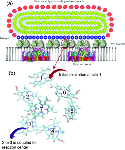
LHC complexes are arrangements of pigments (most importantly chlorophylls) and protein molecules which function as a light gathering ‘antenna’ to absorb photons and become electronically excited [9, 10]. This excitation is then passed to a reaction center, where it is converted into useful chemical energy. One of the most interesting features of this biological machine is the highly efficient transfer mechanism which takes the electronic excitation through the LHC to the reaction center with almost unity quantum yield. In green sulfur bacteria, part of this chain connecting the antenna (which in green bacteria is a large chlorosome) to the reaction center is composed of the Fenna–Matthews–Olson (FMO) pigment-protein complexes [11]; see Fig. 1 for an explanatory schematic. It is this complex which has been most widely studied, and will form part of the focus of this review.
In photosynthetic light harvesting quantum coherence can have different meanings in different circumstances. Quantum coherence can refer to quantum superpositions of localized molecular excitations that occur naturally because electronic couplings between molecular excitations lead to delocalized eigenstates (a.k.a. excitons). Sometimes, coherence in light harvesting alludes to the coherent wave-like dynamics of energy transfer, which actually reflects the superposition of excitonic eigenstates. In the former case, the coherence is represented in the molecular site basis, whereas in the latter case, the coherence is represented in the delocalized exciton basis. Note that the site basis and the exciton basis are special because they are related to the spatial arrangement of chromophores and energy eigenstates of the Hamiltonian, respectively. Although both types of coherence effects play important roles in photosynthetic light harvesting, they must be discussed separately. Quantum coherence manifested in the delocalized eigenstates of photoexcitations in photosynthetic complexes plays a fundamental role in spectral properties, energy tuning, and energy transfer dynamics of photosynthetic light harvesting [12, 13, 14]. The effects of excitonic coherence are more difficult to analyze and have been the subject of intensive research in the past few years. Spectroscopic measurements (termed two-dimensional electronic spectroscopy) on the FMO complex at liquid-nitrogen (77K) [4] and room temperatures [7] have shown time-dependent oscillations, presumed to be quantum beating, in the amplitudes of the spectral signals, which matches the predictions of quantum theory. Inspired by these observations, it has been proposed that a intricate interplay of quantum coherent excitation transfer and environmental dephasing help increase the efficiency of the energy transport process [15, 16, 17, 18, 19].
Apart from the example of photosynthesis it is also believed that other functional biological mechanisms may display, or rely upon, quantum effects. For example, the second part of this review will focus on the singlet and triplet states of spatially-separated electron spins in radical pairs (donor-acceptor molecules), whichh are hypothesized to play a role in avian magnetoreception [20, 21, 22]. If true, this would represent a functional piece of biological “quantum hardware” that could not function in a classical world [23]. More speculative examples we will describe more briefly include long-range quantum tunneling in proteins [24, 25], the sense of smell [26], biological photoreceptors [27, 28], and the flow of ions across a cell membrane [29].
1.1 Functionality defined
Our goal here is to give a brief overview of these different biological mechanisms, summarize some of the theoretical models, and highlight some of the experimental and theoretical evidence both for and against a functional role for quantum effects in biological systems. By “functional” we imply a role where the presence of coherent quantum dynamics achieves something either more efficiently, or otherwise impossible, than could be achieved by a classical mechanism alone. This concept of nature taking advantage of quantum mechanics is an inspiring notion, but the evidence for and against it must be examined carefully. We thus discuss only experimentally-verifiable systems in this review. This topic is growing at a phenomenal pace, and we can only summarize the main points for each system we consider.
1.1.1 Photosynthesis
In the example of energy transfer in photosynthetic complexes, most of the quantum effects observed so far have been at liquid-Nitrogen temperatures ( K) [4], though recent evidence has arisen of persistence quantum coherence at room temperature, both in FMO [7] and in other components of certain light-harvesting complexes [6]. To compliment this a variety of theoretical techniques have been used to show that a quantum model of the FMO complex implies a high transport efficiency even at room temperature [15, 16, 17, 30, 31]. As mentioned, this amalgamation of experimental observation and theoretical modeling suggests that a combination of quantum effects and environmental ‘stimulation’ leads to a high efficiency in the rate of energy transport in the FMO complex. However, alternative points of view still exist in the literature [32], and more work remains to be done to analyze to what extent this environment-assisted transport is really ‘quantum’. For example, in principle one must eliminate all possible classical models [33] and purely thermal/environmental effects [34] that could produce similar efficiencies before unambiguously stating that high efficiency transport itself is an example of functional quantum biology. In addition, it is not yet clear if the dynamics observed in-vitro (in experiment) also occur in-vivo (in nature), due to uncertainty about the nature of the energy transport from the antenna (chlorosome and baseplate) to the FMO complex. We will attempt to summarize all of these issues in this article.
1.2 Avian magnetoreception
In contrast to photosynthesis, the functionality of the proposed radical pair model for avian magnetoreception is entirely dependent on quantum mechanics [23]. In this sense it is different from the possibly more ambiguous role of quantum mechanics in photosynthesis. For example, the proposed mechanism of radical-pair magnetoreception cannot function at all without a large degree of spin-spin entanglement and coherence [23]: if environmental noise is too strong (an effective classical limit of the model) then birds could not navigate via this proposed mechanism. In addition, while ubiquitous in molecular and atomic physics, there is essentially also no classical analogue to the singlet/triplet states of two coupled spins, which is the fundamental element of this model which acts as a sensor of the Earth’s magnetic field.
The open question in this case then is whether such a proposed radical pair really exists and functions as predicted. There is some suggestion it may reside in cryptochromes (a light-sensitive cell found in the eye). However, experiments on a range of possible radical-pair molecules in laboratory conditions have not yet found a radical-pair molecule with the desired properties. Furthermore, evidence must also be found for whether birds really have the biological circuitry in place to process the signal that radical-pair magnetoreception would generate. Later we will summarize the details of this mechanism, explain why it is a strong contender for functional quantum biology, and outline the evidence supporting it as a mechanism for magnetoreception.
2 Photosynthesis
| Biological system | Results | |
|---|---|---|
| Photosynthesis | ||
| Cyrogenic temperature quantum coherence | [5, 4] | |
| Ambient/room temperature quantum coherence (FMO) | [7] | |
| Ambient/room temperature quantum coherence (Algae) | [6] | |
| Environment assisted transport | [15, 16, 35, 19] | |
| Entanglement, Leggett-Garg | [36, 37] | |
| Alternative views | [38, 33] | |
| Radical Pair Magnetoreception | ||
| Early proposals and evidence | [39, 40] | |
| Mathematical models | [39], [41] | |
| Indirect evidence (light dependance, magnetic field) | [42, 43, 44, 45, 46, 47] | |
| Experiments on Radical pairs | [48, 49, 50, 51] | |
| Monarch butterflies, Flys, Humans | [52, 53] | |
| Other examples | ||
| Olfaction | [26, 54] | |
| Ion channels | [29] | |
| Vision | [27, 55] | |
| Long-range electron transfer | [24, 25] | |
| Enzyme catalysis | [56, 57] |
Photosynthesis is one of the most important photochemical processes on Earth. As outlined in the introduction, the primary photosynthetic apparatus (which is typically called a photosynthetic unit, or PSU) can be roughly divided into several parts: a light-harvesting complex antenna, and a reaction center (RC), as shown in Fig. 1 for the example of green sulfur bacteria. The function of the FMO complex in green sulfur bacteria is to transport excitations from the sunlight-harvesting LHC antenna (chlorosome) to the reaction center (RC), where it initiates a charge-separation process that generates chemical potential for biochemistry.
The FMO complex consists of eight bacteriochlorophyll-a (BChl-a) molecules which are bound to a surrounding protein scaffolding. Note that the eighth BChl was not discovered in structural models of the FMO complexes until recently, therefore most studies on the FMO complex so far only considered a model with seven BChls. As we will describe later, in a simplified picture, each BChl molecule can be excited from its ground state into its first singlet excited state, forming a molecular exciton. Electronic couplings between molecular excitons then enable excitation energy transfer between BChl molecules.
After a photon is caught by the chlorosome antenna, it is transferred through the baseplate to the FMO complex (see Fig. 1). This excitation is then passed from one BChl molecule to the next, until it reaches the molecule, or site, closest to the reaction center. It then irreversibly enters the reaction center, and ignites the charge-separation process. In general terms, this apparatus functions in a similar way across a broad range of photosynthetic organisms, though the size and configuration of the antenna and component systems can differ greatly [58] (e.g., see Hu et al [59] for a review of the PSU in purple bacteria).
The most well-known way to describe this excitation-transfer process is the Förster model [60]. In this model the dipole-transition coupling between two chromophores (sites) gives rise to rates for excitation (exciton) hopping between interacting sites. The Förster model is a successful one, but it neglects quantum coherences between different sites. Experiments have shown that the exciton can move coherently among several chromophores, leading to electronic quantum beating [61], and perhaps also coherent collective phenomena like the well-known optical example of superradiance [62, 63, 64, 65].
In addition, some other components of LHCs in other species suggest that coherence and long-range entanglement may exist in larger structures. For example, the LHC of certain types of purple bacteria contain two types of ring-like antenna, LH1 and LH2 [59], which are substantially larger than the FMO complex. Spectroscopic studies on these systems have suggested that photoexcitations may delocalize among 4-5 BChl sites in the strongly coupled rings [62, 63, 65].
Another interesting issue is that the energy transport through the FMO complex is very fast (100’s of femto-seconds), and the efficiency of many LHC systems is very high. The quantum yield of light-to-charge conversion in some photosynthetic units [10] can be up to 95%, and explaining how the excitations navigate the energy landscape of the LHC so successfully is non-trivial. In addition, the need to navigate this landscape quickly must be understood in terms of losses that occur during the transport through the LHC system and its components, and not in terms of the overall energy processing rate of the entire PSU (LHC and RC systems), which is relatively slow in comparison. For example, the ‘reset time’ or ‘turnover’ rate of the RC in purple bacteria [59] is of the order of kHz, and the energy absorption rate of a single BChl in an antenna in bright conditions is Hz. It is thought that the large number of BChl in the antenna in totality provide enough energy to optimally use this kHz turnover rate of the RC. Thus there is a large separation of time-scales. The fast transport through the FMO complex, for example, is needed to “beat” the rate at which excitations are lost due to fluorescence relaxation, not because of the overall “clock rate” of the light harvesting complex. Also, one should note that the “energy” efficiency of photosynthetic bio-mass as, e.g., a fuel source, is relatively low even compared with photovoltaic efficiency [66, 67], because the down-stream biochemical reactions that turn chemical potentials into biomass have extremely low energy efficiency.
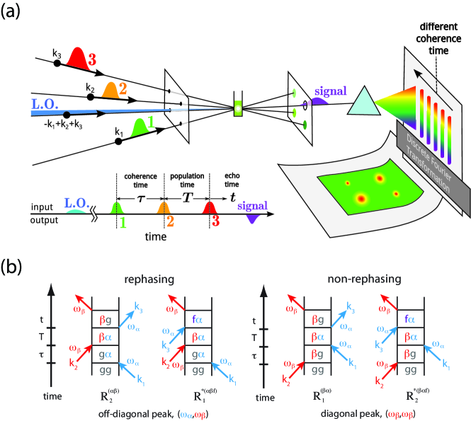
To gain a deeper understanding of these quantum phenomena, powerful spectroscopic tools have been developed, which provide opportunities to study the dynamics of the excitations in these pigment-protein complexes at the time scales of femto-seconds [65, 68]. For example, two-dimensional electronic spectroscopy is a powerful technique that probes the couplings and the dynamics of energy flow on a two-dimensional (2D) map in the frequency domain that allows direct observation of “coherence” between electronic excitations. A full understanding of exactly how much information can be extracted with such techniques is still being discussed and researched [69, 70, 71].
2.1 Experimental signatures of quantumness
As mentioned, the advent of powerful experimental tools have opened-up the ability to probe quantum effects in photosynthetic systems with unprecedented sensitivity. In particular, 2D electronic spectroscopy provides a unique tool that is specifically sensitive to electronic coherence in excitonic systems [74, 18, 75, 76]. A 2D experiment is a four-wave mixing process, in which three laser pulses interact with the sample to create an electronic polarization that generates the signal (Fig. 2a). In the experiment, two periods of time delay can be controlled in the apparatus: the time delay between the first and second pulses (, coherence time) and time delay between the second and third pulses (, population time). At fixed and , the signal field emitted in the photon-echo phase-matching direction is combined with an attenuated local-oscillator pulse for heterodyne-detection and frequency resolved to obtain a spectrum, which can be regarded as the Fourier transform of the oscillating signal fields with respect to the time delay between the third pulse and the signal (, re-phasing time). The measurement can be repeated at varied and Fourier transformed with respect to to obtain a 2D spectrum in frequency domain ( and ) at a fixed population time [75, 74].
Because 2D electronic spectroscopy records the signal at the level of the field rather than the intensity, it is sensitive to the quantum phase evolution of the electronic system during the population time. A broadband pulse can then interact with multiple exciton states to produce superpositions (coherences) of them, and the induced coherence in the exciton basis then undergoes oscillatory phase evolution that leads to beating signals as a function of time [68]. Figure 2b shows the Liouville pathways, i.e. pulse-induced density-matrix dynamics of the system, that contribute to the beating signals. The time evolution of the coherence during the population time has an oscillating phase factor, resulting in quantum beats in the 2D spectra as a function of . The signal provides an incisive tool to probe quantumness of an electronic process.
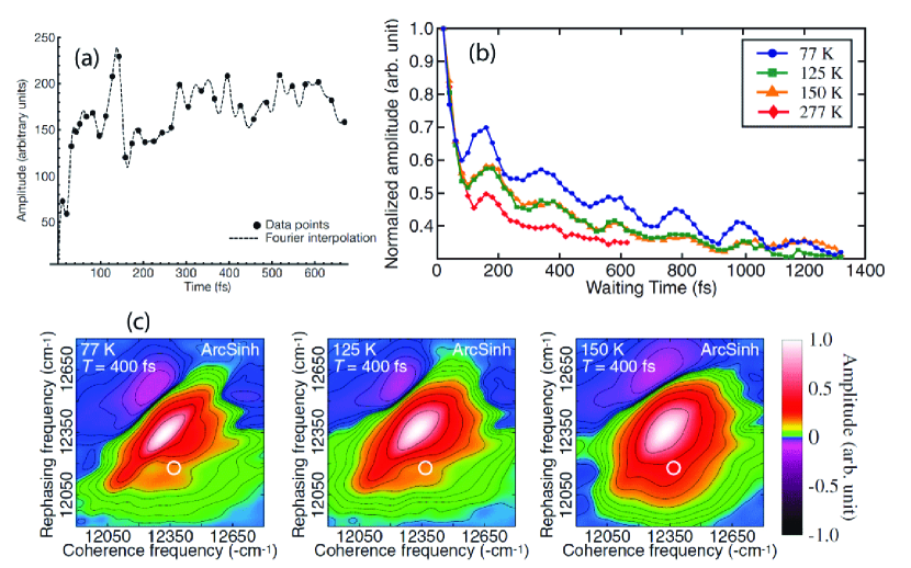
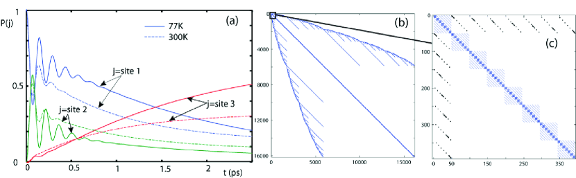
As we have described, the FMO complex [77] is only one component of many types of light-harvesting complexes found in nature, but it is perhaps one of the most well studied and characterized. In the seminal work of Engel et al. [4], 2D electronic spectra of the FMO BChl complex were obtained at K [see Fig. 3(a)], and strong quantum beating in the amplitudes and shapes of the diagonal peaks were revealed. These results have been interpreted [15, 16, 17, 18, 19] as a strong signature of coherent wave-like transfer of excitation energy in the complex. More recently, experimental evidence has shown that quantum coherence survives in the FMO complex at physiological (ambient room) temperatures for at least 300 fs [7]. While much shorter than the coherence observed at cryogenic temperatures [see Fig. 3(b)], it is still thought to be sufficiently long to have an impact on the efficiency of the transport process (which, overall, is of the order of fs). This result is supported by a variety of theoretical models [35] which we will summarize shortly. Similarly, in the experiment performed by Collini et al. [6], two light-harvesting proteins, isolated from marine cryptophyte algae, were found to have relatively long-lasting excitation oscillations at room temperatures. This is a particularly interesting example, as the algae live in exceedingly low-light conditions, and must process the excitation energy transfer extremely efficiently to survive. In addition, the coupling of the LHC systems to their protein environments differs in this case from most other photosynthetic LHCs, and Collini et al. [6] speculate that this may enhance correlated motions of the protein environment, enhancing any “coherence”-preserving effect that may arise.
Because short pulses are used in 2D experiments, it is possible that the quantum beating signals in 2D spectroscopy are results of vibration-coherence instead of electronic-coherence [32, 78]. In principle, this alternative interpretation can be verified by examining the rephasing and non-rephasing spectra separately because vibrational-coherence would cause beating signals in all the rephasing and non-rephasing signals, whereas electronic-coherence would cause beating in the off-diagonal peaks of the rephasing signals or the diagonal peaks of the non-rephasing signals [68]. Recently, Turner and coworkers [79] have carefully examined vibrational coherence effects and electronic effects in 2D spectroscopy and confirmed that quantum beating observed in the PC645 light-harvesting antenna protein of the cryptophyte alga Chroomonas sp. is due to electronic coherence.
In addition to 2D spectroscopy, other nonlinear optical experiments can be made into sensitive tools for quantum coherence phenomena. By using a two-color photon-echo approach, Lee et al. [5] also reveals that electronic coherence between bacteriopheophytin (H) and accessory bacteriochlorophyll (B) in the reaction center from the purple bacterium R. sphaeroides is preserved for much longer time than would be expected from the dephasing of either chromophores both at 77K and 180K [5]. Theoretical modeling revealed that the long-lasting coherence is indicative that the fluctuations of transition energies of B and H are strongly correlated in the reaction center. It was speculated that the “coherence preservation effects” from correlated protein environments also explain the surprisingly long coherence time (of the same order as the time for the excitation to traverse the whole complex) observed in FMO and other light-harvesting complexes.
2.2 Theoretical models
To gain a better understanding of what these various results imply it is helpful to consider a model commonly used to simulate some of these systems. One approach to simulate the quantum behavior of a single excitation in the FMO complex is via an effective Frenkel exciton model [9, 10]. As described earlier, a photon creates an excitation in an antenna molecule, which is transferred to one of the sites in the FMO molecule, which is then transferred along the chain, from chromophore to chromophore via a transition-dipole Coulomb interaction, until reaching the reaction center, where it is then employed for charge separation. The assumption of a single-excitation seems valid for in vivo situations, particularly when the bacteria is living in very low-light conditions. It is exceedingly unlikely for the FMO complex to ever contain two excitations. Although the single-excitation assumption in experimental situations has generated certain controversy [38], rapid exciton-exciton annihilation processes makes multiple-exciton states in a single complex extremely unstable and tend to relax into a single-exciton state before energy transfer occurs. Nevertheless, in principle it is straightforward to construct a multiple-excitation model, e.g., as employed in [80] to describe an artificial photosynthetic system. For the purposes of clarity, however, we restrict ourselves here to the single-excitation model, which can be written as
| (1) |
where the states represent the presence of an excited electron (exciton) at site, or BChl molecule, , where (see Fig. 1(b) for a figurative description of these sites in the FMO complex, and we omit the eighth BChl molecule because it was not discovered in structural models of the FMO complexes until recently). In other words, one can describe the whole FMO complex with a single label defining at which site the excitation is residing. The parameter is the energy of that excitation for a particular site (which is dependent on the surrounding protein structure and varies quite a lot), and is the excitonic coupling between the and sites.
The determination of the various energies and coupling strengths in a given LHC is a complicated and difficult area of research, often involving both spectroscopy and ab initio modeling of the physical structure and environment of the system [18, 81]. In general, accurate values of electronic couplings that are in good agreements with experiments can be calculated with ab initio quantum chemistry methods based on the atomistic models of a LHC [82, 81, 83]. The site energies are more difficult to determine because modeling the protein-pigment interactions is a non-trivial task, therefore experimental inputs are usually required in order to obtain accurate energies. Recently, Renger and coworkers have demonstrated in several photosynthetic complexes [84, 85, 86] that with a careful treatment of the electrostatic interactions between the electronic excitations and the surrounding protein environments, it is possible to calculate site-energy parameters with structure-based theoretical approaches.
Since the FMO LHC is one of the most studied examples, some understanding of the system’s Hamiltonian exists (though often different values for the energies and excitonic coupling amplitudes are given in the literature [87, 88, 84]). We now present the values used in Ref. [89],
| (9) |
where units are cm-1 and the large energy gap relative to the common ground state has been subtracted. The diagonal elements correspond to the energies in Eq. (1), while the off-diagonals correspond to the site-site electronic couplings . We present these numbers here to show that the site-site couplings are of the same order as the energy difference between sites. Already this suggests that site-site coherences could be strong. In addition, the magnitudes of site-site couplings are often distributed broadly, with the larger ones in the scale of 100 cm-1, which is also comparable to the reorganization energy due to exciton-environment couplings.
2.2.1 Approximations used in the models
One of the open questions for this system is the nature of its interaction with its environment. In the simplest model, which we explicitly show below, a variety of approximations are made. The first is to assume that each site is coupled to a bath of oscillators, and that the baths coupled to each site are independent of each other. The second approximation is to assume that this coupling is to the energy of each site [69, 15, 90, 91, 92], which means that the environment causes fluctuations of the energy in the site basis. In the exciton basis, the fluctuations become to have off-diagonal matrix elements that can cause transitions between eigenstates [16]. The third is to assume the environment is Markovian (i.e., without memory). Finally, the fourth assumption is to assume the exciton-bath coupling is weak, and that one can perform a second-order perturbation theory to describe the dynamics of energy transfer.
This gives a master equation that describes the reduced density matrix of the FMO complex that is simple to solve, and has a minimal number of free parameters. Typically there is also a temperature-independent radiative relaxation rate for each site. If the excitation takes too long to traverse its way through the complex, it will be eventually lost due to this relaxation. Fortunately, fluorescence decay of chlorophylls typically occurs in the nanosecond time scale, a slow process compared to the other dynamics, ultimately giving the FMO complex its ability to efficiently transport energy with a high success rate. This simple model has estimated some of the qualitative properties of experiments on FMO, including the long coherence time, and high transport efficiency [17].
As a specific example, the Markovian master equation approach assumes a self-energy,
| (10) |
where denotes the system density matrix. The first term describes irreversible excitation transport between site and the sink [89, 93] (see Fig. 1(a) and (b) for a description of where site and the sink, or reaction center, are in the FMO):
| (11) |
where , and is the sink tunneling rate. Site is coupled to the reaction center because it comprises the lowest energy excitation and lies closest to the reaction center. In contrast, site lies closest to the antenna. Here denotes the empty state with no electronic excitation. The second term, , describes a temperature-dependent dephasing (see Ref. [94] for a detailed discussion of the temperature dependence). The last term, describes the slow fluorescence relaxation process. A variety of insights have been obtained from this simple model [69, 15] which will discuss in the next section, but the essence is that the combination of coherent dynamics from and level-broadening from means there is a high possibility of bringing an excitation from site to site , and thus the RC, before the excitation is lost due to the fluorescence relaxation . The efficiency of the transport is often modelled via the time-dependent sink or reaction-center population
| (12) |
where is the population of the site “”, connected to the sink.
As mentioned before, in most treatments of the FMO system, the coupling to the environment modulates the energy of each site, and in the language of quantum mechanics this is “pure-dephasing”. The radiative relaxation of each site is independent of temperature because the optical transition energy of each site is exceptionally high ( cm-1), leaving only the dephasing processes to be temperature dependent.
In the FMO complex, each state in the above description represents an exciton on a molecule which transfers its energy to its neighbor, but no actual electron transport takes place. This implies that the re-organisation energy (the energy associated with a change in the surrounding protein environment due to the presence of an electron charge) is relatively low compared to processes where electron transport occurs. In general, however, both the assumption of independent Markovian baths and of weak coupling to these baths may not be realistic. There is evidence that the coupling to protein environments around the FMO complex can be of the same order as the electronic couplings [94, 80] ( cm-1). In addition, the bath (e.g., the protein scaffold surrounding the FMO complex) may have structure and dynamics which have a strong correlation with, and back-action on, the dynamics of the excitation in the FMO complex. To understand the effect of this intermediate coupling regime, and complex environment, the system and bath have been treated with a range of non-Markovian and higher-order (in electron-phonon coupling) models [35, 69, 95, 96, 97, 98, 88, 80, 99]. In particular, nonperturbative approaches that provide exact numerical results for certain models of excitation energy transfer have been applied to study coherent quantum dynamics in photosynthetic light harvesting [100, 101, 102, 103]. We will discuss the details of one of these approaches (the hierarchy model) later. Although these nonperturbative methods are computationally too expensive to apply to large photosynthetic complexes, they have provided valuable insight that have shed a different light on the coherence effects in light harvesting in a different light [35]; one where bath memory effects conspire to enhance quantum coherence.
Going beyond the independent bath approximation, initial experiments indicated that the surprising long-lasting quantum coherence between two electronic states may be because of coherence enhancing effects from coupling to common vibrational modes [69, 5, 6]. This has triggered the reexamination of the excitation transfer processes [104, 105] and decoherence effects [106, 107] in the presence of such an unusual environment. In principle, such a strongly coupled “common bath” should contain bath-induced fluctuations that are spatially correlated [108, 109]. As a counter-argument, however, some molecular dynamics simulations for the FMO complex [34] and reaction center [110] show that only weak correlations between the movements (vibrations) of the chromophores appear, and that the uncorrelated bath approximations may be valid.
Very recent work by Shim et al [111] compared some of these master-equation models to a more complex many-body atomistic/molecular modeling scheme and found excellent agreement between the two approaches. This is encouraging as it does suggest that the assumption (based on relatively simple models) that the oscillations seen in experiment have a truly quantum origin might be correct. However, whether the environment surrounding LHC systems has a structure or nature that preserves quantum effects in some way is still a controversial and open question. More experimental work is required to answer this puzzle.
Finally, some authors have argued that it is possible to construct a variety of alternative classical models that can, in principle, produce both classical beating [33] and efficient energy transport [69, 15] without reference to quantum coherence at all. Such alternative descriptions must be eliminated before one can unambiguously state that the FMO complex, or other LHCs, take advantage of quantum mechanics.
2.3 New Insights from Theoretical Modelings
2.3.1 Environment-assisted transport
Apart from the direct observation in experiments of quantum coherent beating, some of the most intriguing insights that have appeared from the study of the FMO complex suggest that a combination of these quantum coherent oscillations of excitations between sites, and interactions with the environment, produce a transport efficiency higher than is possible with a Förster model [60] alone. If correct, this would fit the definition of functional quantum biology. Such a phenomenon was proposed and studied by Plenio et al [15], by Mohseni et al [16], and by Lee et al [19]. In all cases, it is assumed that a single excitation is placed at a particular site in the FMO (usually site one), and that this excitation then propagates through the FMO chain due to a combination of coherent tunnelling and environmental effects. As mentioned earlier, the goal of the FMO complex is to get this excitation efficiently to site three, which is coupled (incoherently) to a reaction center. Thus the typical time, or efficiency, of reaching the reaction center is calculated as a function of environment temperature, site selective-couplings, and so on. In most cases it seems that the full quantum model (e.g., based on Eqs. [1-3]) will give a higher efficiency than a purely classical one. Several physically intuitive explanations have been given for this phenomenon:
-
1.
Local minima avoidance: Refs. [15] and [16] showed that the Markovian quantum model outlined earlier suggests both long-lived coherent oscillations between sites, and an enhanced rate of excitation transfer to reach the “sink” site, over that predicted by the Förster model. Reference [15] argued that essentially the combination of coherent transfer and environmental “noise” causes level broadening, which implies that the excitation can more easily escape local minima in the FMO network (see Fig. [1]). In other words, quantum delocalization can help avoid and overcome local minima, or energy potential traps, in the energy landscape of the FMO complex [35]. It has been suggested that such a phenomena is even more important in higher plants because of possible “up-hill” potential landscapes. These results are related to earlier work by Gaab and Bardeen [112] who outlined how energy transfer through a network can be optimized by carefully choosing the coherence time and the rate of transfer to the sink.
-
2.
Coherence assisted trapping: In a study by Lee et al. [19], it was demonstrated that coherent quantum dynamics, together with rapid incoherent dissipation due to a trapping site can enhance the efficiency of the irreversible energy transfer between the initial energy donor and the sink, or reaction center, over a purely classical model. Effectively, this model suggests that an intricate combination of reversible quantum coherent evolution and incoherent collapsing of exciton wave functions can achieve a greatly enhanced energy-trapping efficiency, effectively using the anti-Zeno effect to promote energy transfer.
However, one should again notice that this “enhanced efficiency” is in comparison to that obtained with the Förster model. There remain several ambiguities; Firstly, is the Förster model the correct classical model to make a comparison to? Secondly, placing a single excitation at site “” may match well certain experiments, but in normal light conditions is this a correct approach?
Quite recently, Briggs and Eisfeld [33] showed that, for realistic coupling strengths, an alternative classical model of the FMO transport process can produce results identical to the quantum one. In addition, a recent analysis by Wu et al. [113] suggests that, as mentioned earlier, the efficiency enhancement gained by coherence in the FMO complex is only a few percent compared even to that predicted by the Förster model. Finally, the true in-vivo conditions are not well understood, and is the subject of current research. Perhaps the only way to really solve this issue is to show that in more complex components of some light harvesting complex, e.g., in LH1 or LH2 [114, 109], there exist significant energy traps which, without the assistance of quantum coherence, drastically impact/reduce the probability of an excitation successfully navigating its way to a reaction center before being lost to fluorescence relaxation.
2.3.2 The Hierarchy model
In an effort to gain a deeper understanding of the interplay between the quantum coherent transport of energy in FMO and how it interacts with its complex protein environment, a variety of non-Markovian and non-perturbative models have been applied to these systems. One of the most successful is the Hierarchy model, originally developed by Tanimura and Kubo [115], which has been applied to both the FMO complex [35] and other LHC components [114, 109]. This model has a had a large impact this field, and thus we will briefly describe its main components here. Full derivations can be found in the literature [116, 117, 115, 118]. Starting with the Hamiltonian we discussed earlier, one explicitly describes the interaction term between site energies and phonon modes,
| (13) |
where , and is the coupling constant of the th site and the th mode in the bath. The phonon modes themselves are described as harmonic oscillators (with a Hamiltonian which we omit here for brevity).
It is typically assumed that at the sites (or pigments) and the phonon modes are separable, so that , and the phonon modes are in a thermal equilibrium state , . After averaging, the correlation function of the bath modes
| (14) |
are sufficient to describe the properties of the bath. With the Hierarchy method one typically uses a Drude spectral density (appropriate for over-damped oscillators)
| (15) |
Here is the “Drude decay constant”, and indicates the memory time of the bath for site (each site is assumed to have its own independent bath, though in general one of the powers of the Hierarchy method is its ability to treat correlated baths [109]). Also, is the reorganisation energy, related to the system-bath coupling strength. This Drude spectral density implies an exponentially-decaying correlation function,
| (16) |
where , , and the coefficients
| (17) |
and
| (18) |
With just this information on the bath properties (the reorganisaiton energy, bath memory time, and bath temperature) one can employ the Hierarchy equations to describe the system-bath dynamics in the strong-coupling and non-Markovian regime,
| (19) | |||||
Here, is the projector on the site , is the Liouvillian described by the Hamiltonian introduced earlier, and for FMO . The various parameters are those we defined above. The Hierarchy is a large set of coupled equations each labelled by , a set of non-negative integers uniquely specifying each equation. The integers are defined as . That is, each site has an additional label , from to , and each of those labels in turn can run from to . The label is special, and refers to the system density matrix. Its properties at any time define those of the system. This is in turn coupled to so called “auxiliary density matrices”, which describe the complex bath fluctuations, by the terms in the equation where (i.e. implies the term in the index defined by and is increased or decreased by ). As a simple example, if the terms in the sum for are truncated at , then the system density matrix is coupled to other auxiliary density matrices directly, , , and so on (see Fig. 5(c)). Each of these auxiliary equations is then in turn coupled to higher-tier equations, and overall the hierarchy forms a high dimensional simplex.
The hierarchy equations are infinite both in the sum of the label (due to the expansion of the correlation function), and in the value of each label itself. Thus both must be truncated in some way. Typically, the label can be truncated if and (where is some characteristic system frequency). The overall truncation of the labels is defined by the largest total number of terms in a label, or the tier, . This concept is important as each ADO in a given tier is only coupled to those ADOS in the tier above or below it. In Yan et al [118] they describe this structure, and the meaning of each tier, as the level of bath self-correlation included in the simulation.
The tier at which the truncation should be made is difficult to predict (there is no small parameter here), though typically truncation should be taken when (though it also depends on the reorganisation energy and temperature). In practice, truncation should be checked by looking for convergence in system dynamics when changing . A systematic and powerful algorithm to find convergence was introduced by Shi et al [119]. They proposed that a renormalization of the hierarchy equations allows a direct inspection of the elements of higher auxiliary equations to be made during the numerical solution, and thus the level of the tier can be truncated as necessary.
In addition, a temperature correction can be added to the equations of motion, which compensates for the truncation of the label at level , and assists in overall convergence. This is made by assuming that all higher-level auxiliary density operators for are uncoupled from each other and that they experience pure Markovian dynamics [116]. This results in an additional term in the equation of motion, sometimes called the “Ishizaki-Tanimura boundary condition” [116], which can be included in the Hierarchy equation of motion;
| (20) |
The double commutator gives a normal Lindblad form, and the summation can be done analytically, e.g., for ,
| (21) |
In other words, this term encapsulates additional environmental dephasing effects neglected by truncating . Including this correction, even for , has been found to substantially assist the convergence of the Hierarchy results. As an example, in Fig. (5), we show the results of an application of this model to the FMO system at K and K, for various and employed in the literature. The beauty of this method is that, to some degree, all parameters can be extracted from experiments and the results correctly predict the long coherence time seen in experiments. Perhaps the most important point here is that long bath memory (implied by small ) helps increase the coherence time of the coherent oscillations [35], though this may not necessarily enhance the overall transport efficiency. In general, for each parameter, there is an optimal value which gives the largest transport efficiency from site to site , and hence to the reaction center [108, 113].
2.3.3 Leggett-Garg inequality and entanglement measures
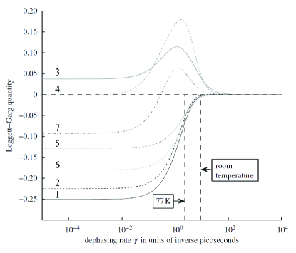
As mentioned earlier, it is important to fully verify whether the experimental observations of coherence truly come from quantum mechanics and not some alternative, classical, model [33]. Wilde et al [37] proposed using the Leggett-Garg inequality as a means to unequivocally ascertain whether the dynamics observed in these experiments are quantum or obey “macroscopic realism”. As yet, however, the measurements needed for such a test cannot be performed in these FMO experiments. However, their results show that if it were possible to perform such measurements, the currently accepted physical parameters for the FMO system suggest a violation of the inequality at room temperatures should occur (see Fig. 4) for an example of the violation of the inequality.
In addition to coherence, it is also interesting to ask whether there is appreciable entanglement in the FMO complex during the exciton transfer. It was recently suggested by Sarovar et al. [36] that a small amount of long-range and multipartite entanglement should exist even at physiological temperatures [36], however, the role of entanglement in the energy transfer process is still not clear (see also [31]).
2.3.4 Quantum process tomography
Quantum state tomography is a well-known technique to reconstruct the full density matrix or state of a system. Recently, there has been increasing attention on what is termed quantum process tomography [120, 121], which attempts to fully characterize a given quantum process (i.e., a systems dynamics) in an open environment (e.g., in contact with Markovian and non-Markovian baths). Simply speaking, quantum process tomography is a systematic procedure to characterize an unknown quantum system, or black box, by systematically analyzing the functional relationships between inputs and outputs as a function of time. Very recent works [70, 71] showed that the 2D electronic spectra used to investigate light-harvesting complexes is equivalent to a kind of quantum process tomography, elucidating the meaning of the results found with these techniques in earlier experiments. In principle this means that the spectroscopic experiments of excitonic systems can be studied from a quantum information theory approach, and that perhaps the full nature of the dynamics in FMO and other systems can be understood on a deeper level. In the future perhaps it may be possible to discuss important questions such as the existence of non-Markovian environments, entanglement, and decoherence during the transfer process using this tool. However, their proposal still requires steps that are experimentally challenging.
2.4 Functionality
One of the arguments used to justify the presence of “functional quantum mechanics” is that if there is any advantage, or improvement in efficiency, to be had from taking advantage of quantum mechanics, evolution will inevitably do so. Quantum coherence, manifested in the delocalized eigenstates of photoexcitations in photosynthetic complexes, plays fundamental roles in the spectral properties and energy-transfer dynamics of photosynthetic light harvesting [12, 14]. In structures of strongly-coupled clusters of pigments, coherence is essential for shaping the energy landscapes and energy flow towards the reaction center. This has been demonstrated clearly in the LH2 antenna complex of purple bacteria [59, 122, 65], the FMO complex [123, 89], the reaction centers [124], and the major light-harvesting complex of higher plants [65, 125]. For example, in the LH2 antenna complex, energy transfer from a B800 BChl to coherently delocalized B850 states is an order of magnitude faster than that to a single B850 BChl [124, 122]. Such a significant enhancement in energy transfer rate clearly contributes to the highly efficient light-harvesting process, hence the functionality, in purple bacteria.
A separate issue is whether or not quantum coherent dynamics, i.e. excitonic coherence, plays a role in the functionality of photosynthesis. This issue has become the subject of intensive research recently. However, in reality the calculated gain in efficiency (as a function of time, for example) seen in Refs.[15, 16, 19, 108, 113] is relatively small. In some species (e.g., the algae living in low-light conditions studied by Collini et al [6]) this small advantage may be useful. But in general, (e.g., plants in high-level light conditions), is there still a significant advantage provided by coherence? As mentioned earlier, some studies of “higher plants” suggest the energy landscape of their LHCs is even more rugged than that in the FMO complex [6], and thus coherence may become even more important in efficiently navigating this terrain before other processes (like fluorescence relaxation) steal away the excitation. Interestingly, a recent study by Hoyer et al. [126] indicates that the effect of “quantum speedup” is rather small in the time scales of photosynthetic energy transfer, and they propose that quantum coherent effects in light-harvesting systems are more likely to be optimized for robustness or total efficiency.
Finally, very recent results by Ritschel et al [88] paint an even more complex picture; they model the full FMO trimer, including an additional eighth bacteriochlorophyll (BChl) molecule which was recently found to exist in the FMO complex. On a positive note, they find that the subunits of the trimer act as separate transport channels from the antenna to the reaction center, thus the assumption of studying the individual FMO monomers individually seems safe. However, if they use this new eighth site as the source of initial excitation (which may be the case, as it is located close to the antenna), they find that no coherent exciton dynamics occur; only exponential decay. In addition, they find discrepancies in the overall transport efficiency, depending on which energies and couplings (i.e., which Hamiltonian) are chosen from the literature. In contrast, Olbrich et al. [87] used a Molecular Dynamics approach to derive a time-dependent equation of motion for the excitation transfer, and found that the eighth BChl did not form a stable complex with the other seven, and that, while thermal effects in total were stronger than assumed in other studies, the coherent effects were enhanced when considering the trimer system in totality. This is attributed to a corresponding increase of the site coupling within a monomer due to the influence of the other monomers in the trimer. However, they found that the quantum coherent effects were still quite small in their model, and Olbrich et al. [87] argue that the fast thermal fluctuations of the site energies are the main contributing factor to the efficient transport of energy.
Inevitably a broader picture is needed before we can fully understand the role of quantum coherence in natural conditions. Is the coherence observed in experiments contributing significantly to the efficient transfer of energy in natural conditions? In-vitro experiments and theoretical models suggest a positive answer, but ultimately experiments into other types and classes of LHC systems (e.g., LH-1 and LH-2) are required, and a broader understanding of the transport process in vivo is needed.
3 Avian Magnetoreception: a tale of two spins
Magnetoreception is a unique animal ability to detect either the polarity or inclination of the earths magnetic field as a navigation tool. As with electro-reception (the ability to detect electrical fields, e.g., sharks hunting their prey), it represents a sense slightly alien to human experience. Or so it was thought, until recently. Experiments on manipulating a specific gene in flies and monarch butterflies have indicated this gene plays a significant role in magnetoreception, and since humans share this gene in a dormant form it suggests that even we may harbor this unique ability [52, 53]. However, some evidence [53] suggests it is not driven by the radical-pair mechanism we discuss here.
The epitomic example of magnetoreception however is in migrating birds. They apparently use this powerful ability, alongside other biological geo-mapping tools, to navigate large distances. A complete and satisfying description of the mechanism for the magnetoreception ability of many species remains a mystery. Some evidence suggests that a magnetite-based (deposits of magnetic iron minerals) mechanism describes well this ability in some animals [43] (though recent reports suggest that, at least in pigeons, these deposits are in fact macrophages and play no role in the magnetic sense [127]). In other avian species, particularly robins, several intriguing aspects of their behavior rule out magnetite-based mechanisms. Here we focus on the radical-pair model for magnetoreception that represents another potential functional quantum biological system. Precise details will be given later, but summary radical pair is a pair of molecules bound together, with each has an unpaired electron. These electrons form naturally into singlet or triplet states, the state of which, in turn, affects the disassociation, or reaction products, of these radical pairs. It is proposed that the relative weights of these reaction products which are biologically detectable. Thus, if the relative probabilities of having singlet or triplet pairs is effected by the Earth’s magnetic field, this in turn affects the distributions of the two different reaction products. As a result, a biologically detectable signal can be generated to function as a compass.
3.1 Behavioral studies
The hypothesis that migrating birds can make use of the earth’s magnetic field for orientation was first proposed by von Middendorff [128]. Later, European robins were found to utilize a magnetic compass when migrating [129]. Since then, many experimental tests on the behavior of migrating birds have been performed. In particular, it was found that for some species their magnetic sense acts as an inclination compass, but is insensitive to parity [40]. In other words, they can detect the relative angle of the local earth’s magnetic field lines, but not whether they are going north or south. There is some benefit to this in that the compass would not be effected by geomagnetic reversal [129]. Typically, however, most models of magnetite-based magnetoreception act as a parity compass [43].
With advances in technology and technique, many more delicate behaviorial tests have been performed. For example, it was found that the compass was: (a) dependent on the frequency and intensity of ambient light [42, 43]; (b) dependent on the intensity of the external magnetic field [43]; (c) disrupted when repeated magnetic pulses were applied to the migratory birds [44], and (d) disrupted when an external oscillating field was applied at certain angles to the geomagnetic field [45]. In addition, lateralization in terms of the the birds’ visual system associated with magnetoreception has also been reported [130], suggesting the mechanism operates primarily in the right eye.
All of this data lends support to a photo-activated radical-pair mechanism. In particular, the experiments showing (d), the disruption of the navigation mechanism by an external oscillating field in the megahertz range (as seen in migratory robins and directionally-trained chickens as well as zebra finches [43, 45, 47, 46]), match exactly what is predicted by simple models of radical-pairs, which we will describe shortly.
Very recent evidence (see Ref. [43] for a detailed review) suggests that both an inclination compass and a polarity compass are present in certain avian species, suggesting that a magnetite and radical-pair compass can coexist. We also point out, as mentioned earlier, that in some species of flies, butterflies, and even perhaps humans, there may be a third unknown mechanism at work [52, 53] that has some of the same behavioral properties of the radical-pair model, but not others. Here we will focus on describing the evidence for the radical-pair mechanism, and in what way that mechanism represents “functional quantum biology”.
3.2 Radical-pair model: evidence and motivation
Based on findings of behavioral studies, it is obvious that a successful model should contain a light-dependent factor. Another feature is that because the geomagnetic field is very weak ( T), the magnetic compass mechanism must be very sensitive. In addition, this mechanism must transduce the response to the Earth’s field into a biologically-detectable signal. Finally, as we mentioned earlier, in some species the compass is only an inclination compass.
To help satisfy some of these criteria, the radical-pair mechanism (RPM) for magnetoreception was proposed by Schulten et al. [39] in 1978. Within the radical-pair model there exists the possibility of a photo-sensitive element [41, 131], i.e., absorption of light triggers an electron transfer from a donor to an acceptor molecule to form the initial radical pair (biologically this is proposed to occur within the cryptochrome proteins within the eye). This process can be made to satisfy most of the frequency and intensity dependencies observed in experiments on Robins [43].
How then is this donor-accepter system sensitive to the Earth’s field? Most known radicals contain atoms (hydrogen and nitrogen) with nuclear spins which act like an effective internal magnetic field affecting the electron spin via the nuclear hyperfine interaction. To sense the weak magnetic field of the Earth, the combination of anisotropic internal nuclear hyperfine fields and external geomagnetic fields causes a mixing of the electron singlet and triplet states, the strength of which is dependent on the angle of the external geomagnetic field. Ritz et al [131] assumed a simplistic internal magnetic environment with only one anisotropic nucleus (i.e., one radical is devoid of internal magnetic fields, and the other radical has very strong internal magnetic fields) and showed that this maximizes the singlet-triplet mixing compared with other designs [131, 132]. Whether this occurs in nature is unknown, but their extreme example helps to present a simple model of how this mechanism works (see later).
For the last stage, as mentioned, this radical pair can decay into different reaction products at different rates [41, 133] depending on the spin state (singlet or triplet). If this radical-pair is located in the cryptochrome proteins within the eye, this could lead to a vision-based compass where the bird actually sees a directional signal [41]. Finally, this model requires that the many molecules forming the radical-pair-based magnetic compass must be ordered in some way so that the directional effects will not be averaged out [134, 135]. This may require that the radical-pairs exist in a regular ordered biological structure, or lattice, which has not yet been identified.
3.2.1 The Schulten model
A simple model to calculate the influence of the Earth’s field on the reaction products of the radical pair was outlined originally by Schulten [39, 41]. Here we present specific details of this model, to help clarify the physical picture described in the previous section. As discussed above, the radical pair is formed when an electron is transferred from a donor to an acceptor molecule, typically via an optically-excited process. This forms a singlet state for the combined donor-acceptor system. The, ideally the subsequent dynamics of this state is primarily affected by an anisotropic hyperfine interaction between the electron spin and the nuclear spin within the molecules. This must occur faster than any decay (recombination) or decoherence of the radical-pair. The interaction is described by a hyperfine coupling Hamiltonian (assuming only one of the two spins is affected by its internal nuclear spin, for the most sensitive possible compass [131])
| (22) |
where is the operator of electron spin , is the nuclear spin operator, and in its diagonal basis is the anisotropic hyperfine coupling tensor for a cigar-shaped molecule with and MHz, for high sensitivity [23] (though recent results have suggested the weak-hyperfine coupling regime can produce a stronger sensitivity, given the right combination of parameters [136]). The Zeeman splitting of the electron spins is
| (23) |
where is the magnetic field vector describing the external geomagnetic field (or any other applied fields), , and is the Bohr magneton of the electron. The magnitude of the geomagnetic field is typically of the order G ( T). For the axial symmetry of the hyperfine tensor, the magnetic field vector is explicitly written as , for .
In addition to the coherent evolution driven by and , the radical-pair dynamics feels several incoherent effects. Both singlet () and triplet () states undergo spin-selective relaxation or recombination into the singlet product state and the triplet product state . This occurs when, e.g., there is electron back-transfer from the acceptor radical to the donor radical, which is described by the self-energy
| (24) |
where , , , and , for the up and down nuclear spin states . Here we assume that the singlet and triplet products have the same recombination rates . The final incoherent term is the environmental noise. The simplest noise model is where both phase and amplitude of both electron spins are equally perturbed with self-energy
| (25) |
where for the electron spins .
The dynamics of the electron and nuclear spins are then determined by the Hamiltonian together with the incoherent contributions for the generalized master equation: , where the Liouvillian is .
The biologically-detectable signal is the integrated triplet or singlet yield. For example, the triplet yield is,
| (26) |
where, , and is the projector onto the triplet states. The interplay between the external geomagnetic field and the anisotropic nuclear hyperfine interaction results in a dependence of the triplet yield on the relative angle between the geomagnetic field and the radical-pair molecule.
This simple model is sufficient to describe the destructive effect of additional radio frequency fields, as observed in experiments, and enables one to estimate the most ideal properties of the radical-pair system to maximize the sensitivity of the magnetic compass.
3.2.2 Radical-pair molecules: experimental tests
As discussed earlier, a variety of behaviorial experiments support the radical-pair mechanism; e.g., the disorientation of the magnetoreception ability in Robins when external magnetic fields of around a megahertz frequency are applied [46, 137, 47]. In addition, comparison of the frequency-dependence of this behavior to theoretical models indicates that the radical-pair system does have the optimal design mentioned above; i.e., one of the radicals may contain almost no nuclear spin polarization.
To further bolster evidence supporting the radical-pair model, there are also experimental tests at the molecular level on a variety of candidate molecules that could be at the heart of the mechanism. There have been a large amount of studies on radical-pairs in solution, and on the effect of external fields on the reaction products (see a review of early experiments by Steiner and Ulrich [48]) though only under magnetic field strengths much stronger than Earth’s. In recent work, Woodward et al [49] developed a technique for studying the properties of such candidate reaction-pair systems. As an example they studied the photochemical reaction of pyrene (Py) and N,N-dimethylaniline (DMA) in solution under a very weak (500 T) radio-frequency magnetic field. When pyrene is continuously irradiated by UV light, in the presence of DMA, the transient radical ion pair Py⋅- DMA⋅- is produced in a spin-correlated singlet state and is found to interconvert with the corresponding triplet state, as expected. An exciplex (an electronically-excited complex of Py⋅- and DMA⋅-) is then produced due to the electron-hole recombination of the singlet radical pair. Their technique uses the fluorescence of the exciplex to monitor the concentration of singlet pairs, and estimate the reaction products. They found that even this very weak magnetic field induced changes of up in the reaction products.
Rodgers et al. [50] also studied the spin-selective recombination of Py⋅- and DMA⋅- radicals under the affect of – mT magnetic fields. It was found that weak magnetic field effects become most pronounced when the ratio of hyperfine coupling strengths for the two radicals is large. Another recent example is the triad composed of linked carotenoid (C), porphyrin (P) and fullerene (F) groups considered by Maeda et al. [51]. By using spectroscopic measurements they found this particular radical-pair’s lifetime was changed by the application of magnetic fields less than T (as needed to detect the Earth’s weak geomagnetic field). In addition, they found this effect responded anisotropically to the angle between the molecule and the external field, which is of course an essential feature of a radical-pair compass. However the anisotropy was only manifest at stronger fields ( mT), and no anisotropy could be observed for T, even at low temperatures ( K). It is hoped that future work will show whether a radical pair formed by “reduced flavin cofactor and an oxidized tryptophan residue in a cryptochrome flavoprotein” (Maeda et al [51]) will have the long spin coherence lifetime and low recombination time to achieve the T sensitivity at room temperature needed for this mechanism to function.
3.3 Functionality and significance of quantum coherence
Attention has recently focused on the functionality of quantum coherence in the radical-pair model because, as we noted in the introduction, spin singlet and triplet states are inherently quantum. This has led to a great degree of interest in this phenomena from researchers in condensed matter and quantum information theory. For example, an analysis of the entanglement and decoherence effects of this mechanism [23] suggests that, in their words, for this model to function as desired, “superposition and entanglement are sustained in this living system for at least tens of microseconds, exceeding the durations achieved in the best comparable man-made molecular systems”
This implies that the radical-pair model is, if it really exists and functions as described, a piece of functional quantum biological hardware. From the perspective of condensed matter physicists, it is very akin to man-made artificial molecule systems, e.g., the singlet and triplet states of spin-blockaded electrons in double quantum dots [138]. However, further work remains to be done on identifying the exact radical pair which sustains this mechanism, to show that it has the properties necessary to act as a compass, and to verify this with further in vitro experiments.
4 Other possible quantum biological systems
4.1 Tunneling in biological systems
In many biological processes, tunneling of light particles (hydrogen or electrons) provides an alternative route to classical over-the-barrier reactions, resulting in new channels for chemical reactions that are forbidden in classical mechanics. Tunneling in biological systems was first reported in long-range electron-transfer processes in proteins [24, 25, 139], and then in the transfer of a proton, hydride, or hydrogen atom in various enzymatic catalytic reactions [57, 56]. Furthermore, the simultaneous transfer of an electron and a proton from different sites (so-called proton-coupled electron transfer) also play an important role in many biological functions [140]. A significant increase in the enzyme catalytic efficiency or long-range electron transfer through proteins due to tunneling have been observed in many biological systems. This class of quantum phenomena in biology clearly fits into our description of functional quantum biology.
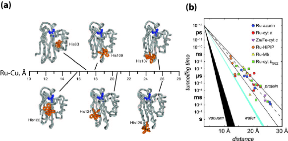
Electron transfer between redox centers separated by distances of the order of 15-30 plays important roles in respiration and photosynthesis [24, 141]. In the past three decades, Gray and coworkers [24, 142] have yielded a remarkably detailed description of the distance- and driving-force dependencies of long-range electron tunneling rates in ruthenium-modified proteins. Particularly, Ru-azurin has proven to be an excellent model system for electron tunneling through folded proteins (Fig. 7a) [143]. Experimentally observed electron transfer rates that exhibit exponential decay as distance increases and weak temperature dependence indicate a single-step tunneling mechanism (Fig. 7b). It is remarkable that such long-distance electron transfer in biology occurs via quantum mechanical tunneling. Tunneling over such long distances would be impossible in vacuum, however, through bonded as well as non-bonded interactions in the protein, electron tunneling barriers can be significantly reduced, leading to significantly higher electron tunneling rates in proteins.
A major open question remains in this field is whether or not the electron conducting proteins have evolved specific pathways for electron tunneling [141, 24, 144]. Theoretical analysis has demonstrated that an empirical model treating the protein as a structureless random medium explains the experimental data [144, 145]. However, structure-based analysis reveals that specific channels through covalent bond, hydrogen bond or even van der Waals contacts form electron tunneling pathways that facilitate electron tunneling over a long distance [146, 147, 24, 25, 148]. Are these redox proteins specifically wired to perform efficient electron tunneling? How much do the functions of these biological systems depend on the quantumness of the process? Again, these are not questions that can be answered easily, and interpretation of current experimental results often involves ambiguities.
Recently, Skourtis and coworkers have suggested a proposal that is likely to provide an incisive test for the pathways model. Quantum theory predicts that electron transfer pathways could interfere with each other [147], and it has been suggested that electron transfer through the azurin protein depends critically by quantum interferences between multiple distinct pathways [147]. Motivated by these earlier theoretical studies, Skourtis and coworkers suggested a molecular which-way interferometer experiment in which localized and distinguishable normal modes coupled to bridge atoms can be vibrationally excited to control inelastic scattering of electrons and interferences between transfer pathways [149, 148]. Experiments have demonstrated that electron transfer in a a small donor-bridge-acceptor organic molecule can be controlled by excitation of vibrational modes localized on the bridge moiety [150]. Such experimental probes of pathway coherence in electron-transfer proteins in the single-protein level would provide decisive proof for the quantumness and functionality of the electron tunneling process.
Tunneling of a proton, hydride, or hydrogen atom also play an important role in a wide range of biological enzymatic catalytic reactions [57, 56]. Measurements of the intrinsic kinetic isotope effects (KIE) in enzymatic reactions have clearly demonstrated nuclear quantum effects in enzymes [57, 56]. For example, in soybean lipoxygenase [151] and methane mono-oxygenase [152], large KIEs with close to 100 have been observed, and the sizes of the KIEs clearly indicate quantum-tunneling effects. In enzyme catalysis, a large portion of the quantum improvement over the classical catalytic rates can be attributed to the energy shift due to the zero-point energy that gives a quantum correction to the barrier height and the H-tunneling rates [57]. Note that semi-classical models including environmentally coupled H-tunneling have been shown to adequately describe H-tunneling in enzymes [153, 154]. Such nuclear quantum effects in enzymes might represent a class of quantum phenomena in biological systems that depends only on trivial quantum effects, not quantum coherence.
4.2 Smell
Our sense of smell allows us to discriminate between small molecules in very low concentrations via scent molecules interacting with receptors in the nose. At this time, the biomolecular processes of olfaction are not fully understood, and some evidence suggests that a mechanism based solely on the size and shape of odorant molecules is inadequate. For example, it has been noted that molecules with very similar shapes and sizes have a remarkably different scent [26]. Thus traditional models of a “docking”-type mechanism, where the size and shape of an odorant molecule actuates the receptor in some way, are thought to be insufficient. Turin [26] proposed a mechanism which, in addition to “docking”, gives a further level of selectivity (or sensitivity) by a process of inelastic electron tunnelling. In this case the odorant molecule both docks with a receptor and then mediates phonon-assisted inelastic tunnelling of an electron from a donor to an acceptor (i.e., donor and acceptor electronic states differ in energy by , and thus transport only occurs when energy is conserved by emission of an odorant phonon, the vibrational degree of freedom of the molecule one is “smelling”, of the right energy).
A recent model proposed by Brookes [54] expanded on this idea, and presented evidence that such a mechanism fits the observed features of smell, and is at least “physically” credible (see Fig. [8] for an overview and details of the mechanism). Whether such a mechanism ultimately exists in nature has yet to be determined. While not specifically requiring “coherence” to function, this mechanism requires inelastic phonon-assisted tunnelling of electrons, and is certainly more “microscopic”, and sensitive, than previously thought.
4.3 Quantum coherence in ion channels
Ion channels, which regulate the flow of ions across the membrane of a cell, are a vital component of many biological processes [155], e.g., neuronal communication (potassium channels), muscle contraction, etc. In many cases the flow of ions through the channel is controlled by a gate that can be activated by a voltage, a chemical signal, incident light or mechanical stress. In addition the channels often feature a filter which controls which types of ions may pass. This selectivity filter is only a few angstroms wide, which forces the ions to pass through it one-by-one [156].
A recent conjecture by Vaziri and Plenio [29] is that the selectivity filter of ion channels, and the transport of ions through the channel, may exhibit quantum coherence. They are motivated by the large selectivity found in the bacterial KcsA potassium channel, which can select potassium over sodium with a ratio of . They argue that the energy scale of the thermal fluctuations of the atoms which form the channel suggest two possible quantum coherent phenomena. Firstly, there could be a coherent diffraction of a 1-D potassium “matter wave” off an effective grating formed by modulations of the potential energy landscape inside the channel. Secondly, since the channel forms a 1-D array of trapping sites, the potassium ions could take advantage of quantum tunnelling to process through the channel in an efficient manner. They calculate a tunnelling rate which is of the same order as the decoherence time, and thus argue that an “environment assisted” quantum tunnelling phenomena, akin to that seen the energy transport in photosynthesis, is responsible for the high selectivity and throughput of these channels. However, unlike in the transport of energy in FMO, this process involves the movement of charges through a complex environment, with a correspondingly large re-organization energy, which implies the coupling to the environment may be too strong for coherence to thrive.
This conjecture is intriguing, but at this time lacks experimental support (apart from the already observed remarkable performance of the channels). Vaziri and Plenio [29] propose several ways to verify the presence of this “functional quantum effect”, focusing on collective transport resonances which, they argue, are the functionality provided by the quantum coherent transport in this case. Furthermore, a nitrogen-vacancy (NV) probe in proximity to the ion channel [157] might be a promising tool to observe any possible quantum coherent effects.
4.4 Photoreceptors
Biological photoreceptors, i.e. light-sensing proteins, are ubiquitous in the functions of life forms on Earth. These proteins contain chromophores that upon excitation, undergo ultrafast chemical transformation on their excited-state potential energy surface and eventually lead to light-induced signal transduction [28, 158]. Because of the quantum nature of the electronically excited state and nuclear quantum coherence that often accompany excited-state molecular dynamics in the femtosecond time scale, quantum mechanics are required to describe these photo-activated dynamics in biological photoreceptors [159].
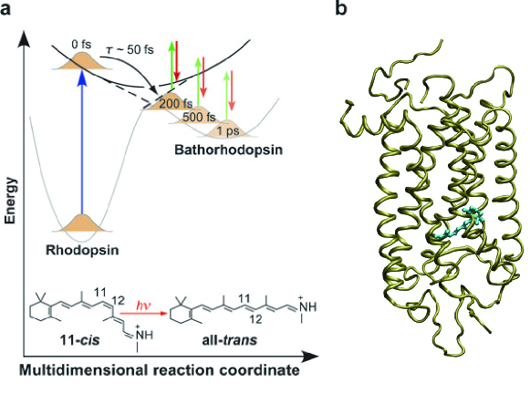
For example, the primary event in vision involves the photoisomerization of a retinal molecule in the trans-membrane protein called rhodopsin (Fig. 9), which is the most well studied and characterized photoreceptor. Following light absorption, this photoisomerization reaction proceeds with remarkable rate (200 fs, one of the fastest chemical reactions known) and high specificity (quantum yield of ) [27]. This isomerization triggers a protein conformational change that is subsequently amplified through protein-protein interactions with signal-transduction proteins, leading to the visual signal. The efficient photoisomerization process makes the retinal molecule an effective and reliable photo-switch. The quantum mechanical positioning of electronic states and their symmetries enables ultrafast coherent wave-packet dynamics that are responsible for the rapid photoisomerization reaction [28, 158, 160]. In addition, the unique reactivity of rhodopsin has often been attributed to a conical intersection between its electronic excited state and ground state [161, 162], which has recently been demonstrated experimentally [55]. Nuclear quantum coherence clearly plays an important role in the remarkably rapid and efficient photo-activated one-way reaction dynamics in rhodopsin [160]. Intriguingly, coherent optical control of retinal isomerization yields in bacteriorhodopsin, a protein closely related to vertebrate rhodopsins, has been demonstrated experimentally [163, 164]. The frontiers of the investigations of biological photoreceptors are thus to further elucidate the quantum nature that controls the high yield and specificity of natural photoreceptors in order to design and direct chemical dynamics using coherent light [165].
5 Open issues
5.1 Open problems in photosynthetic light harvesting
5.1.1 Protein Environment and Spectral Density
The protein environments surrounding pigments in photosynthetic complexes play crucial roles in light harvesting [167, 168]. On the one hand, the protein scaffolding provides a rigid structure that fixes photosynthetic pigments in space to form an antenna network, and pigment-protein interactions also shift the transition energies of individual pigments to yield the energy landscape that directs energy flow towards the reaction center [89, 81]. These aspects of proteins’ roles are static and well recognized, and as a result structure-based studies have formed a large component in this research field [169].
On the other hand, dynamical fluctuations of pigment-protein couplings are directly responsible for the energy transfer dynamics. However, these dynamical effects are often overlooked and treated without details, as the simple models of spectral densities described in Sec. 2.2. Does the popular diagonal exciton-phonon coupling model completely capture the physical reality in photosynthetic complexes? How important (or unimportant) are off-diagonal exciton-phonon couplings? Are the simplified spectral density functions used in the literature adequate to describe the real system? Can correlated baths significantly influence exciton dynamics? And if yes, can the correlated-bath effects be directly observed in real systems? What are the importance of non-Markovian effects? Can we directly measure non-Markovianity and the memory kernel of system-bath interactions? All these questions are still unresolved and require a combination of experimental and theoretical studies in order to provide adequate answers. Experimentally, probes more specific to exciton-phonon interactions and bath dynamics must be developed. In this regard, the rich information in the peak shapes and spectral dynamics of 2D electronic spectra must not be overlooked [75, 18, 74, 103]. Theoretically, simulations of exciton transfer dynamics in photosynthetic systems must be expanded to investigate more general models including different types of exciton-phonon couplings and bath spectral densities [168]. Recently, molecular dynamics simulations combined with quantum chemistry calculations have been applied to study fluctuations in the FMO complex [34, 87] and reaction center [170, 110]. This type of study might lead to new insights on the dynamical role of the protein environments and provide atomistic resolution to the spectral densities; however, the interpretation of the hybrid-quantum/classical results should be treated with caution [171, 170].
5.1.2 Consequences of Coherence
As we have discussed in Sec. 2, quantum coherence effects in photosynthesis have drawn intense research interest recently. So far, experimental and theoretical works have focused on the question of how quantum coherence effects contribute to improve the efficiency of energy transfer, and a measure of efficiency such as the quantum yield defined in Eq. (4) is often adopted as the target to benchmark the effects of quantum coherence. It is clear that site-basis quantum coherence, i.e. coherent delocalization of excitons, plays crucial roles in determining the energy landscape and spectral properties of photosynthetic complexes [12, 13, 14, 172, 173]. However, the consequences of excitonic quantum coherence responsible for the reversible wave-like exciton dynamics are less clear. As we have detailed in Sec. 2.3, it has been speculated that excitonic coherence can improve functions such as robustness against energetic disorder or dynamical perturbations. In addition, Ishizaki and Fleming [35] argued that excitonic coherence can be utilized to form energy “ratchets” for energy transfer that avoid trapping by local energy minima. However, these results are often model-dependent and a unifying view of excitonic coherence effects in photosynthesis seems to be still lacking.
Furthermore, to establish excitonic coherence as a general and essential phenomena in photosynthetic light harvesting, measurements on larger photosynthetic complexes such as LH2, LH1, and LHCII must be carried out to confirm the general existence of long-lasting quantum coherence. It is also necessary to consider more general initial conditions beyond the coherent laser-excitation condition suitable for the interpretation of laser experiments [174]. Does quantum coherence play a role when excitations are generated by incoherent sunlight or energy transfer from another antenna complex? Another open question that follows naturally after the observations of long-lasting quantum coherence in photosynthetic complexes is whether or not quantum effects (quantum coherence and entanglement) in photosynthetic systems can be used as a quantum resource? Here, a crucial experimental development is the capability to prepare and measure an excitation localized on a single chlorophyll site, which would allow the quantum mechanical manipulation of the wavefunction of excitons.
5.1.3 Biological Relevance
So far excitonic quantum coherence has been observed only in in vitro experiments, and theoretical studies have only established that quantum coherence could affect efficiency of energy transfer within a single complex. However, a connection between excitonic quantum coherence effects and biological function in vivo has yet to be established [167, 175, 126]. In particular, the functionality of light harvesting systems depends on assemblies with a length-scale beyond a single complex; the whole antenna could contain tens of light-harvesting complexes. Does the single-complex efficiency provide enough information to determine the energy transfer efficiency of the whole antenna? Moreover, what is the system optimized for? It is conceivable that in the larger length scales and longer time scales that determine the functionality of photosynthetic light harvesting, biological systems actually strive to achieve robustness and safety, instead of maximal efficiency. It is therefore necessary to investigate and model dynamics of light harvesting in larger assemblies including the whole antenna. Specifically, experimental probes for the combined temporal and spatial dynamics of exciton motions on the antenna have to be developed [176]. In addition, evidence of long-lasting quantum coherence has yet to be obtained in vivo. Theoretically, models of quantum coherence effects should be considered for larger structures, and it is necessary to develop measures of quantum coherence that can be linked with in vivo observables that can be determined and tested in experiments.
5.1.4 Design Principles
It is surprising that after decades of research we still do not know how to build artificial light-harvesting systems that can rival the efficiency achieved in natural photosynthesis [14, 177]. We know that: (1) chromophores with strong absorbance must be used, (2) the antenna must contain high-density of light absorbers yet avoiding the formation of exciton quenching charge-transfer species, and (3) a energy gradient can help to funnel excitations towards the reaction center [14]. However, the knowledge required to implement these principles in artificial systems is lacking. Moreover, certainly there exist other design principles in the working of natural photosynthesis. If quantum coherence can help the efficiency of light harvesting, then what are the molecular architectures that can utilize this advantage to make a more efficient antenna? It is crucial that theoretical studies focused on how chromophores should be arranged are carried out and tested by experiments, maybe with the help of synthetic organic chemists, to eventually formulate the design principles that can help us build more efficient solar cells.
5.2 Open problems in the radical-pair model
5.2.1 Radical Pair Candidate Molecules
The discovery of cryptochromes as a possible candidate to be the light-sensitive host of the magnetoreception radical pair mechanism gives hope to the radical-pair model. However, in-vitro experiments have yet to show that cryptochromes have the exquisite and anisotropic magnetic field sensitivity needed to function as a magnetic compass. Ideally cryptochromes, or other candidate molecules, need to have some of the following properties (see [133] and [178] for a more in-depth review of these issues):
-
1.
Spin-correlation and coherence time on the scale of geomagnetic B-field effects. The rate of coherent spin oscillations induced by the geomagnetic field must exceed that of any environmental losses. A T field induces oscillations on the time scale of ns. This coherence, or correlation, must be protected from environmental losses on this time scale for significant change in the reaction rates to occur. Such correlation times have been seen in “photolyases” radical pair systems.
-
2.
Spin-correlation time and coherence on the same time-scales as weak disruptive radio frequency fields. In behavioral experiments it was found that the magnetic sense was disrupted for oscillating fields with very small magnitudes. As with the geomagnetic sensitivity this implies the spin correlation time must survive for an exceptionally long time. For example, in Ref. [47], Robins were found to be sensitive to fields as weak as nT. Ritz [178] discusses this point and argues that the effect of a nT field induces a change in the reaction yield of % at s, and % at s (a similar effect was shown in the calculation in [23]). This implies both an exceptionally long spin correlation time and extreme sensitivity of the magnetoreception sense to changes in the reaction yield (see Fig. (6)).
-
3.
Optimization of the hyperfine field. Recent work by Cai et al [136] suggests that a highly anisotropic but weak nuclear hyperfine coupling can give a stronger angular dependance on the yield than a strong one. Whether this occurs in candidate radical pairs needs to be determined.
5.2.2 Behavioral Tests
There are a variety of further behavioral tests that have yet to be done [133, 178], and which might provide even stronger proof for the radical pair model:
-
1.
Functionality window: For static fields it was found that changes in the intensity by more than 25% disrupted the magnetic compass mechanism. However, after a period of time the sense returned. Ritz [178] has raised the possibility that a behavioral test for a similar “re-adjustment over time” to the disruptive effect of oscillating fields will reveal if the disruption is related to the functionality window seen with static fields.
-
2.
Magnetoreception via oscillating fields: If the oscillating field amplitude is of similar intensity to the Earth’s geomagnetic static field (in an environment where the geomagnetic field is suppressed), some of the simple radical pair models predict that the oscillating field with also induce a directionally sensitive change in the reaction yields. Rodgers et al [133] argue that observation of animals responding in a directional way to this effect (essentially navigating by the oscillating field) would be stronger proof of the radical pair mechanism than the disruption effects observed so far (which in principle could be attributed to some unknown causal effect on the motivation of the animals).
Apart from these directly testable issues, there remains a lot to be learned about the transduction of the radical pair yield to a “compass” that is biologically useful. The chain of events connecting changes in concentrations of molecules, into nerve impulses, and then into a signal of directionality is completely unknown. Indeed, as we have mentioned earlier, a large range of information is probably combined to ultimately form the final relevant directional cues (or instincts). This also has an impact on an understanding of why very small changes in the radical pair yields can apparently disrupt the magnetoreception sense. Finally, there have been proposals for using quantum control techniques, in the form of magnetic field pulses, to coherently control the spin states [179]. If this induced a directional response in test animals this would be extremely difficult to explain via any alternative magnetoreception mechanism.
Finally, a very recent experiment on pigeons has identified a neural substrate responsible for processing magnetic sense information [180]. This substrate was shown to respond to the intensity, direction, and polarity of the magnetic field, perhaps ruling out a radical pair mechanism in this case. For pigeons this is particularly interesting as other recent evidence [127] has contradicted the previously held notion that magnetite deposits in their beaks were responsible for their magnetic sense. Perhaps a third, unknown, mechanism plays a role in this particular species. Similar experiments on neural response in species in which the radical pair model is thought to reside (e.g., european robins) are vital.
6 Concluding remarks
At the atomic level, the demesne of quantum mechanics, one can always say that of course we exist in a quantum world (or at least a non-classical one), and that life itself is thus, in some manner, quantum. However, since the birth of quantum mechanics, the extent to which the features of quantum mechanics, such as quantum coherence or entanglement, can operate on biological time and energy scales in a “hot and wet” environment, and provide a biological advantage, has been seen mostly as a curiosity.
The recent development of quantum information theory has revealed that quantum mechanics can, in some situations, do things better than an equivalent classical theory (such as speed up computation and provide secure communication). Thus, as we have described here, the goal of “functional quantum biology” is in some sense similar; to enhance the efficiency of transport in a molecular chain, or to provide an exquisite sensor of geomagnetic fields. A very extreme example is the idea proposed by Penrose [181]: that quantum computation may occur in microtubule assemblies within the neurons of the brain. This idea has attracted much attention, criticism and debate [182, 183] because it portrays the fascinating possibility of quantum mechanical effects playing a central role in cognitive function. Evidence so far is lacking [182, 183], but this idea has helped stir up a broader interest in the role of quantum effects in biological processes, and the hunt for other candidates of functional quantum biology.
Thus, at this time, the most promising candidates for function quantum biology remain photosynthetic units [5, 4, 7, 6], and magnetoreception [41, 43]. The evidence so far is in favor of both these systems not only containing quantum coherence in a hot and wet biological environment, but also that it is used to gain a biological advantage. There is a great possibility that further examples of functional quantum biology remain to be found. After all, as in a great number of technological breakthroughs, we usually find that nature has been there before us.
7 Acknowledgements
We wish to thank Jian Ma, A. Y. Smirnov and S. De Liberato for helpful discussions. NL is supported by the RIKEN FPR Program. YCC thanks the National Science Council, Taiwan (Grant No. NSC 100-2113-M-002-004-MY2), National Taiwan University (Grant No. 10R80912-5), and Center for Quantum Science and Engineering (Subproject: 10R80914-1) for financial support. YN thanks the National Science Council, Taiwan (Grant No. NSC 98-2112-M-006-002-MY3) for financial support. FN acknowledges partial support from the NSA, LPS, ARO, NSF grant No. 0726909, JSPS-RFBR contract No. 09-02-92114, MEXT Kakenhi on Quantum Cybernetics, and the JSPS-FIRST Program.
References
- [1] E. Schrodinger, What is Life?, Cambridge University Press, 1992.
- [2] P. C. W. Davies, Quantum Aspects of Life, Imperial College Press, London, 2008.
- [3] H. C. Longuet-Higgins, Quantum mechanics and biology, Biophys. J. 2 (1962) 207.
- [4] G. S. Engel, T. R. Calhoun, E. L. Read, T.-K. Ahn, T. Mancal, Y.-C. Cheng, R. E. Blankenship, G. R. Fleming, Evidence for wavelike energy transfer through quantum coherence in photosynthetic systems, Nature 446 (2007) 782.
- [5] H. Lee, Y.-C. Cheng, G. R. Fleming, Coherence Dynamics in Photosynthesis: Protein Protection of Excitonic Coherence, Science 316 (2007) 1462.
- [6] E. Collini, C. Y. Wong, K. E. Wilk, P. M. G. Curmi, P. Brumer, G. D. Scholes, Coherently wired light-harvesting in photosynthetic marine algae at ambient temperature, Nature 453 (2010) 644.
- [7] G. Panitchayangkoon, D. Hayes, K. A. Fransted, J. R. Caram, E. Harel, J. Wen, R. E. Blankenship, , G. S. Engel, Long-lived quantum coherence in photosynthetic complexes at physiological temperature, PNAS 107 (2010) 12766.
- [8] G. Panitchayangkoon, D. V. Voronine, D. Abramavicius, J. R. Caram, N. H. C. Lewis, S. Mukamel, G. S. Engel, Direct evidence of quantum transport in photosynthetic light-harvesting complexes, PNAS 108 (2011) 20908.
- [9] H. van Amerongen, L. Valkunas, R. van Grondelle, Photosynthetic excitons, World Scientific, 2000.
- [10] R. E. Blankenship, Molecular Mechanisms of Photosynthesis, Blackwell Sci, London, 2002.
- [11] J. M. Olson, The FMO Protein, Photosyn. Res. 80 (2004) 181.
- [12] T.-C. Yen, Y.-C. Cheng, Electronic coherence effects in photosynthetic light harvesting, Procedia Chemistry 3 (2011) 211.
- [13] G. R. Fleming, G. D. Scholes, Y.-C. Cheng, Quantum effects in biology, Procedia Chemistry 3 (2011) 38.
- [14] G. D. Scholes, G. R. Fleming, A. Olaya-Castro, R. van Grondelle, Lessons from nature about solar light harvesting, Nat. Chem. 3 (2011) 763.
- [15] M. B. Plenio, S. F. Huelga, Dephasing-assisted transport: quantum networks and biomolecules, New J. Phys. 10 (2008) 113019.
- [16] M. Mohseni, P. Robentrost, S. Lloyd, A. Aspuru-Guzik, Environment-assisted quantum walks in photosynthetic energy transfer, Jour. Chem. Phys. 129 (2008) 176106.
- [17] F. Caruso, A. W. Chin, A. Datta, S. F. Huelga, M. B. Plenio, Highly efficient energy excitation transer in light-harvesting complexes: The fundamental role of noise-assisted transport, J. Chem. Phys. 131 (2009) 105106.
- [18] Y.-C. Cheng, G. R. Fleming, Dynamics of light harvesting in photosynthesis, Annual review of physical chemistry 60 (2009) 241–262.
- [19] H. Lee, Y.-C. Cheng, G. R. Fleming, Quantum Coherence Accelerating Photosynthetic Energy Transfer, Springer Series in Chemical Physics 92 (2009) 607.
- [20] S. Johnsen, K. Lohmann, The physics and neurobiology of magnetoreception, Nature Reviews Neuroscience 6 (9) (2005) 703–712.
- [21] T. Ritz, M. Ahmad, H. Mouritsen, R. Wiltschko, W. Wiltschko, Photoreceptor-based magnetoreception: optimal design of receptor molecules, cells, and neuronal processing, Journal of The Royal Society Interface.
- [22] S. Johnsen, K. J. Lohmann, Magnetoreception in animals, Phys. Today 61 (2008) 29.
- [23] E. M. Gauger, E. Rieper, J. J. L. Morton, S. C. Benjamin, V. Vedral, Sustained Quantum Coherence and Entanglement in the Avian Compass, Phys. Rev. Lett. 106 (2011) 040503.
- [24] H. B. Gray, J. R. Winkler, Electron tunneling through proteins, Q. Rev. Biophys. 36 (2003) 341.
- [25] A. A. Stuchebrukhov, Long-distance electron tunneling in proteins, Theor. Chem. Acc. 110 (2003) 291.
- [26] L. Turin, A Spectroscopic Mechanism for Primary Olfactory Reception, Chem. Senses 21 (1996) 773.
- [27] R. W. Schoenlein, L. Peteanu, R. A. Mathies, C. Shank, The first step in vision: femtosecond isomerization of rhodopsin, Science 254 (1991) 412.
- [28] M. A. van der Horst, K. J. Hellingwerf, Photoreceptor proteins, ”star actors of modern times”: A review of the functional dynamics in the structure of representative members of six different photoreceptor families, Acc. Chem. Res. 37 (2004) 13.
- [29] A. Vaziri, M. B. Plenio, Quantum coherence in ion channels: resonances, transport and verification, New J. Phys. 12 (2010) 085001.
- [30] A. W. Chin, A. Datta, F. Caruso, M. B. Plenio, S. F. Huelga, Noise-assisted energy transfer in quantum networks and light-harvesting complexes, New J. Phys. 12 (2010) 065002.
- [31] M. B. Plenio, S. F. Huelga, Entanglement and entangling power of the dynamics in light-harvesting complexes, Phys. Rev. A 81 (2010) 062346.
- [32] T. Mancal, A. Nemeth, F. Milota, V. Lukes, H.-F. Kauffmann, J. Sperling, Vibrational wave packet induced oscillations in two-dimensional electronic spectra. II. theory, J. Chem. Phys. 132 (2010) 184515.
- [33] J. S. Briggs, A. Eisfeld, Equivalence of quantum and classical coherence in electronic energy transfer, Phys. Rev. E 83 (2011) 051911.
- [34] C. Olbrich, J. Strumpfer, K. Schulten, U. Kleinekathofer, Quest for Spatially Correlated Fluctuations in the FMO Light-Harvesting Complex, J. Phys. Chem. B 115 (2011) 758.
- [35] A. Ishizaki, G. R. Fleming, Theoretical examination of quantum coherence in a photosynthetic system at physiological temperature, PNAS 106 (2009) 17255.
- [36] M. Sarovar, A. Ishizaki, G. R. Fleming, K. B. Whaley, Quantum entanglement in photosynthetic light-harvesting complexes, Nature Phys. 6 (2010) 462.
- [37] M. M. Wilde, J. M. McCracken, A. Mizel, Could light harvesting complexes exhibit non-classical effects at room temperature?, Proc. R. Soc. A 446 (2010) 1347.
- [38] M. Tiersch, S. Popescu, H. J. Briegel, A critical view on transport and entanglement in models of photosynthesis, (Preprint) (2010) arXiv:1104.3883.
- [39] K. Schulten, C. E. Swenberg, A. Weller, A biomagnetic sensory mechanism based on magnetic field modulated coherent electron spin motion, Z. Phys. Chem. 111 (1978) 1.
- [40] W. Wiltschko, R. Wiltschko, Magnetic Compass of European Robins, Science 176 (1972) 62.
- [41] T. Ritz, S. Adem, K. Schulten, A model for photoreceptor-based magnetoreception in birds, Biophys. J. 78 (2000) 707.
- [42] W. Wiltschko, R. Wiltschko, U. Munro, Light-dependent magnetoreception in birds: does directional information change with light intensity?, Naturwissenschaften 87 (2000) 36.
- [43] R. Wiltschko, K. Stapput, P. Thalau, W. Wiltschko, Directional orientation of birds by the magnetic field under different light conditions, Journal of The Royal Society Interface 7 (2010) 163.
- [44] W. Wiltschko, J. Traudt, O. G. n, H. P. R., Wiltschko, Lateralization of magnetic compass orientation in a migratory bird, Nature 419 (2002) 467.
- [45] T. Ritz, P. Thalau, J. B. Phillips, R. Wiltschko, W. Wiltschko, Resonance effects indicate a radical pair mechanism for avian magnetic compass, Nature 429 (2004) 177.
- [46] W. Wiltschko, R. Freire, U. Munro, T. Ritz, L. Rogers, P. Thalau, R. Wiltschko, The magnetic compass of domestic chickens, Gallus gallus., Gallus gallus. J. Exp. Biol. 210 (2007) 2300.
- [47] T. Ritz, R. Wiltschko, P. J. Hore, C. T. Rodgers, K. Stapput, P. Thalau, C. R. Timmel, W. Wiltschko, Magnetic compass of birds is based on a molecule with optimal directional sensitivity., Biophys. J. 96 (2009) 3451.
- [48] U. Steiner, T. Ulrich, Magnetic field effects in chemical kinetics and related phenomena, Chem. Rev. (1989) 51.
- [49] J. R. Woodward, C. R. Timmel, K. A. McLauchlan, P. J. Hore, Radio Frequency Magnetic Field Effects on Electron-Hole Recombination, Phys. Rev. Lett. 87 (2001) 077602.
- [50] C. T. Rodgers, S. A. Norman, K. B. Henbest, C. R. Timmel, P. J. Hore, Determination of Radical Re-encounter Probability Distributions from Magnetic Field Effects on Reaction Yields, J. AM. CHEM. SOC. 129 (2007) 6746.
- [51] K. Maeda, K. B. Henbest, F. Cintolesi, I. Kuprov, C. T. Rodgers, P. A. Liddell, D. Gust, C. R. Timmel, P. J. Hore, Chemical compass model of avian magnetoreception, Nature 453 (2008) 387.
- [52] L. E. Foley, R. J. Gegear, S. M. Reppert, Human cryptochrome exhibits light-dependent magnetosensitivity, Nature Communications 2 (2010) 1.
- [53] R. J. Gegear, L. E. Foley, A. Casselman, S. M. Reppert, Animal cryptochromes mediate magnetoreception by an unconventional photochemical mechanism, Nature 463 (2010) 804.
- [54] J. C. Brookes, F. Hartoutsiou, A. P. Horsfield, A. M. Stoneham, Could Humans Recognize Odor by Phonon Assisted Tunneling?, Phys. Rev. Lett. 98 (2007) 038101.
- [55] D. Polli, P. Altoe, O. Weingart, K. M. Spillane, C. Manzoni, D. Brida, G. Tomasello, G. Orlandi, P. Kukura, R. A. Mathies, M. Garavelli, G. Cerullo, Conical intersection dynamics of the primary photoisomerization event in vision, Nature 467 (2010) 440.
- [56] Z. D. Nagel, J. P. Klinman, Tunneling and dynamics in enzymatic hydride transfer, Chem. Rev. 106 (2006) 3095.
- [57] R. K. Allemann, N. S. Scrutton, Quantum tunnelling in enzyme-catalysed reactions, RSC Publishing, Cambridge, UK, 2009.
- [58] R. J. Cogdell, A. T. Gardiner, H. Hashimoto, T. H. P. Brotosudarmo, A comparative look at the first few milliseconds of the light reactions of photosynthesis, Photochem. Photobio. Sci. 7 (2008) 1150.
- [59] X. Hu, T. Ritz, A. Damjanovic, F. Autenrieth, K. Schulten, Photosynthetic apparatus of purple bacteria, Q. Rev. Biophys. 35 (2002) 1–62.
- [60] T. Förster, Zwischenmolekulare Energiewanderung und Fluoreszenz, Ann. Phys. (Berlin) 437 (1948) 55.
- [61] S. Savikhin, D. R. Buck, W. S. Struve, Oscillating anisotropies in a bacteriochlorophyll protein: Evidence for quantum beating between exciton levels, Chem. Phys. 223 (1997) 303.
- [62] R. Monshouwer, M. Abrahamsson, F. van Mourik, R. van Grondelle, Superradiance and exciton delocalization in bacterial photosynthetic light-harvesting systems, J. Phys. Chem. B 101 (1997) 7241.
- [63] Y. Zhao, T. Meier, W. Zhang, V. Y. Chernyak, S. Mukamel, Superradiance coherence sizes in single-molecule spectroscopy of LH2 antenna complexes, J. Phys. Chem. B 103 (1999) 3954.
- [64] D. Leupold, D. Stiel, K. Teuchner, F. Nowak, W. Sandner, B. Ucker, H. Scheer, Size enhancement of transition dipoles to one and twoexciton bands in a photosynthetic antenna, Phys. Rev. Lett. 77 (1996) 4675.
- [65] R. van Grondelle, V. I. Novoderezhkin, Energy transfer in photosynthesis: experimental insights and quantitative models, Phys. Chem. Chem. Phys. 8 (2006) 793.
- [66] J. Barber, Photosynthetic energy conversion: natural and artificial, Chem. Soc. Rev. 38 (2009) 185.
- [67] R. E. Blankenship, D. M. Tiede, J. Barber, G. W. Brudvig, G. Fleming, M. Ghirardi, M. R. Gunner, W. Junge, D. M. Kramer, A. Melis, T. A. Moore, C. C. Moser, D. G. Nocera, A. J. Nozik, D. R. Ort, W. W. Parson, R. C. Prince, R. T. Sayre, Comparing Photosynthetic and Photovoltaic Efficiencies and Recognizing the Potential for Improvement, Science 332 (2011) 805.
- [68] Y.-C. Cheng, G. R. Fleming, Coherence quantum beats in two-dimensional electronic spectroscopy, J. Phys. Chem. A 112 (2008) 4254.
- [69] Y.-C. Cheng, G. S. Engel, G. R. Fleming, , Chem. Phys. 341 (2007) 285.
- [70] J. Yuen-Zhou, A. Aspuru-Guzik, Quantum process tomography of excitonic dimers from two-dimensional electronic spectroscopy. I. General theory and application to homodimers, J. Chem. Phys. 134 (2011) 134505.
- [71] J. Yuen-Zhou, J. J. Krich, M. Mohseni, A. Aspuru-Guzik, Quantum process tomography of excitonic dimers from two-dimensional electronic spectroscopy. I. General theory and application to homodimers, Proc. Nat. Ac. Sci. 108 (2011) 17615.
- [72] G. D. Scholes, Biophysics: Green quantum computers, Nat. Phys. 6 (2010) 402.
- [73] Y. Xin, S. Lin, G. A. Montano, R. E. Blankenship, Purication and characterization of the B808 866 light-harvesting complex from green lamentous bacterium Chloro?exus aurantiacus, Photosynth. Res. 86 (2005) 155.
- [74] D. Abramavicius, B. Palmieri, D. V. Voronine, F. Sanda, S. Mukamel, Coherent multidimensional optical spectroscopy of excitons in molecular aggregates; quasiparticle versus supermolecule perspectives, Chem. Rev. 109 (2009) 2350.
- [75] N. S. Ginsberg, Y.-C. Cheng, G. R. Fleming, Two-dimensional electronic spectroscopy of molecular aggregates, Acc. Chem. Res. 42 (2009) 1352.
- [76] G. S. Schlau-Cohen, A. Ishizaki, G. R. Fleming, Two-dimensional electronic spectroscopy and photosynthesis: Fundamentals and applications to photosynthetic light-harvesting, Chem. Phys. 386 (2011) 1.
- [77] R. E. Fenna, B. W. Matthews, Chlorophyll arrangement in a bacteriochlorophyll protein from Chlorobium limicola, Nature 258 (1975) 573.
- [78] A. Nemeth, F. Milota, T. Mancal, V. Lukes, J. Hauer, H.-F. Kauffmann, J. Sperling, Vibrational wave packet induced oscillations in two-dimensional electronic spectra. i. experiments, J. Chem. Phys. 132 (2010) 184514.
- [79] D. B. Turner, K. E. Wilk, P. M. G. Curmi, G. D. Scholes, Comparison of electronic and vibrational coherence measured by two-dimensional electronic spectroscopy, J. Phys. Chem. Lett. 2 (2011) 1904.
- [80] P. K. Ghosh, A. Y. Smirnov, F. Nori, Quantum effects in energy and charge transfer in an artificial photosynthetic complex, J. Chem. Phys. 134 (2011) 244103.
- [81] T. Renger, Theory of excitation energy transfer: from structure to function, Photosynth. Res. 102 (2009) 471.
- [82] C.-P. Hsu, The electronic couplings in electron transfer and excitation energy transfer, Acc. Chem. Res. 42 (2009) 509.
- [83] B. Mennucci, C. Curutchet, The role of the environment in electronic energy transfer: a molecular modeling perspective, Phys. Chem. Chem. Phys. 13 (2011) 11538.
- [84] J. Adolphs, F. Müh, M. E.-A. Madjet, T. Renger, Calculation of pigment transition energies in the fmo protein : From simplicity to complexity and back, Photosynth. Res. 95 (2008) 197.
- [85] F. Mueh, M. E.-A. Madjet, T. Renger, Structure-based identification of energy sinks in plant light-harvesting complex II, J. Phys. Chem. B 114 (2010) 13517.
- [86] J. Adolphs, F. Mueh, M. E.-A. Madjet, M. S. A. Busch, T. Renger, Structure-based calculations of optical spectra of photosystem i suggest an asymmetric light-harvesting process, J. Am. Chem. Soc. 132 (2010) 3331.
- [87] C. Olbrich, T. L. C. Jansen, J. Liebers, M. Aghtar, J. Strumpfer, K. Schulten, K. Knoester, U. Kleinekathofer, From Atomistic Modeling to Excitation Transfer and Two-Dimensional Spectra of the FMO Light-Harvesting Complex, J. Phys. Chem B 115 (2011) 8509.
- [88] G. Ritschel, J. Roden, W. T. Strunz, A. Aspuru-Guzik, A. Eisfeld, On the suppression of quantum oscillations and the choice of site energies in electronic excitation transfer in the Fenna-Matthews-Olson trimer, arXiv:1108.3452.
- [89] J. Adolphs, T. Renger, How proteins trigger excitation energy transfer in the FMO complex of green sulphur bacteria, Biophysical J. 91 (2006) 2778.
- [90] B. Palmieri, D. Abramavicius, S. Mukamel, Lindblad equations for strongly coupled populations and coherences in photosynthetic complexes, J. Chem. Phys. 130 (2009) 204512.
- [91] W. T. Pollard, A. K. Felts, R. A. Friesner, The Redfield equation in condensed-phase quantum dynamics, Adv. Chem. Phys. 93 (1996) 77.
- [92] M. Yang, G. R. Fleming, Influence of phonons on exciton transfer dynamics: comparison of the Redfield, Forster, and modified Redfield equations, Chem. Phys. 275 (2002) 355.
- [93] J. Wen, H. Zhang, M. L. Gross, R. E. Blankenship, Membrane orientation of the FMO antenna protein from Chlorobaculum tepidum as determined by mass spectrometry-based footprinting, Proc. Natl Acad. Sci. 106 (2006) 6134.
- [94] P. Rebentrost, M. Mohseni, I. Kassal, S. Lloyd, A. Aspuru-Guzik, Environment-assisted quantum transport, New. J. Phys. 11 (2009) 033003.
- [95] S. Jang, Theory of multichromophoric coherent resonance energy transfer: A polaronic quantum master equation approach, J. Chem. Phys. 135 (2011) 034105.
- [96] S. Jang, Y.-C. Cheng, D. R. Reichman, J. D. Eaves, Theory of coherent resonance energy transfer, J. Chem. Phys. 129 (2008) 101104.
- [97] A. Ishizaki, T. R. Calhoun, G. S. Schlau-Cohen, G. R. Fleming, Quantum coherence and its interplay with protein environments in photosynthetic electronic energy transfer, Phys. Chem. Chem. Phys. 12 (2010) 7319.
- [98] P. Nalbach, A. Ishizaki, G. R. Fleming, M. Thorwart, Iterative path-integral algorithm versus cumulant time-nonlocal master equation approach for dissipative biomolecular exciton transport, New J. Phys. 13 (2011) 063040.
- [99] A. Kolli, A. Nazir, A. Olaya-Castro, Electronic excitation dynamics in multichromophoric systems described via a polaron-representation master equation, J. Chem. Phys. 135 (2011) 154112.
- [100] A. Ishizaki, G. R. Fleming, Unified treatment of quantum coherent and incoherent hopping dynamics in electronic energy transfer: Reduced hierarchy equation approach, J. Chem. Phys. 130 (2009) 234111.
- [101] J. Jin, X. Zheng, Y. Yan, Exact dynamics of dissipative electronic systems and quantum transport: Hierarchical equations of motion approach, J. Chem. Phys. 128 (2008) 234703.
- [102] P. Nalbach, D. Braun, M. Thorwart, Exciton transfer dynamics and quantumness of energy transfer in the fenna-matthews-olson complex, Phys. Rev. E 84 (2011) 041926.
- [103] L. Chen, R. Zheng, Y. Jing, Q. Shi, Simulation of the two-dimensional electronic spectra of the Fenna-Matthews-Olson complex using the hierarchical equations of motion method, J. Chem. Phys. 134 (2011) 194508.
- [104] A. Nazir, Correlation-Dependent Coherent to Incoherent Transitions in Resonant Energy Transfer Dynamics, Phys. Rev. Lett. 103 (2009) 146404.
- [105] X. Chen, R. J. Silbey, Effect of correlation of local fluctuations on exciton coherence, J. Chem. Phys. 132 (2010) 204503.
- [106] H. Hossein-Nejad, G. D. Scholes, Energy transfer, entanglement and decoherence in a molecular dimer interacting with a phonon bath, New J. Phys. 12 (2010) 065045.
- [107] A. Ishizaki, G. R. Fleming, On the Interpretation of Quantum Coherent Beats Observed in Two-Dimensional Electronic Spectra of Photosynthetic Light Harvesting Complexes, J. Phys. Chem. B 115 (2011) 6227.
- [108] J. Wu, F. Liu, Y. Shen, J. Cao, R. J. Silbey, Efficient energy transfer in light-harvesting systems, I: optimal temperature, reorganization energy, and spatial-temporal correlations, New Journal of Physics 12 (2010) 105012.
- [109] J. Strümpfer, K. Schulten, The effect of correlated bath fluctuations on exciton transfer, J. Chem. Phys. 134 (2011) 095102.
- [110] Y. Jing, R. Zheng, H.-X. Li, Q. Shi, Theoretical Study of the Electronic-Vibrational Coupling in the Q(y) States of the Photosynthetic Reaction Center in Purple Bacteria, J. Phys. Chem. B 116 (2012) 1164.
- [111] S. Shim, P. Rebentrost, S. Valleau, A. Aspuru-Guzik, Microscopic origin of the long-lived quantum coherences in the Fenna-Matthew-Olson complex, arXiv:1104.2943.
- [112] K. M. Gaab, C. J. Bardeen, The effects of connectivity, coherence, and trapping on energy transfer in simple light-harvesting systems studied using the Haken-Strobl model with diagonal disorder, J. Chem. Phys. 121 (2004) 7813.
- [113] J. Wu, F. Liu, J. Ma, R. J. Silbey, J. Cao, Efficient Energy Transfer in Light-Harvesting Systems, II: Quantum-Classical Comparison, Flux Network, and Robustness Analysis, http://arxiv.org/abs/1109.5769.
- [114] J. Strümpfer, K. Schulten, Light havesting complex II B850 excitation dynamics, J. Chem. Phys. 131 (2009) 225101.
- [115] Y. Tanimura, R. Kubo, Time Evolution of a Quantum System in Contact with a Nearly Gaussian-Markoffian Noise Bath, J. Phys. Soc. Jpn. 58 (1989) 101.
- [116] A. Ishizaki, Y. Tanimura, Quantum Dynamics of System Strongly Coupled to Low-Temperature Colored Noise Bath: Reduced Hierarchy Equations Approach, J. Phys. Soc. Jpn. 74 (2005) 3131.
- [117] Y. Tanimura, Nonperturbative expansion method for a quantum system coupled to a harmonic-oscillator bath, Phys. Rev. A. 41 (1990) 6676.
- [118] Y. L. Y. A. Tan, F. Yang, J. S. Shao, Hierarchical approach based on stochastic decoupling to dissipative systems, Chem. Phys. Lett. 395 (2004) 216.
- [119] Q. Shi, L. Chen, G. Nan, R.-X. Xu, Y. Yan, Efficient hierarchical Liouville space propagator to quantum dissipative dynamics, J. Chem. Phys. 130 (2009) 084105.
- [120] J. F. Poyatos, J. I. Cirac, P. Zoller, Complete Characterization of a Quantum Process: The Two-Bit Quantum Gate, Phys. Rev. Lett. 78 (1997) 390.
- [121] M. Mohseni, D. A. Lidar, Direct Characterization of Quantum Dynamics, Phys. Rev. Lett. 97 (2006) 170501.
- [122] S. Jang, M. D. Newton, R. J. Silbey, Multichromophoric forster resonance energy transfer, Phys. Rev. Lett. 92 (2004) 218301.
- [123] R. Louwe, J. Vrieze, A. J. Hoff, T. J. Aartsma, Toward an integral interpretation of the optical steady-state spectra of the FMO-complex of Prosthecochloris aestuarii. 2. Exciton simulations, J. Phys. Chem. B 101 (1997) 11280.
- [124] X. Jordanides, G. D. Scholes, G. R. Fleming, The mechanism of energy transfer in the bacterial photosynthetic reaction center, J. Phys. Chem. B 105 (2001) 1652.
- [125] G. S. Schlau-Cohen, T. R. Calhoun, N. S. Ginsberg, E. L. Read, M. Ballottari, R. Bassi, R. van Grondelle, G. R. Fleming, Pathways of Energy Flow in LHCII from Two-Dimensional Electronic Spectroscopy, J. Phys. Chem. B 113 (2009) 15352.
- [126] S. Hoyer, M. Sarovar, K. B. Whaley, Limits of quantum speedup in photosynthetic light harvesting, N. J. Phys. 12 (2010) 065041.
- [127] C. D. Treiber, M. C. Salzer, J. Riegler, N. Edelman, C. Sugar, M. Breuss, P. Pichler, H. Cadiou, M. Saunders, M. Lythgoe, J. Shaw, D. A. Keays, Clusters of iron-rich cells in the upper beak of pigeons are macrophages not magnetosensitive neurons, Nature 484 (2012) 367.
- [128] A. von Middendorff, Die Isepiptesen Russlands, Mem. Acad. Sci. St. Petersbourg VI, Ser. Tome. 8 (1859) 1.
- [129] W. Wiltschko, F. Merkel, Orientierung zugunruhiger Rotkehlchen im statischen Magnetfeld, Verh. dt. zool. Ges. 59 (1966) 362.
- [130] W. Wiltschko, U. Numro, R. Wiltschko, J. Kirschvink, Magnetite-based magnetoreception in birds: the effect of a biasing field and a pulse on migratory behaviour, J. Exp. Biol. 205 (2002) 3031.
- [131] T. Ritz, M. Ahmad, H. Mouritsen, R. Wiltschko, W. Wiltschko, Photoreceptor-based magnetoreception: optimal design of receptor molecules, cells, and neuronal processing, J. R. Soc. Interface 7 (2010) S135.
- [132] C. T. Rodgers, P. J. Hore, Chemical magnetoreception in birds: The radical pair mechanism, PNAS 106 (2009) 353.
- [133] C. T. Rodgers, P. J. Hore, Chemical magnetoreception in birds: the radical pair mechanism., Proc. Natl Acad. Sci. USA 106 (2009) 353.
- [134] E. Hill, T. Ritz, Can disordered radical pair systems provide a basis for a magnetic compass in animals?, J. R. Soc. Interface 7 (2010) S265.
- [135] I. A. Solovyov, H. Mouritsen, , K. Schulten, Acuity of a Cryptochrome and Vision-Based Magnetoreception System in Birds, Biophys. J. 99 (2010) 40.
- [136] J. Cai, F. Caruso, M. B. Plenio, Quantum limits for the magnetic sensitivity of a chemical compass, Phys. Rev. A. 85 (2012) 040304(R).
- [137] N. Keary, T. Ruploh, J. Voss, P. Thalau, R. Wiltschko, W. Wiltschko, H.-J. Bischof, Oscillating magnetic field disrupts magnetic orientation in zebra finches, Taeniopygia guttata. Front. Zool. 6 (2009) 25.
- [138] W. G. Van der Wiel, S. D. Franceschi, J. M. Elzerman, T. Fujisawa, S. Tarucha, L. P. Kouwenhoven, Electron transport through double quantum dots, Rev. Mod. Phys. 75 (2002) 1–22.
- [139] H. B. Gray, J. R. Winkler, Long-range electron transfer, PNAS 102 (2005) 3534.
- [140] S. Hammes-Schiffer, A. A. Stuchebrukhov, Theory of coupled electron and proton transfer reactions, Chem. Rev. 110 (2010) 6939.
- [141] A. A. Stuchebrukhov, Long-distance electron tunneling in proteins: A new challenge for time-resolved spectroscopy, Laser Phys. 20 (2010) 125.
- [142] H. B. Gray, J. R. Winkler, Electron flow through metalloproteins, BBA-Bioenergetics 1797 (2010) 1563.
- [143] R. Langen, I. Chang, J. Germanas, J. Richards, J. R. Winkler, H. B. Gray, Electron-tunneling in proteins - coupling through a beta-strand, Science 268 (1995) 1733.
- [144] C. C. Moser, J. L. R. Anderson, P. L. Dutton, Guidelines for tunneling in enzymes, BBA - Bioenergetics 1797 (2010) 1573.
- [145] C. Page, C. C. Moser, X. Chen, P. L. Dutton, Natural engineering principles of electron tunnelling in biological oxidation-reduction, Nature 402 (1999) 47.
- [146] J. Onuchic, D. Beratan, J. Winkler, H. Gray, Pathway analysis of protein electron-transfer reactions, Ann. Rev. Biophys. Biomol. Struct. 21 (1992) 349.
- [147] J. Regan, J. N. Onuchic, Electron-transfer tubes, Adv. Chem. Phys. 107 (1999) 497.
- [148] S. S. Skourtis, D. H. Waldeck, D. N. Beratan, Fluctuations in biological and bioinspired electron-transfer reactions, Annu. Rev. Phys. Chem. 61 (2010) 461.
- [149] D. Xiao, S. S. Skourtis, I. V. Rubtsov, D. N. Beratan, Turning charge transfer on and off in a molecular interferometer with vibronic pathways, Nano Lett. 9 (2009) 1818.
- [150] Z. Lin, C. M. Lawrence, D. Xiao, V. V. Kireev, S. S. Skourtis, J. L. Sessler, D. N. Beratan, I. V. Rubtsov, Modulating unimolecular charge transfer by exciting bridge vibrations, J. Am. Chem. Soc. 131 (2009) 18060.
- [151] K. Rickert, J. P. Klinman, Nature of hydrogen transfer in soybean lipoxygenase 1: Separation of primary and secondary isotope effects, Biochemistry 38 (1999) 12218.
- [152] J. Nesheim, J. Lipscomb, Large kinetic isotope effects in methane oxidation catalyzed by methane monooxygenase: Evidence for c-h bond cleavage in a reaction cycle intermediate, Biochemistry 35 (1996) 10240.
- [153] R. A. Marcus, H and other transfers in enzymes and in solution: Theory and computations, a unified view. 2. applications to experiment and computations, J. Phys. Chem. B 111 (2007) 6643.
- [154] S. Hammes-Schiffer, A. V. Soudackov, Proton-coupled electron transfer in solution, proteins, and electrochemistry, J. Phys. Chem. B 112 (2008) 14108.
- [155] H. B, Ion Channels of Excitable Membranes 3rd ed., Sunderland, Massachusetts.
- [156] B. Roux, K. Schulten, Computational Studies of Membrane Channels, Structure 12 (2004) 1343.
- [157] L. T. Hall, C. D. Hill, J. H. Cole, B. Stadler, F. Carusoa, P. Mulvaney, J. Wrachtrup, L. C. L. Hollenberg, Monitoring ion-channel function in real time through quantum decoherence, PNAS 107 (2010) 18777.
- [158] V. Sundstrom, Femtobiology, Annu. Rev. Phys. Chem. 59 (2008) 53.
- [159] J. Onuchic, P. Wolynes, Classical and quantum pictures of reaction dynamics in condensed matter - resonances, dephasing, and all that, J. Phys. Chem. 92 (1988) 6495.
- [160] M. Ben-Nun, F. Molnar, H. Lu, J. C. Phillips, T. J. Martinez, K. Schulten, Quantum dynamics of the femtosecond photoisomerization of retinal in bacteriorhodopsin, Faraday Discuss. (1998) 447.
- [161] M. Abe, Y. Ohtsuki, Y. Fujimura, W. Domcke, Optimal control of ultrafast cis-trans photoisomerization of retinal in rhodopsin via a conical intersection., J. Chem. Phys. 123 (2005) 144508.
- [162] M. Garavelli, P. Celani, F. Bernardi, M. A. Robb, M. Olivucci, The C5H6NH2+ protonated Shiff base: An ab initio minimal model for retinal photoisomerization, J. Am. Chem. Soc. 119 (1997) 6891.
- [163] V. I. Prokhorenko, A. M. Nagy, S. A. Waschuk, L. S. Brown, R. R. Birge, R. J. D. Miller, Coherent control of retinal isomerization in bacteriorhodopsin, Science 313 (2006) 1257.
- [164] V. I. Prokhorenko, A. Halpin, P. J. M. Johnson, R. J. D. Miller, L. S. Brown, Coherent control of the isomerization of retinal in bacteriorhodopsin in the high intensity regime, J. Chem. Phys. 134 (2011) 085105.
- [165] A. Nagy, V. I. Prokhorenko, R. J. D. Miller, Do we live in a quantum world? Advances in multidimensional coherent spectroscopies refine our understanding of quantum coherences and structural dynamics of biological systems, Curr. Opin. Struct. Bio. 16 (2006) 654.
- [166] P. Kukura, D. W. McCamant, R. A. Mathies, Femtosecond stimulated raman spectroscopy, Annu. Rev. Phys. Chem. 58 (2007) 461.
- [167] J. B. Gilmore, R. H. McKenzie, Quantum dynamics of electronic excitations in biomolecular chromophores: role of the protein environment and solvent, J. Phys. Chem. A 112 (2008) 2162.
- [168] A. Ishizaki, T. R. Calhoun, G. S. Schlau-Cohen, G. R. Fleming, Quantum coherence and its interplay with protein environments in photosynthetic electronic energy transfer, Phys. Chem. Chem. Phys. 12 (2010) 7319.
- [169] A. Warshel, W. W. Parson, Dynamics of biochemical and biophysical reactions: insight from computer simulations, Q. Rev. Biophys. 34 (2001) 563.
- [170] A. Damjanovic, I. Kosztin, U. Kleinekathoefer, K. Schulten, Excitons in a photosynthetic light-harvesting system: A combined molecular dynamics, quantum chemistry, and polaron model study, Phys. Rev. E 65 (2002) 031919.
- [171] M. C. Zwier, J. M. Shorb, B. P. Krueger, Hybrid molecular dynamics-quantum mechanics simulations of solute spectral properties in the condensed phase: Evaluation of simulation parameters, J. Comp. Chem. 28 (2007) 1572.
- [172] J. Struempfer, M. Sener, K. Schulten, How Quantum Coherence Assists Photosynthetic Light-Harvesting, J. Phys. Chem. Lett. 3 (2012) 536.
- [173] A. Ishizaki, G. R. Fleming, Quantum Coherence in Photosynthetic Light Harvesting, Annu. Rev. Conden. Matt. Phys. 3 (2012) 333.
- [174] T. Mancal, L. Valkunas, Exciton dynamics in photosynthetic complexes: excitation by coherent and incoherent light, N. J. Phys. 12 (2010) 065044.
- [175] P. G. Wolynes, Some quantum weirdness in physiology, PNAS 106 (2009) 17247.
- [176] J. M. Dawlaty, D. I. G. Bennett, V. M. Huxter, G. R. Fleming, Mapping the spatial overlap of excitons in a photosynthetic complex via coherent nonlinear frequency generation, J. Chem. Phys. 135 (2011) 044201.
- [177] G. R. Fleming, G. S. Schlau-Cohen, K. Amarnath, J. Zaks, Design principles of photosynthetic light-harvesting, Farad. Discuss. 155 (2012) 27.
- [178] T. Ritz, Quantum effects in biology: Bird navigation, Procedia Chemistry 3 (2011) 262.
- [179] J. Cai, G. G. Guerreschi, H. J. Briegel, Quantum Control and Entanglement in a Chemical Compass, Phys. Rev. Lett. 104 (2010) 220502.
- [180] L.-Q. Wu, J. D. Dickman, Neural Correlates of a Magnetic Sense, Science DOI: 10.1126/science.1216567.
- [181] R. Penrose, The Large, the Small and the Human Mind, Cambridge University Press, Cambridge,England.
- [182] M. Tegmark, Importance of quantum decoherence in brain processes, Phys. Rev. E 61 (2000) 4194.
- [183] L. K. McKemmish, J. R. Reimers, R. H. McKenzie, A. E. Mark, N. S. Hush, Penrose-Hameroff orchestrated objective-reduction proposal for human consciousness is not biologically feasible, Phys. Rev. E 80 (2009) 021912.