Physical descriptions of the bacterial nucleoid at large scales, and their biological implications.
Abstract
Recent experimental and theoretical approaches have attempted to quantify the physical organization (compaction and geometry) of the bacterial chromosome with its complement of proteins (the nucleoid). The genomic DNA exists in a complex and dynamic protein-rich state, which is highly organised at various length scales. This has implications for modulating (when not directly enabling) the core biological processes of replication, transcription, segregation. We overview the progress in this area, driven in the last few years by new scientific ideas and new interdisciplinary experimental techniques, ranging from high space- and time-resolution microscopy to high-throughput genomics employing sequencing to map different aspects of the nucleoid-related interactome. The aim of this review is to present the wide spectrum of experimental and theoretical findings coherently, from a physics viewpoint. In particular, we highlight the role that statistical and soft condensed matter physics play in describing this system of fundamental biological importance, specifically reviewing classic and more modern tools from the theory of polymers. We also discuss some attempts towards unifying interpretations of the current results, pointing to possible directions for future investigation.
I Introduction
The demarcation line between a “physical” and a “biological” system is rapidly becoming anachronistic, to the point that it possibly hinders research in both disciplines. This is particularly true in the context of gene expression, which is a fundamental process in biology at the molecular level, common to all life on earth: the genetic code is read out (“transcribed”) from DNA and written into RNA, which, in the case of messenger RNA, is then translated into proteins. In order for the right number of proteins to be produced in response to changes in environment, internal states, and stimuli, all cells are capable of tightly regulating this sequence of events Alberts et al., (2008). The physico-chemical implementation of this gene regulation process takes place primarily through specific DNA-binding interactions of transcription factors, which repress or promote transcription.
It is becoming very clear that the genome’s conformational properties as a polymer come into play in the processes involved in the regulatory fine-tuning of gene expression, in particular its topological, chemical, geometric and mechanical properties. These properties can influence the activity of the transcription factors and can play a role in the coordination of a large scale cellular response. Evidence for this level of regulation has been found in the different kingdoms of life. We focus here on the efforts to describe the genome’s physical state in the case of bacteria, where it is (perhaps surprisingly) less explored than for higher life forms.
Understanding this problem requires stringent, quantitative experiments with the standards of physics, together with up-to-date physical models and arguments. Since considerable knowledge has been developed in polymer science over the last 50 years, it is very tempting to try to apply this knowledge to understand the energy and time scales involved in maintaining the DNA at the same time compact and accessible inside a cell, and the role of its geometry, structure and compaction in gene regulation.
This review discusses challenges that arise in the biological arena, in which mature experimental and theoretical tools from the physical sciences might now allow significant progress. The review is primarily aimed at our colleagues in the physical sciences: it should communicate a feel for the main questions and the main challenges, and our understanding that a comprehensive physical approach is possible and necessary at this point. We will not explain the physical models in great technical detail, and we hope this work will also be of interest to biologists who could take the references given here as a starting point and a “compass” in order to evaluate different modelling approaches. Ultimately, we believe that optimal progress in this area will take place in collaboration, and this review might contribute to establishing a common ground and language.
BOX 1: Main factors affecting nucleoid organization.
Supercoiling by topoisomerases. These enzymes affect the winding of DNA, and play a role in the creation of the observed branched structure of plectonemic loops along the genome. Nucleoid associated proteins (NAPs). These proteins bind to DNA with different specific modes, each responsible for a different aspect of organization, ranging from double-strand bridging to nucleoprotein filament formation. Confinement. Millimeters of genomic DNA are confined within the small cell volume of a bacterium. Molecular crowding. There is a high concentration of macromolecules present in the cytoplasm, This factor could affect nucleoid organization at different levels, for example creating a general effective self-attraction favoring collapse, and strong depletion attraction between large objects, such as ribosomes. Replication and transcription. These are the non-equilibrium processes of DNA and mRNA production that continuously take place in dividing cells. The first affects the sheer amount of genome present and supercoiling and makes both highly nonsteady, the second can create uneven ribosome concentrations. Both contribute to non-thermal force and displacement fluctuations of the chromosome.
II Background
Bacteria are single cell organisms of fundamental importance in nature and to mankind. They are “prokaryotes”, in the etymological sense that they lack a nucleus as a compartment enclosed by a membrane (in contrast to the “eukaryotic” cells that make up animals and plants). Indeed, all the DNA, RNA, and proteins in the bacterial cell are always present together in the same single compartment. While the classical picture of bacteria (still implicitly adopted by many theoretical and experimental investigators) views them as little more than a bag (or “well-stirred reactor”) of proteins and DNA, it is now clear that this tenet is flawed on many different levels. Despite their lack of membrane-bound organelles, bacteria have a high degree of intracellular spatial organization, related to most cellular processes. Perhaps the highest organized structure of the bacterial cell is the genome itself.
In most bacteria, the chromosome is a single circular DNA molecule confined by the cell membrane. In E. coli (the best studied bacterial system), the chromosome consists of about million base pairs (bp) and has a total length of mm Trun & Marko, (1998); Stavans & Oppenheim, (2006). Bacterial DNA is organized into a specific structure called the nucleoid, which is composed of DNA, RNA and proteins, and occupies a well-defined region of the cell Sherratt, (2003). The nucleoid is organized by a set of nucleoid associated proteins, or “NAPs” (such as Dps and transcription factors Fis, H-NS, IHF, HU), which can modify the shape of the DNA both at local and global levels Luijsterburg et al., (2006); Ohniwa et al., (2011). Since the linear size of the genomic DNA is orders of magnitude larger than the length of the cell, it must be packaged and organized in such a way that the resulting structure is compact, while still allowing the primary information processing, genome replication and gene expression Thanbichler et al., (2005). In fact, recent evidence strongly suggests that changes in chromosome architecture can directly affect the accessibility and activity of the regulatory proteins at the local level as well as at larger scales. In addition, the genome can efficiently control gene expression by changing the way DNA-binding regulatory proteins can access their target sites via the chromosome architecture Dillon & Dorman, (2010). Historically, the nucleoid has been visualized by transmission electron microscopy (TEM), phase-contrast microscopy and confocal scanning light microscopy; an overview of early microscopic visualization of the nucleoid can be found in Robinow & Kellenberger, (1994). These studies showed that the nucleoid mostly occupies a separate subcellular region, without being bound by a membrane, and that thin DNA threads extrude from this region.
The double-stranded genomic DNA is generally torsionally constrained in bacteria, typically in such a way that its linking number (the number of times each single strand of DNA winds around the other) is lower than in the relaxed configuration. In biological words this is referred to as “negatively supercoiled”, and the specific difference in linking number is called superhelical density. The name is due to the fact that supercoiled DNA develops nonzero writhe, or “supercoils”, i.e. it is wrapped around itself in the manner of a twisted telephone cord. This torsional constraint has important consequences for gene expression, as the mechanical stress carried by a negatively supercoiled configuration can locally weaken the interaction between the two strands. The resultant breaking of base-pair bonds between the two helices is required for the initial steps of transcription, as well as DNA replication and recombination. Since most DNA-binding proteins bind to DNA sensitively to the arrangement of the two strands, and in particular to their average distance, negative supercoiling usually also facilitates protein binding Dillon & Dorman, (2010). The level of supercoiling is tightly regulated by the cell, and it can be changed by the action of specific enzymes such as topoisomerases and gyrases.
The average supercoiling is generally negative in bacterial cells (except for thermophilic bacteria Confalonieri et al., (1993)). In the case of E. coli, the supercoiling superhelical density is maintained around the value , where the supercoil density is defined as the relative change in linking number due to the winding (or unwinding) of the double helix, where negative values of correspond to an unwound helix Stavans & Oppenheim, (2006). This value is set by the constraining action of DNA-binding proteins and the combined activity of topoisomerases. Deviations larger than to either side of are detrimental to cell growth. For example, overwinding causes the formation of DNA structures that impede transcription and replication, while excessive underwinding leads to poor chromosome segregation Stavans & Oppenheim, (2006).
The most important features linking nucleoid organization and cell physiology are summarized here, and discussed further in the review. See also BOX 1 on page I for a brief description of the main factors affecting nucleoid organization.
-
(i)
At large scales, it is seen from in vitro experiments that the nucleoid is composed of topologically unlinked dynamic domain structures; these are due to supercoiling forming plectonemes and toroids Marko & Cocco, (2003), and stabilized by nucleoid-associated proteins, for example Fis. This combination of effects gives the chromosome the shape of a branched tree-like polymer visible from TEM Postow et al., (2004); Skoko et al., (2006); Kavenoff & Bowen, (1976). Topological domains are thought to be packaged during replication, otherwise it appears that their boundaries are “fluid” and randomly distributed Postow et al., (2004); Thanbichler & Shapiro, (2006).
-
(ii)
Strong compaction is experimentally observed in vivo, and possibly arises from confinement within the cell boundaries, but also from various factors such as molecular crowding de Vries, (2010) and supercoiling Stuger et al., (2002). The degree of compaction changes with the cell’s growth conditions and in response to specific kinds of stress. The E. coli chromosome, with a linear size of mm, occupies a volume of 0.1-0.2 m3 (the bare DNA volume is about a factor 20-30 smaller), which brings the need to study the folding geometry of the nucleoid Stavans & Oppenheim, (2006);
-
(iii)
Supercoiling and nucleoid organization play an important role in gene expression Dillon & Dorman, (2010). For example, during rapid growth, while several chromosome equivalents are present in the cell due to multiple ongoing replication cycles, an increased production of some nucleoid-associated proteins is observed, compacting the chromosome and probably giving rise to specific transcription patterns in a way that is not yet fully characterized.
- (iv)
-
(v)
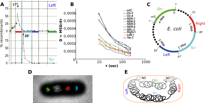
For cells that are not replicating the genome, the position of genetic loci along the chromosome is linearly correlated with their position in the cell Viollier et al., (2004); Breier & Cozzarelli, (2004); Wiggins et al., (2010). The exact subcellular positioning of different loci varies in different bacteria Toro & Shapiro, (2010).
-
(vi)
During replication, daughter chromosomes demix and segregate before cell division. In E. coli, the two arms of the chromosome are segregated in an organized left-right-left-right asymmetric fashion from the center of the cell Wang et al., (2006); Nielsen et al., (2007); Toro & Shapiro, (2010). Replicated chromosomal loci are thought to be immediately recondensed, as they appear to preserve the linear arrangement while they are moved in opposite directions to assume their final position in the incipient daughter cell Thanbichler et al., (2005); Thanbichler & Shapiro, (2006).
In short, the nucleoid’s physical organization plays a major role in the most important cellular processes, such as cell division, DNA replication, and gene expression. Explaining these links is a long-standing open problem in microbiology. Today, it can be revisited with new quantitative experiments, including both the “-omics” approaches and more sophisticated and controlled experimental techniques allowing the analysis of nucleoids both in vitro and in vivo. This is paralleled by a renewed interest in the quantitative characterization of bacterial physiology Scott & Hwa, (2011); Scott et al., (2010); Zaslaver et al., (2009), of which the nucleoid constitutes a fundamental part. Also note that the relative chromosomal positioning and the orientation of genes are subject to natural selection on evolutionary time-scales as shown by the comparison of the genetic maps of different bacteria Ochman & Groisman, (1994); Rocha, (2008).
Consequently, the field is blossoming with a new wave of studies, hypotheses and findings. While many new pieces of evidence are available, the challenge of building coherent pictures for physical nucleoid organization and its role in the different cell processes remains open. Our scope here is to present the main hypotheses and the experimental and modeling tools that have been put forward in order to understand the physical aspects of nucleoid organization, and give the interested reader an ordered account of the known facts.
III Measurements
We start by reviewing some of the salient experimental findings at the scale of whole-nucleoids, with a particular emphasis on the more recent results. The evidence that we will discuss emerges from a combination of experimental biology, biophysics, high throughput biology and bioinformatic approaches (see BOX 2 on page III). Most of the studies reviewed here are based on E. coli or Caulobacter.
BOX 2: Several available experimental techniques can probe the nucleoid at large scales.
![[Uncaptioned image]](/html/1204.3518/assets/x1.png)
Advanced microscopy together with cell-biological techniques yield information about structure and dynamics of the nucleoid (Right). High-resolution tracking of tagged loci allows measurement of the local viscoelastic properties of the nucleoid. Static configurations of chromosomal loci in fixed cells allow determination of their spatial arrangement within nondividing cells and following cell division. Dynamic tracking at long time scales gives information on the chromosome’s “choreography” followed over a cell cycle and on the macrodomain subcellular arrangement. Fusions of NAPs with fluorescent proteins also enable evaluation of their localization within the nucleoid.
Nucleoids can be purified and manipulated outside of a cell in order to access more directly their biophysical properties (Left). This procedure implies release of confinement and crowding, and dilution of binding proteins. As a result, purified nucleoids are several times larger in radius than the size of a cell. Traditionally, purified nucleoids were imaged by electron microscopy, showing a ramified plectonemic structures. More recently, investigators have concentrated on characterizing their organization as polymers tracking of labelled loci, fluorescence correlation spectroscopy(FCS) and probing them mechanically (AFM).
In addition, information on nucleoid organization can be obtained from high-throughput experimental datasets (Bottom). Transcriptomics (using sequencing or microarrays) can be used to probe transcriptional response to nucleoid perturbations, such as NAP deletions, changes in the average level of supercoiling, or local release of a plectonemic loop. Another important source of data is protein occupancy, for example by NAPs, and its correlation with gene expression. This information is obtained both by microarray (CHiP-chip) and by next-generation sequencing techniques (ChIP-seq). Recombination has been used to define macrodomains, as compartments within which recombination between chromosomal segments was more likely than recombination with segments laying outside of the compartment. Finally, Chromosome Conformation Capture (3C) techniques probe the spatial vicinity of pairs of chromosomal loci in the (average) cell. They can be combined with sequencing in order to produce high-throughput data sets (Hi-C).
Chromosome spatial arrangement and compartmentalization.
Strikingly, the intracellular localization of a given chromosomal locus in a cell is remarkably deterministic, as revealed by fluorescent tagging of chromosomal loci on E. coli and Caulobacter crescentus Niki et al., (2000); Viollier et al., (2004); Wang et al., (2006); Liu et al., (2010); Nielsen et al., (2006); Wiggins et al., (2010). The series of chromosome segments is localized along the long axis of the cell in the same order as their positions along the chromosome map, with the interlocus distance typically linearly proportional to (arclength) genomic distance. The precise location of individual loci varies in the known bacteria, and might depend on DNA-membrane tethering interactions Toro & Shapiro, (2010). A recent microscopy study on E. coli Meile et al., (2011) considered the positioning of the chromosome in the short-axis section of the cell. They found that the Ter region occupies the periphery of the nucleoid, at a larger distance from the longitudinal axis with respect to the rest of the chromosome. In newborn or non-replicating cells, the two chromosome arms are spatially arranged such that loci on the left arm of the chromosome lie in one half of the cell and loci on the right arm lie in the opposite half, with the replication origin between them. It is tempting to interpret the resulting sausage-shaped structure as a chromosomal “fiber.” In a recent study Wiggins et al., (2010), the cell-to-cell variability of loci positioning in non-replicating cells was used to estimate an internal elasticity, which, perhaps not surprisingly, appears to be much higher than expected from a naive estimate for a linear polymer.
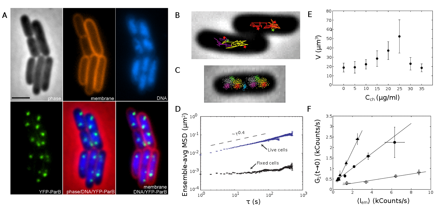
Even more recently Umbarger et al., (2011), high-throughput Chromosome Conformation Capture (3C) techniques have been used in combination with live-cell fluorescent tagging of loci, in order to determine the global folding architecture of the Caulobacter crescentus swarmer cell genome. These data indicate that a chromosomal fiber exists also in this case, spanning the whole chromosomal ring. Additionally to the linear spatial arrangement of loci according to their chromosomal coordinate, loci of the left chromosomal arm tend to be very proximal to symmetric loci on the right arm. The resulting structure is a compressed ring-like fiber, which, the authors argue, typically takes an eight shape, free to roll around the long cell axis. They also find that the symmetry in the cross-chromosomal arm interactions is determined by the protein-dense attachment point to the cell membrane at the old pole of the cell, triggered by the binding of the ParB protein to its target parS binding sites. Moving the parS pole-anchoring site by 400 Kb along the chromosome (but not the replication origin) determines a sliding of the whole interaction structure, as in a tank crawler. This sliding is slightly asymmetric, suggesting the presence of supplementary attachment points between chromosomal arms or between the chromosome and the cell body.
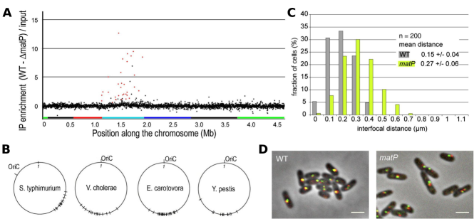
Another important recent discovery, consistent with the mentioned correlation between chromosome arms and cell halfs, is the existence of “macrodomains” Valens et al., (2004); Moulin et al., (2005); Espéli & Boccard, (2006) often described as chromosomal isolated compartments. The first evidence in this direction Valens et al., (2004) came from measurements of the recombination frequency between loci, see figure 1A. All else being equal, this should be proportional to the probability that the two chromosomal segments come into contact within the cell. For a well mixed polymer, the recombination frequency should be uniform. However, experiments show a highly non uniform pattern, compatible with a compartmentalized structure with clear boundaries. Four macrodomains of a few hundred Kb in size have been identified, corresponding to regions surrounding the replication origin and terminus, and to two symmetric regions at the edges of the Ter macrodomain, see box and figure 1C,D,E. The remaining “non-structured regions” appear to have different physical properties. Subsequent studies have confirmed the presence of macrodomains and measured their dynamics using fluorescently labelled loci Espéli et al., (2008); Espéli & Boccard, (2006); Lesterlin et al., (2005), see figure 1B,D. While the molecular mechanisms responsible for this level of organization are not yet clear, the same authors also found that the Ter macrodomain appears to be condensed by a single DNA-binding protein (MatP, figure 2) with a small set of specific binding sites Mercier et al., (2008). Other proteins with macrodomain-specific DNA-binding properties have recently been identified Dame et al., (2011); Sánchez-Romero et al., (2010); Tonthat et al., (2011); Cho et al., (2011), and appear to be conserved in related bacteria. In view of the results from this thread of work, macrodomains might be seen as a process of microphase separation triggered by specific protein binding. Interestingly, as mentioned above macrodomain-like regions also emerge from independent large-scale genomic data Mathelier & Carbone, (2010); Berger et al., (2010); Scolari et al., (2011).
During replication, the chromosomes segregate following a well-defined “choreography,” which has been the subject of multiple studies Berlatzky et al., (2008); Toro & Shapiro, (2010); Jun & Wright, (2010). While segregation is not the main focus here, it is useful to discuss it briefly, as the existing approaches to this problem (both experimental and theoretical) are intimately linked with chromosome organization and will be mentioned in the following. Specifically, spatial reorganization of the segregating chromosome arms appears to preserve qualitatively the relationship between loci distance along the chromosome and in the cell. Moreover, reorganization ensures that the two replication forks remain in opposite halves of the cell during replication and that the relative orientation of the two reorganized nucleoids in pre-division cells is not random. Quite interestingly, the spatial separation of sister chromosomes is not a continuous process, but has been observed to proceed through “snaps” Joshi et al., (2011); Espéli et al., (2008), suggesting the existence of energetic or entropic barriers for separation, possibly overcome by active processes.
There is debate on what is the main driver for chromosome segregation: Entropic repulsion forces due to strong confinement into a box of linear polymers have been proposed to explain this behavior at least in part Jun & Wright, (2010); in experiments on replicating B. subtilis the chromosome compaction and spatial organization have been hypothesized to result from non-equilibrium dynamics Berlatzky et al., (2008), rather than from an entropic repulsion process; in Caulobacter crescentus, a contribution from bidirectional extrusion of the newly synthesized DNA from the transcription complex has been postulated to contribute to chromosome segregation Jensen et al., (2001); Toro & Shapiro, (2010). Experiments using inhibition of protein synthesis by chloramphenicol show that this produces nucleoids with a more rounded shape and induces fusion of separate nucleoid bodies van Helvoort et al., (1996). Upon release from protein synthesis inhibition, the two nucleoids reoccupy the DNA-free cell independently of cell elongation Helvoort et al., (1998). The control of the segregation mechanism by protein synthesis processes could be indirect: ongoing protein synthesis can affect nucleoid compactness and segregation at multiple levels, including the decrease in the amount of enzymes modifying the topology of the DNA or carrying out transcription and DNA replication. The idea that membrane protein synthesis activates nucleoid segregation directly has also been proposed Norris, (1995) (in bacteria the presence of cotranscriptional translation creates a direct physical link between the genome and the membrane Woldringh & Nanninga, (2006)) Also note that current arguments based on segregation by entropy and confinement might turn out to be inconsistent with interpreting macrodomains as the result of a microphase separation, since a variety of interactions, and specifically protein-DNA binding processes could play an important role in defining the free energy of the nucleoid.
Supercoil domains and nucleoid associated proteins.
At smaller scales, the circular chromosome of E. coli is organized in plectonemic loops, or “supercoil domains.” Those regions are separated by topological barriers formed by nucleoid associated proteins (NAPs) such as Fis and H-NS, see figure 3. These proteins bridge two strands of DNA by binding to both, and prevent the propagation of torsional energy. As a result, they also prevent the spreading of uncontrolled effects on gene expression in case of accidental DNA breaks or mechanical strain, caused for example by advancing replication forks Postow et al., (2004); Skoko et al., (2006); Stavans & Oppenheim, (2006).
Multiple NAPs have been identified, each with its specific binding properties (reviewed in Luijsterburg et al., (2006)). Besides bridging double strands, they can change the local shape of DNA inducing bends or hinges and form nucleoprotein filaments. Additionally, NAPs often have multiple DNA binding modes which might be dependent on physiological factors. A good example of this behavior is H-NS. Its binding results in DNA—H-NS—DNA bridges, but also forms a rigid nucleoprotein filament which could act as zipper in vitro Amit et al., (2003); Dame et al., (2006); Wiggins et al., (2009). It seems that the nucleoprotein filament formation may be important, as it has been found to be a structure shared by other NAPs including HU van Noort et al., (2004) and StpA Lim et al., (2011). Biologically, this could hypotetically provide a mechanism for environmental sensing by NAPs. It is thus possible that our current understanding of NAP binding, being based on the limited number of conditions tested, is incomplete and generalization of a DNA binding property to other solution conditions may be dangerous.
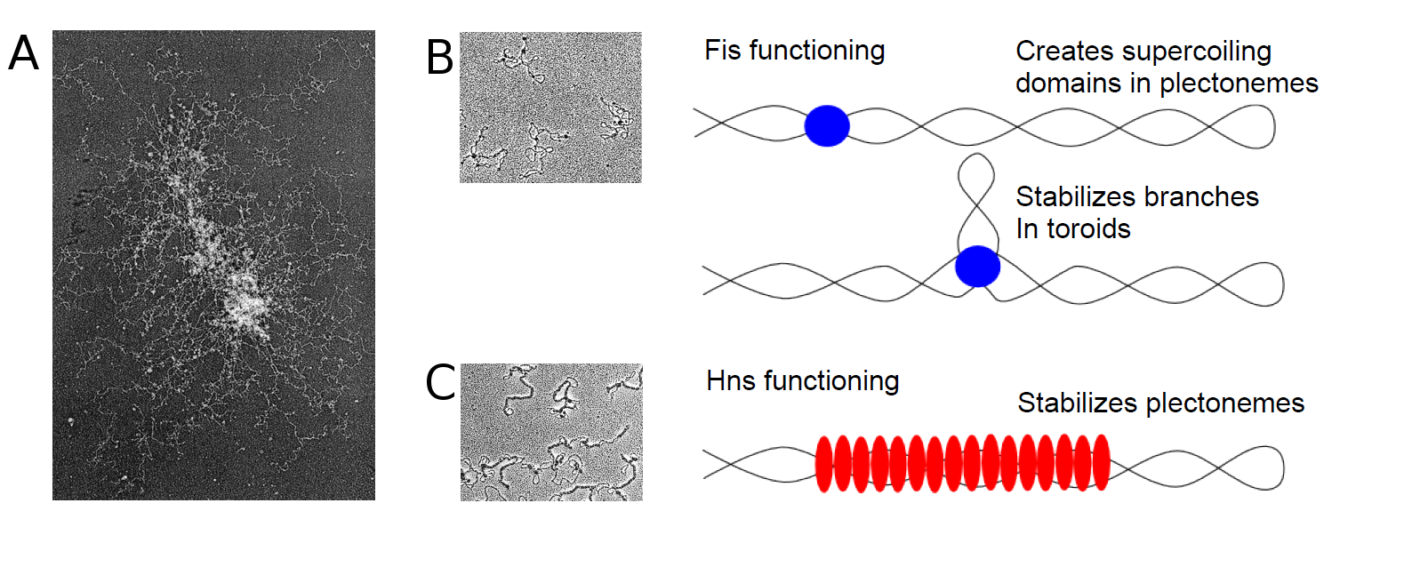
The structure of supercoil domains was studied in E. coli by Postow and coworkers, through analysis of the supercoiling-sensitive transcription of more than genes following relaxation by restriction enzymes in vivo, and by electron microscopy Postow et al., (2004). They concluded that domain barriers may vary dynamically and/or across a population, but they follow an exponential length distribution. The average domain size is Kb, implying the existence of about domains Postow et al., (2004); Stavans & Oppenheim, (2006). Branches of the same typical length are visible directly from electron micrographs of purified nucleoids Postow et al., (2004); Kavenoff & Bowen, (1976). Thus, the genome topology may be visualized as a branched structure with supercoiled domains that are subject to modulation by nucleoid-associated proteins, and active processes such as DNA transcription and DNA replication.
The factors responsible for establishing the boundaries of supercoiled domains and the determinants of domain size and number are still largely unknown. While H-NS and Fis, along with MukB, the analogue of the eukaryotic SMC (Structural Maintenance of Chromosomes) protein, could be involved in stabilizing plectonemic conformations, their precise roles and importance in this context have not yet been established in a definitive fashion, either in vitro or in vivo Dillon & Dorman, (2010); Skoko et al., (2006); Maurer et al., (2009); Grainger et al., (2006).
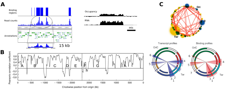
By contrast, a consistent amount of information on the binding of NAPs in different conditions and its effects on the cell state is available from high-thoughput experimental techniques (figure 4.) Nucleoid associated proteins can modulate the nucleoid conformation structure in response to changes in environmental conditions Luijsterburg et al., (2006). This can result in large-scale changes in gene expression Dillon & Dorman, (2010). The local mechanical action of NAPs on DNA is often well-characterized by single molecule experiments Luijsterburg et al., (2006), which also lead to the observation of chromatin-like nucleoprotein “fibers” Kim et al., (2004). Large-scale NAP binding data in specific growth conditions was obtained from high-throughput experiments involving microarrays (CHiP-chip) or sequencing (CHiP- seq) Grainger et al., (2006, 2008); Oshima et al., (2006); Wade et al., (2007); Grainger et al., (2007); Kahramanoglou et al., (2011). Furthermore, transcriptomics studies profiled the changes in gene expression upon different nucleoid perturbations, such as NAP deletion and/or altered supercoiling Blot et al., (2006); Marr et al., (2008); Bradley et al., (2007); Berger et al., (2010). Many of these data sets show linear regions of dense binding that often correspond to macrodomain boundaries, and associate with global or NAP-dependent transcriptional response and its correlation with codon bias Scolari et al., (2011); Mathelier & Carbone, (2010).
The physical origin of this preference in binding and the gene expression changes at the boundaries of macrodomains is not precisely clear. One possibility is that macrodomain boundaries might be co-localized by NAP structures. A recent interesting experimental study Vora et al., (2009) looked at protein occupancy along the genome regardless of protein identity. This work uncovered extended polymer-like domains rich in bound proteins (including NAPs) with an average length of 1.6Kb, associated with transcriptionally silent or transcriptionally enhanced regions (and also with high intrinsic DNA curvature.)
When cells enter the stationary phase a radical, global condensation of the nucleoid occurs. It is believed that this is a mechanism via which the cell can protect its DNA in harsh conditions Kim et al., (2004). AFM studies have shown that the structure of the DNA differs at the supercoiling level Kim et al., (2004) and that action of Dps and CbpA, the NAPs that replace Fis in this growth phase, is quite different. The Dps and CbpA proteins produce compact aggregates (which can protect DNA from degradation by nucleases) rather than binding to distributed sites as Fis does Cosgriff et al., (2010). Interestingly, the action of Fis counters Dps-induced compaction through a transcriptional response affecting the expression of topoisomerase and gyrase Ohniwa et al., (2006).
Compaction by molecular crowding, specific proteins, transcription factories, and confinement.
A different question concerns identifying the main factors contributing to nucleoid compaction and organization. The main candidates are macromolecular crowding, electrostatic self-attraction, supercoiling and nucleoid proteins.
Macromolecular crowding, or the high concentration of macromolecules present in the cytoplasm, is generally believed to be an important determinant on the basis of theoretical arguments de Vries, (2010); Odijk, (1998), which predict a possible phase separation mechanism between nucleoid and cytoplasm. Note that the generic term “macromolecular crowding” might include depletion interactions (see below), together with a number of additional effects of entropic and energetic nature.
DNA condensation by crowders can be observed in vitro under very controlled and well-understood conditions, for example by experiments using dextran or PEG, demonstrating that naked DNA can be directly condensed by these crowders Zhang et al., (2009); Estevez-Torres & Baigl, (2011); Huang et al., (2007); Xu & Muller, (2012) Similar (but less controlled) behavior, is shown by purified nucleoids Zimmerman, (2004). At the same time, experimental studies on isolated nucleoids obtained from mutants lacking various NAPs Zimmerman, (2006a) suggest that the effects of crowding on compaction are substantial and independent of the NAP composite background. It has also been suggested that the action of NAPs could be aimed at antagonizing compaction rather than compacting the nucleoid Zimmerman, (2006b). However, this must be a complicated, combined effect involving forces of different nature. In the absence of crowding and confinement, it is obvious that the radius of gyration of the genome would be smaller if it were organized, e.g. in a branched structure of plectonemic loops stabilized by DNA-bridging NAPs Postow et al., (2004); Trun & Marko, (1998).
As a particular case of crowding effect, it has been proposed that the (attractive) depletion interactions, well known in colloid science, might play an important role in chromosome organization Marenduzzo et al., (2006a) This force is due to reduction in total solvent excluded volume upon formation of a molecular complex. Depletion interactions are consequential when large molecular assemblies are formed in presence of smaller particles. In fast-growth conditions, genome-bound RNA polymerase is localized into a few transcriptionally active foci or “transcription factories,” Jin & Cabrera, (2006); Cabrera et al., (2009), analogous to the eukaryotic case Marenduzzo et al., (2006a, b). Depletion interactions were suggested to explain the formation of these macromolecular assemblies Marenduzzo et al., (2006b). Interestingly, the formation of these foci has been associated with the presence of the NAP protein HU Berger et al., (2010). We add that a similar argument might hold for the local compaction of the surroundings of OriC, rich in ribosomal RNA producing regions, in a macrodomain-microphase. In other words, ribosome-rich ribosomal RNA transcripts, attached to the genome through RNA polymerase, could help compact the Ori region. If this should be the case, the compaction properties of this region would change with the number of ribosomes being synthesized, i.e. with growth rate and translation efficiency Scott & Hwa, (2011) (One can also speculate that, for the same reason, newly-replicated DNA could be sequentially aggregated during replication.) On one hand, experiments show that transcription of ribosomal RNA operons (which are generally located in the Ori macrodomain) is related to the compaction of nucleoids observed upon inhibition of translation Cabrera et al., (2009). On the other hand, these processes must be intersected with the (non entropic) binding of NAPs, given the evidence connecting specific NAPs to the compaction of the Ter macrodomain (MatP) and indirectly to the effect of the HU protein on organization of the Ori macrodomain.
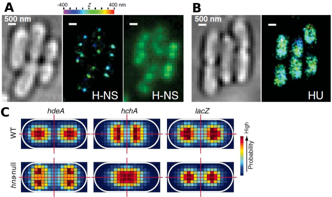
A very recent study Wang et al., (2011a) systematically addressed the chromosomal localization and role in spatial organization of the nucleoid of five major NAPs (HU, Fis, IHF, Stpa, H-NS) using fluorescent protein fusions and super-resolution fluorescence microscopy. In the growth conditions tested all the proteins showed scattered distributions in the nucleoid, except for H-NS, which seemed to form (on average) two distinct foci per chromosome copy, bringing together different (even distant) H-NS targets, see figure 5. Thus, H-NS should be added to the list of NAPs with a compacting action on the nucleoid associated with the formation of specific foci. The long-range interactions between H-NS binding targets were validated by 3C, and show no apparent coherence with the macrodomain structure; loci pairs that are near to H-NS targets but are not targets show no 3C signal and wider distributions of subcellular distances (evaluated with microscopy). In general, DNA associated with many NAPs has a much larger surface, which should enhance the depletion interactions. For instance, DNA coated with an H-NS nucleoprotein filament will have a diameter of about 20 nm instead of the 2 nm of naked DNA. H-NS nucleoprotein filament formation could strongly enhance the depletion attractions with respect to other NAPs not forming filaments, and filaments formed at remote locations on the contour of the chromosomal DNA could find each other in a crowded environment. This may explain the results from super-resolution imaging mentioned above Wang et al., (2011a). We note again that this is one of the counter-intuitive aspects of molecular crowding, since one might think that it would be easier to encounter another filament if there were no “obstacles”. The confusion can come from thinking about diffusion, which is hindered in a crowded environment due to the higher effective viscosity, versus thermodynamics, where the depletion interactions increase the probability of observing two nucleofilaments in contact at equilibrium. Adopting a simple Kramers-type model for the kinetics, it is not obvious whether the rate of association would go down due to the prefactor or go up due to the lower free energy minimum. Implicit in the entire discussion implicating crowding in aggregation in this cellular context is the idea that the distribution of nucleoprotein filaments is at near-equilibrium even though the cell is dividing rapidly.
Finally, depending on the degree of autonomous compaction, the confinement exerted by the cell wall might play a relevant role in nucleoid organization and segregation Jun & Wright, (2010). This is expected to be particularly significant in fast-growth conditions, where the genome needs to be highly accessible for transcription (and thus will not be condensed) and the amount of genome per cell is higher due to overlapping replication rounds Nielsen et al., (2007).
To summarize, the degree of condensation and the geometry of the nucleoid are strongly dependent on the growth phase and growth rate of the bacteria. Such changes may be modeled by means of equilibrium states, slowly evolving in accordance to “external” control parameters (e.g. the concentration of the various NAPs). However, it seems likely that nonequilibrium processes are also important, and relevant aspects of the problem might be lost in attempting to describe the nucleoid condensation process purely in terms of an approach to thermodynamic equilibrium. For example, on the other side of the spectrum in terms of biological complexity, the recently found scale-invariant structure of the human genome Mirny, (2011); Lieberman-Aiden et al., (2009) was suggested not to be the result of an equilibrium state, but similar in nature to the so-called “crumpled globule,” or “fractal globule,” Grosberg et al., (1993) since loci that are near along the genome arclength coordinate are also physically proximal in three-dimensional space. This proximity contrasts with what happens in an (equilibrium) collapsed polymer, where the linear structure is completely mixed in the globule.
Let us quote a few experimental findings, some of which are very recent, supporting the role of nonequilibrium processes, specifically for bacteria. The initial conditions, i.e. the choreography of DNA replication, appear to play a central role in defining the final structure of the nucleoid Daube et al., (2010). In artificial E. coli strains with two distant origins instead of just one Wang et al., (2011b), the two origins initiate replication synchronously at the expected separate positions of the genetic loci associated with them. Replication forks move independently, indicating that replication does not occur in a single replication factory and that the replication machinery is recruited to origins rather than vice versa. Most importantly, in these experiments progression of replication plays a major role in determining the space-time pattern of locus segregation. The large scale structure of the B. subtilis nucleoid Berlatzky et al., (2008) has been observed at various stages of the replication process. The newborn portions of the chain are compacted and sequentially conveyed towards the poles, resulting in an ordered, spiraling structure. A strong correlation between space coordinate and genomic coordinate is preserved, similar to the linear behavior observed in E. coli Wiggins et al., (2010). A choreography of this sort is found also in Caulobacter Jensen et al., (2001). Finally, the large scale spiral structure of the nucleoid of Bdellovibrio bacteriovorous Butan et al., (2011) also suggests a metastable steady state, sustained by cooperative motion and/or energy exchanges. It seems difficult to disregard the deterministic replication-segregation dynamics in describing such phenomenology Breier & Cozzarelli, (2004); Toro & Shapiro, (2010).
Viscoelasticity and structural units.
Tracking studies of fluorescently labeled chromosomal loci have evaluated in vivo dynamic properties of the nucleoid with fairly high time resolution, measuring for example the mean-square displacement (MSD) of the loci or the time autocorrelation function Espéli et al., (2008); Weber et al., (2010a); Meile et al., (2011). In general, these measurements give information on the local relaxation time scales of the nucleoid and its viscoelastic behavior, see figure 6. On one hand, for large time-scales Espéli et al., (2008), loci mobility correlates well with macrodomain structure. In particular, the MSD saturates at the spatial scale of the macrodomain size. On the other hand, especially for smaller time scales, the mean square displacement of a locus is seen to follow a power law: (where is its arclength genomic coordinate) and the exponent seems to be universally close to , independent of Weber et al., (2010a). Perhaps surprisingly, an extra-chromosomal RK-2 plasmid showed the same behavior, while smaller RNA particles had a higher subdiffusive exponent. It must be mentioned that the localization of RK-2 plasmids appears to be highly regulated Kolatka et al., (2008); Derman et al., (2008). The underlying viscoelasticity of the bacterial cytoplasm surrounding the nucleoid is still poorly understood. Some characterisation has been approached in vivo via FRAP measurements of diffusing GFP Konopka et al., (2006).
The observed anomalous diffusion has been modeled using phenomenological approaches. There are a variety of dynamical models exhibiting anomalous diffusion, such as Langevin equations with time dependent viscosity (also equivalent to fractional Langevin equations), continuous time random walks, and random walks over a fractal object Burov & Barkai, (2008); He et al., (2008); Condamin et al., (2008). Weber et al. Weber et al., (2010a, b) compared their data with the results obtained on the basis of the first two approaches, and the fractional Langevin equation gave a more satisfactory agreement.
The anomalous diffusion exponent per se does not give quantitative information on the geometry of the nucleoid. Physical measurements, possible on purified nucleoids Cunha et al., (2001, 2005); Romantsov et al., (2007), can enrich the scenario. In particular, Romantsov and coworkers Romantsov et al., (2007) have obtained, using fluorescence correlation spectroscopy, the time-dependent coarse-grained density distribution of purified nucleoids. They conclude that the polymer appears to be composed of a set of “structural units” defined by a measurable correlation length. The measurements were performed at varying degrees of supercoiling, induced by different concentrations of the chloroquine drug. The size of the structural units was found to vary from 50-100Kb in high (positive or negative) supercoiling to 3Kb at zero supercoiling. The diameter of the purified nucleoid varied from 2.5m in high supercoiling to 3.5m in low supercoiling. The authors also estimated the typical diameter of the structural units from the diffusion constant (obtained from the decay of the fluorescence autocorrelation function) and Stokes-Einstein’s relation. Perhaps surprisingly, the resulting size of structural units was near 70-80 nm regardless of supercoiling. Thus the emerging picture for the unconfined genome is that of a string of highly dense “beads”, each containing Kuhn lengths (effective independently jointed elementary polymer segments) of DNA each. These values apply in the presence of supercoiling but in the absence of crowding and confinement effects, as for purified nucleoids most of the cytoplasmic (and probably a considerable part of the DNA-binding) proteins are probably diluted away.
IV Models
We will now review a few modeling approaches put forward in recent years, and point to some more classic work in polymer physics that we believe could be relevant in this context. As the reader will have observed, the wealth of existing experimental results is appealing on one hand, but, on the other hand, does not offer any clear grasp on a small set of relevant ingredients necessary for building coherent physical descriptions of the nucleoid. Consequently, the approaches adopted in the literature are highly diverse and heterogenous in terms of premises, methods, ingredients, and points of view.
The notable efforts to understand entropic aspects of chromosome segregation (reviewed in Jun & Wright, (2010)) have revived the studies on entropic forces of single and multiple confined polymers dating back to Edwards and DeGennes Jun & Mulder, (2006); DeGennes, (1979); Edwards & Freed, (1969); Jung & Ha, (2010). As we have pointed out above, the role of bound proteins on nucleoid entropy is typically disregarded in these arguments.
A comprehensive review of the literature on confined DNA in different contexts is provided in Marenduzzo et al., (2010). Considering this approach, there remains the open issue that the observed segregation and compaction times might be too short to be compatible with an entropic process. For linear polymers this time has been evaluated by Molecular Dynamics simulations to scale like the square of the number of monomers, , which, for sufficiently large , will be smaller than the chain diffusion time Arnold & Jun, (2007). However, it is not straightforward to use these scaling relations for empirically relevant estimates. Moreover, these estimates will depend on the model used to represent the structure of the nucleoid and its correlation with the replication process, linking this problem to other unanswered questions. Overall, it seems likely that entropy is only part of the story, and active / nonequilibrium processes of different kinds might play a role in chromosome segregation.
We will now turn our attention to work concerning nucleoid organization and cellular arrangement. Buenemann and Lenz have attempted to understand the linear arrangement of chromosomal loci in terms of a purely geometrical model Buenemann & Lenz, (2010), where a linear polymer in the form of a string of blobs, is confined within a cylinder and locked at one or at a few loci. This constrained geometry obviously provides an ordering mechanism, as long as the blobs are large enough with respect to the cylinder’s diameter. This model makes predictions on the spatial arrangement of the chromosome in mutants of C. crescentus Viollier et al., (2004) and on the cell-cycle dependent ordering in E. coli. A very recent simulation study Fritsche et al., (2011) explains the linear ordering observed in E. coli as a product of confinement and entropic repulsion of a string of linearly arranged chromosomal loops. In order to show this, they represent the chromosome as a confined circular self-avoiding chain under the constraint that consecutive loops, identical in size, are distributed along the arclength coordinate, while the Ter region does not contain such loops. Their simulations show both linear ordering along the cell axis and Ter region occupying the outer periphery of the nucleoid Meile et al., (2011), as properties of the equilibrium states. Intriguingly, the same study suggests that this linear-loop ordering could be a consequence of the transcription network organization. To support this point, they simulate a polymer where transcription factor-target pairs are coupled by attractive harmonic interactions, and show that the linear ordering is recovered. It is well-known that transcription factor-target distances have a statistical tendency to be short along the chromosome Warren & ten Wolde, (2004).
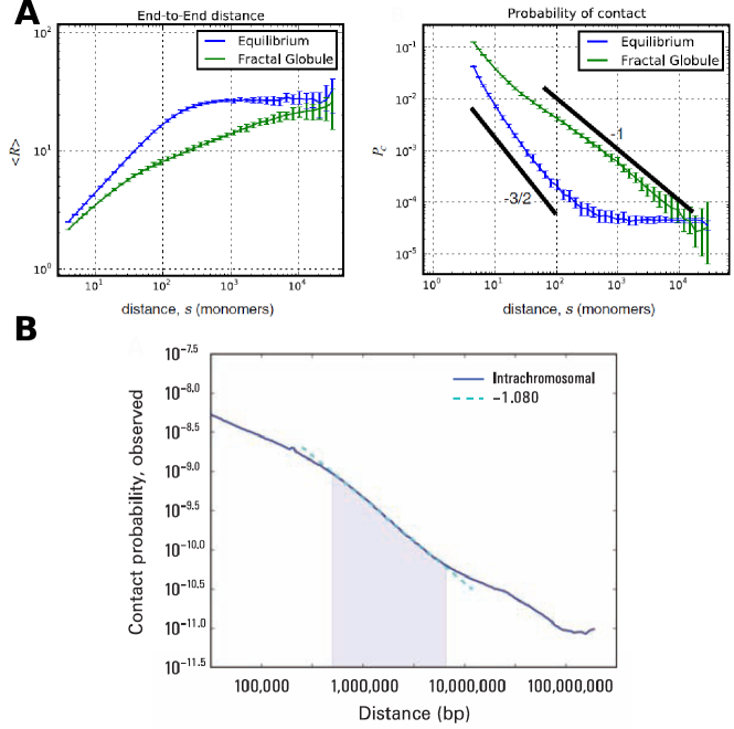
Vettorel and coworkers Vettorel et al., (2009) performed an abstract study motivated by the possible nature of the compartmentalization and the structural units of a generic (eukaryotic or prokaryotic) chromosome forming a crumpled or fractal globule mentioned in section III (figure 7). Specifically, they explore metastable collapsed states of polymers, where the total size scales as as in an equilibrium compacted globule, but a much higher degree of compartmentalization is present. At odds with the intrinsic disorder of the equilibrium globule, in a fractal globule, for any pair of loci separated by a chain length , the distance has the scaling behavior Mirny, (2011). In other words, both the equilibrium globule (i.e. the equilibrium collapsed structure emerging from polymer self-attraction or unfavorable entropy of mixing) and the fractal globule have mass fractal dimension , but a generic volume (and in particular a blob in the DeGennes sense) inside the equilibrium globule can include non-sequential segments, while in the fractal globule it contains a single sequential segment Grosberg et al., (1993). This structure can be understood as resulting from a process where condensation sequentially involves larger and larger scales in , so that the genomic proximity is preserved in a scale invariant fashion. As a consequence, distant portions of the chromosome will occupy different compartments within the globule.
In order to obtain a fractal globule in a simulation, a constraint preventing entanglement must exist Vettorel et al., (2009). To obtain this condition, Vettorel and coworkers considered a semi-dilute or concentrated solution of mutually disentangled (unconcatenated) rings. Also note that because of the constraint preventing entanglement during collapse, a fractal globule is generally larger in size than an equilibrium globule of equal chain length (while for both the total size scales as ). The fractal globule configuration has proven to be relevant for the description of genomic DNA organization in non-dividing eukaryotic (human) cells Lieberman-Aiden et al., (2009); Mirny, (2011). In particular, it has been found that the human chromosomes display this structure from Kb to Mb. This range of sizes might be relevant to bacteria that have genomes spanning a few Mb (also because the lower cutoff might be related to the fiber organization of chromatin, which in bacteria is different). Thus, as the authors speculate, the fractal globule description might be useful for the nucleoid as well. We will further discuss this point in the light of the available data in section V.
We have already mentioned above the theoretical work on the role of macromolecular crowding on compaction de Vries, (2010); Odijk, (1998), and of depletion interactions on loop formation (and possibly on macrodomain organization Marenduzzo et al., (2006b)). A recent simulation study Junier et al., (2010) has concentrated on the chromosome-shaping role of transcription factories. Considering a self-avoiding worm-like chain with a fixed hard-core repulsion radius, and short-ranged bridging protein complexes, they show that this system can take “micro-structured” collapsed globule configurations, where bridging complexes cluster and regions of high and low densities of interacting sites coexist in a microphase separated thermodynamic state. More abstract analytical studies could provide a useful context for understanding the role of NAP binding Diamant & Andelman, (2000); Kantor & Kardar, (1996).
Finally, some attention has been devoted recently to the role played by the branched plectonemic structure of the nucleoid Odijk & Ubbink, (1998); Ubbink & Odijk, (1999), which is believed to have relevant implications for transcription Dillon & Dorman, (2010); Postow et al., (2004). Provided the correct questions are formulated, this topic has the advantage of being placed in a strong framework developed in the past 30 years, building on the classical calculations of Zimm and Stockmayer Zimm & Stockmayer, (1949) for considering arbitrarily ramified ghost chains as gaussian networks, and obtaining their equilibrium properties Sommer & Blumen, (1995); Farago & Kantor, (2000); Graessley, (1980). For example, some recent work has focused on the induction of loops involving multiple polymer segments Sumedha & Weigt, (2008). In this framework, reliable estimates for the self-avoiding case can be obtained with Flory-like arguments, taking into account the fact that branched polymers have higher internal topological complexity, which makes the repulsive interactions stronger than between linear or circular chains. The same approach is also possibly relevant for studying dynamic aspects of loci mobility Jasch et al., (2003); Dolgushev & Blumen, (2009), which can be compared with simulations and more phenomenological models Weber et al., (2010b). Finally, a number of very refined results based on renormalization group / field theoretical methods have been obtained in a more abstract context using the “randomly branched polymers” model (or “lattice animals”), typically defined as the ensemble of all the clusters of connected sites (monomers) on a regular lattice. While this ensemble is probably too general, it is possible that these results have implications for questions related to the structure of the nucleoid. For example, recent work based on Langevin dynamics Janssen & Stenull, (2011) analyzes their collapsed regime, obtaining a fractal dimension , intermediate between the swollen chain and the fully compacted globular state .
V Discussion. Hypotheses and paradoxes concerning nucleoid geometry and dynamics.
We would like to discuss here some speculations on the possible links between the experimental and theoretical results discussed above.
Let us start with an analogy to the geometrical organization of eukaryotic chromatin, where different geometrical features are observed at different scales, ranging from the known fiber organization up to the arrangement in preferred “chromosome territories” within the nucleus. In this case, experimental techniques such as Chromosome Conformation Capture (3C) and its high-throughput variants Lieberman-Aiden et al., (2009), or FISH (fluorescence in situ hybridisation, where fluorescent tags are attached to pairs of DNA loci) allow, for instance, to measure the mean absolute distance, , and the contact probability, , of two genomic loci at arclength distance . For bacteria, these techniques entail a number of specific difficulties, but the data are starting to be available, as already mentioned Umbarger et al., (2011). However, the study by Umbarger and coworkers focuses on the large-scale nucleoid 3D architecture, rather than on more detailed properties of the interaction map. Apart from saturation at large genomic distances, is typically found to increase with Mateos-Langerak et al., (2009) with an approximate power-law behavior, . Here, is a scaling exponent, interpretable as , which empirically varies with scale of observation and cell type in eukaryotes. If a stretch of length of a polymer spans a region of size , in dimensions, it can occupy a volume . Thus, heuristically, one expects that the probability of one end of the stretch meeting the other scales as . Experimentally, for loci on the same chromosome, decreases as a power-law , for a set of length scales in the approximate interval 0.5-7Mb for Lieberman-Aiden et al., (2009), which provides evidence for a fractal globule-like organization, . This is confirmed by FISH data for on a smaller range of genomic lengths Mateos-Langerak et al., (2009).
However, experimental data on for chromatin are complex, as the exponent is cell-type specific and varies with genomic length, reflecting different degrees and modes of chromatin compaction. At short genomic distances is found in the range at short distances, and reaches a plateau (i.e., ) at order 10 Mb genomic distances, because of chromosome territories Shopland et al., (2006); Mateos-Langerak et al., (2009); Barbieri et al., (2011). For the E. coli nucleoid, the only available quantitative data Wiggins et al., (2010) indicate that might scale like for non-replicating chromosomes at scales of 0.3-2 Mb.
In principle, a link between nucleoid geometry and the measured nucleoid local dynamics is expected. The anomalous diffusion of chromosomal loci in E .coli has been modeled in terms of fractional Langevin equations Weber et al., (2010b); such an approach correctly reproduces the temporal behavior of the loci, but disregards the geometry of the structure containing them. Very likely, for any polymer, this structure has a fractal character, and one would like to understand how this influences the motion of the loci. A minimal model would consider a relaxation equation over the fractal. For a purely self-avoiding polymer one has . In the case of a self-attracting polymer where attraction is screened by self-avoidance, i.e. at the point DeGennes, (1979); Grosberg & Khokhlov, (1994), it is reasonable to assume a mass fractal dimension , as for a ghost chain. When the tendency towards compaction increases, one expects that will increase accordingly.
In order to illustrate this point with an example, we can consider protein structures. Data from the Protein Data Bank Berman et al., (2000) for 200 proteins with a number of amino acids ranging from to give values of from 2.3 up to 2.6 Enright & Leitner, (2005). On a larger scale, high resolution X-ray spectroscopy has resolved the ribosome structure at the atomic level. Such data indicate Lee, (2006) that the heavier 50S unit is fully compacted, with , while the lighter 30S unit has ; it has been argued that the sparser structure of 30S is compatible with a dynamic geometry, as required in the translation process. Folded proteins are generally described as an harmonic network, by the so-called Gaussian Network Model Reuveni et al., (2010), and we can try to apply a similar reasoning to the nucleoid. Close to the fully collapsed regime of a very long polymer such as a bacterial chromosome, where one expects , the harmonic approximation strictly does not apply, because hard-core repulsion acts against chain compression. The total energy can then be written as an harmonic “Rouse” term, describing waves propagating along the chain, plus a self-attraction term and a self-avoidance term. The relaxation dynamics in such conditions (neglecting hydrodynamic interactions) has been studied within a continuum model approach by Pitard and Orland Pitard & Orland, (1998). They find that the relaxation time of the globule scales with the polymer length as . Taking into account that the globule size scales as (i.e. it fills space), this implies that (since ). This scaling appears to coincide with the experimental value for the anomalous diffusion exponent measured by Weber and coworkers Weber et al., (2010a), previously quoted in the text. In other words, the Rouse subdiffusive dynamics of a collapsing (and thus off-equilibrium) globule follows the same scaling law as the observed local dynamics of nucleoid loci within a range of time scales. While this might be simply a coincidence, it leads us to speculate that the measured dynamic exponent for the mean-square displacement might be the consequence of a fractal-globule-like nature of the nucleoid, at least within a range of length scales.
This argument can be recast in more generic terms. Rouse polymer relaxation dynamics was originally explored by De Gennes DeGennes, (1976), who obtained the scaling relation , where is the so-called dynamical exponent. At the point DeGennes, (1979); Grosberg & Khokhlov, (1994) where one has , the DeGennes’s relation gives , the Rouse result for non-interacting chains. As the polymer dimension increases, a smaller value of is to be expected; in the compacted configuration, where , the relation gives , which is the result reported above. DeGennes’s work is based on scaling arguments, but is confirmed by field-theoretical methods Wiese, (1998) for two-body interactions.
To our knowledge, in the collapsed regime the relation has been proved only at the level of mean field Pitard & Orland, (1998) by modeling, in the spirit of a virial expansion, the effective interaction with an attractive two-body term and a repulsive three-body term.
In conclusion, if the nucleoid behaves like a fractal globule (i.e. an off-equilibrium polymer collapsing because of self attraction, but where entanglement is prevented by topological constraints), or more in general if it has fractal dimension , from mean field theory one expects the subdiffusion exponent . Conversely, if the DeGennes relation is valid, the experimental result , observed in E. coli and in large plasmids Weber et al., (2010a), implies that the nucleoid fractal dimension could be .
To our knowledge the available experimental result that comes closer to a direct measurement of deals with the mean square displacement of fluorescently tagged replisomes Reyes-Lamothe et al., (2008). In the approximation of constant replication fork velocity along the mother DNA, replication time in this experiment and genomic arclength distance have equal scaling. Hence, neglecting the global movements due to chromosomal segregation, the replisome’s anomalous diffusion exponent measures the effective fractal dimension of the replicating DNA. Specifically, the scaling implies . Obviously, this measured in principle contains errors due to fluctuations of a stationary background as well as large scale effects associated with coherent restructuring of the nucleoid. The latter processes are relevant for segregation, but in the initial phases of replication one can assume that they can be disregarded, as we are in the presence of a “weak perturbation” of the stationary (non-replicating) structure. In such a case, the time fluctuations of the fork velocity and the steady state anomalous diffusion of genetic loci would be the main corrections to be taken into account in order to estimate from the measured . Quite interestingly the experimental estimates of the exponent from Reyes-Lamothe et al., (2008) are and for experiments with s and min time lapses respectively. If these numbers were confirmed by further measurements and accurate data analysis, they would support the hypothesis that for a range of chromosomal scales, independently on the reasoning presented above, based on the DeGennes scaling relation .
Clearly, for the nucleoid one expects a structure with a range of fractal dimensions in different scale regimes, as suggested by the case of chromatin and the ribosome. The existence of a range of fractal dimensions is also supported by the microscopy results for fluorescent pairs of loci reporting a linear correlation between loci distance in the cell and along the chromosome Wiggins et al., (2010). The linear correlation could result from any sequentially ordered segregation process generating a uniform mass density. For example, one might consider a periodic winding of the chromosome Képès & Valliant, (2003); Mathelier & Carbone, (2010), that could be produced by a segregation choreography of the type observed in B. subtilis as well as in E. coli. However, this needs to be reconciled with the observed subdiffusion. As for the case of chromatin, a hierarchical structure is very logical, since some chromosomal functions, such as transcription, replication and DNA repair, require a certain degree of plasticity, and are not compatible with full compaction at all scales and at all times. Thus, the linear correlation of loci subcellular position and genomic distance can be consistent with , but further work is needed to determine the range of spatial scales where these properties apply.
The main objection against the fractal-globule as a long-lasting transient state supported by topological constraints is the ubiquitous presence of topoisomerases, DNA enzymes able to cut and paste strands and thus easily resolve these constraints. An interesting theoretical study has focused on the entanglement of tethered rings Marko, (2009); it is argued that entanglements would “condense”, i.e. aggregate in space, in physically relevant situations, which, in presence of enzymes, would facilitate further the resolution of topological constraints. This kind of objection holds for both the eukaryotic and the prokaryotic case. It cannot be excluded that the non-equilibrium constraints leading to the fractal structure observed in Hi-C experiments are caused by something else, or more in general, and more plausibly, that a different physical process than simple topological constraints leads to the observed phenomenology. However, the generic reasoning presented above for connecting and might be robust with respect to these considerations.
Other approaches to chromatin organization aim at reproducing the interlocus distance and the distribution of interacting loci with alternative polymer models, such as a collapsing self-avoiding walk in a solution of organizing proteins which can bind and act as discrete self-attraction points, representing organizing proteins Nicodemi & Prisco, (2009). The spirit of this kind of study is to go beyond a dominant role of entropy, and take more seriously the “energetic part” of the free energy, and in particular the organizing proteins. The consequences of this hypothesis are explored by analyzing the resulting equilibrium structures for the polymer. Nicodemi and coworkers have recently found that such a model polymer could be close the point for empirically relevant protein concentrations, and small variation of the concentration of binding proteins around this state could recapitulate a considerable part of the observed phenomenology of nuclear eukaryotic DNA Barbieri et al., (2011).
Finally, as mentioned above, it is also worthwhile to consider whether equilibrium statistical mechanics is even the proper starting point to understand the structure of the bacteria nucleoid. Cells expend a considerable amount of energy maintaining steady-state, non-equilibrium environments. A classic example is the membrane potential, whose existence requires an elaborate mechanism for pumping protons through the membrane. Given that the genomic information is arguably the most important part of the cell, containing both the instructions for the current cell and the inheritable information for the next generation, it seems unlikely that bacteria have evolved such that the structure of the nucleoid is resigned to equilibrium.
VI Conclusions.
To conclude, we briefly review some of the main features of the partial and emerging picture of the nucleoid, from the physics viewpoint. All these problems still need to be understood in a quantitative framework for bacterial physiology Scott & Hwa, (2011), and in particular for varying growth rates (and subcellular compositions) and during adaptation to different growth conditions Muskhelishvili et al., (2010).
A first problem that can be isolated is the explanation of its compaction/condensation properties. Likely mechanisms that can influence (positively or negatively) nucleoid condensation (and more than one can be at play) include (i) supercoiling, bending and looping, in interplay with binding of NAPs, which can cause punctual or polymer-like links building aggregation foci (H-NS, MatP) and stabilize a ramified plectonemic loop structure (Fis), (ii) consequences of molecular crowding, in the form of both phase separation and depletion interactions, and (iii) (nonequilibrium) segregation after replication, which could be induced by different physical processes.
A second, related problem is the geometry of the nucleoid, which requires understanding how the subunits are arranged in the cell at different scales and times. It is likely that the organization principles in a given cell state (determined e.g. by growth rate and growth phase) are different at different scales. At the micron scale, the experiments seem to converge towards a linearly-arranged sausage-shaped structure, sometimes wrapped by the Ter region, and the main outstanding questions seem to relate to the physical mechanisms behind segregation and its choreography. Below this scale, the existence of macrodomains and transcription foci still elude a physical explanation, which could be microphase separation stabilized by short-ranged attractions of chemical (organizing proteins such as MatP) or of entropic (ribosome-induced depletion interactions) origin. At an even smaller scale, an organization in blobs or fibers seems to be equally elusive, despite the existence of numerous pieces of evidence for different aspects of NAP binding and plectonemic loop formation and stabilization.
Finally, it is important to point out that within the layered information given here there lies more than one unresolved question. For example, if macrodomains are microphases structured by protein binding, then certainly these proteins must play an important role in the configurational entropy of the nucleoid, which is not considered in the arguments concerning entropy-driven chromosome segregation. Also, if the genome is compacted (at least in a range of scales) in a fractal or conventional globule configuration by attractive interactions of entropic or energetic origin, this will greatly affect its entropy, and thus its mechanical properties, loci dynamics and the interactions between segregating chromosomes. Equally important, the supercoiling-independent size of structural units measured for purified nucleoids (whose size varies with supercoiling) appears challenging for theoretical explanations. While we are certainly far from a coherent and consistent physical description of the nucleoid, there is a clear abundance of existing data and many ongoing experiments merging quantitative biophysics and high-throughput molecular biology. These emerging results, together with the fragmented but partially successful modeling approaches, make us believe that we might be on the verge of resolving at least some of the existing issues regarding the physics of the bacterial nucleoid.
Acknowledgements.
We are very grateful to Bianca Sclavi for discussions, feedback, and help with the revision of this manuscript, and to Mario Nicodemi, Andrea Parmeggiani, Eric Siggia, Georgi Muskhelishvili, Ivan Junier, Zhicheng Long, Avelino Javer, Matteo Osella, Matthew Grant, and Eileen Nugent for useful discussions. We also thank Christine Hardy for kindly allowing us to reprint Fig. 3A. This work was supported by the International Human Frontier Science Program Organization, grant RGY0069/2009-C.References
- Alberts et al., (2008) Alberts, B., Bray, D., Lewis, J., Raff, M., Roberts, K., & Watson, J.D. 2008. Molecular Biology of the Cell. 5th edn. Garland.
- Amit et al., (2003) Amit, Roee, Oppenheim, Amos B., & Stavans, Joel. 2003. Increased bending rigidity of single DNA molecules by H-NS, a temperature and osmolarity sensor. Biophys J, 84(4), 2467–2473.
- Arnold & Jun, (2007) Arnold, Axel, & Jun, Suckjoon. 2007. Time scale of entropic segregation of flexible polymers in confinement: implications for chromosome segregation in filamentous bacteria. Phys Rev E Stat Nonlin Soft Matter Phys, 76(3 Pt 1), 031901.
- Barbieri et al., (2011) Barbieri, M, Chotalia, M, Lavitas, L-M, Fraser, J, Dostie, J., Pombo, A, & Nicodemi, M. 2011. Complexity of Chromatin Folding: the Strings and Binders Switch Model. submitted.
- Berger et al., (2010) Berger, Michael, Farcas, Anca, Geertz, Marcel, Zhelyazkova, Petya, Brix, Klaudia, Travers, Andrew, & Muskhelishvili, Georgi. 2010. Coordination of genomic structure and transcription by the main bacterial nucleoid-associated protein HU. EMBO Rep, 11(1), 59–64.
- Berlatzky et al., (2008) Berlatzky, Idit Anna, Rouvinski, Alex, & Ben-Yehuda, Sigal. 2008. Spatial organization of a replicating bacterial chromosome. Proc Natl Acad Sci U S A, 105(37), 14136–14140.
- Berman et al., (2000) Berman, H. M., Westbrook, J., Feng, Z., Gilliland, G., Bhat, T. N., Weissig, H., Shindyalov, I. N., & Bourne, P. E. 2000. The Protein Data Bank. Nucleic Acids Res, 28(1), 235–242.
- Blot et al., (2006) Blot, N., Mavathur, R., Geertz, M., Travers, A., & Muskhelishvili, G. 2006. Homeostatic regulation of supercoiling sensitivity coordinates transcription of the bacterial genome. Embo Rep., 7(7), 710–715.
- Bradley et al., (2007) Bradley, M. D., Beach, M. B., de Koning, A. P. J., Pratt, T. S., & Osuna, R. 2007. Effects of Fis on Escherichia coli gene expression during different growth stages. Microbiology, 153, 2922–2940.
- Breier & Cozzarelli, (2004) Breier, Adam M, & Cozzarelli, Nicholas R. 2004. Linear ordering and dynamic segregation of the bacterial chromosome. Proc Natl Acad Sci U S A, 101(25), 9175–9176.
- Buenemann & Lenz, (2010) Buenemann, Mathias, & Lenz, Peter. 2010. A geometrical model for DNA organization in bacteria. PLoS One, 5(11), e13806.
- Burov & Barkai, (2008) Burov, S., & Barkai, E. 2008. Fractional Langevin equation: overdamped, underdamped, and critical behaviors. Phys Rev E Stat Nonlin Soft Matter Phys, 78(3 Pt 1), 031112.
- Butan et al., (2011) Butan, Carmen, Hartnell, Lisa M, Fenton, Andrew K, Bliss, Donald, Sockett, R. Elizabeth, Subramaniam, Sriram, & Milne, Jacqueline L S. 2011. Spiral architecture of the nucleoid in Bdellovibrio bacteriovorus. J Bacteriol, 193(6), 1341–1350.
- Cabrera et al., (2009) Cabrera, Julio E, Cagliero, Cedric, Quan, Selwyn, Squires, Catherine L, & Jin, Ding Jun. 2009. Active transcription of rRNA operons condenses the nucleoid in Escherichia coli: examining the effect of transcription on nucleoid structure in the absence of transertion. J Bacteriol, 191(13), 4180–4185.
- Cho et al., (2011) Cho, Hongbaek, McManus, Heather R, Dove, Simon L, & Bernhardt, Thomas G. 2011. Nucleoid occlusion factor SlmA is a DNA-activated FtsZ polymerization antagonist. Proc Natl Acad Sci U S A, 108(9), 3773–3778.
- Condamin et al., (2008) Condamin, S., Tejedor, V., Voituriez, R., Bénichou, O., & Klafter, J. 2008. Probing microscopic origins of confined subdiffusion by first-passage observables. Proc Natl Acad Sci U S A, 105(15), 5675–5680.
- Confalonieri et al., (1993) Confalonieri, F., Elie, C., Nadal, M., de La Tour, C., Forterre, P., & Duguet, M. 1993. Reverse gyrase: a helicase-like domain and a type I topoisomerase in the same polypeptide. Proc Natl Acad Sci U S A, 90(10), 4753–4757.
- Cosgriff et al., (2010) Cosgriff, Sarah, Chintakayala, Kiran, Chim, Ya Tsz A, Chen, Xinyong, Allen, Stephanie, Lovering, Andrew L, & Grainger, David C. 2010. Dimerization and DNA-dependent aggregation of the Escherichia coli nucleoid protein and chaperone CbpA. Mol Microbiol, 77(5), 1289–1300.
- Cunha et al., (2001) Cunha, S., Odijk, T., Suleymanoglu, E., & Woldringh, C. L. 2001. Isolation of the Escherichia coli nucleoid. Biochimie, 83(2), 149–54.
- Cunha et al., (2005) Cunha, S., Woldringh, C. L., & Odijk, T. 2005. Restricted diffusion of DNA segments within the isolated Escherichia coli nucleoid. J Struct Biol, 150(2), 226–32.
- Dame et al., (2006) Dame, Remus T., Noom, Maarten C., & Wuite, Gijs J L. 2006. Bacterial chromatin organization by H-NS protein unravelled using dual DNA manipulation. Nature, 444(7117), 387–390.
- Dame et al., (2011) Dame, Remus T, Kalmykowa, Olga J, & Grainger, David C. 2011. Chromosomal macrodomains and associated proteins: implications for DNA organization and replication in gram negative bacteria. PLoS Genet, 7(6), e1002123.
- Daube et al., (2010) Daube, Shirley S, Bracha, Dan, Buxboim, Amnon, & Bar-Ziv, Roy H. 2010. Compartmentalization by directional gene expression. Proc Natl Acad Sci U S A, 107(7), 2836–2841.
- de Vries, (2010) de Vries, Renko. 2010. DNA condensation in bacteria: Interplay between macromolecular crowding and nucleoid proteins. Biochimie, 92(12), 1715–1721.
- DeGennes, (1979) DeGennes, P. G. 1979. Scaling concepts in Polymer Physics. Cornell University Press.
- DeGennes, (1976) DeGennes, P.G. 1976. Dynamics of entangled polymer solutions I. The Rouse model. Macromolecules, 9, 587.
- Derman et al., (2008) Derman, Alan I, Lim-Fong, Grace, & Pogliano, Joe. 2008. Intracellular mobility of plasmid DNA is limited by the ParA family of partitioning systems. Mol Microbiol, 67(5), 935–946.
- Diamant & Andelman, (2000) Diamant, H., & Andelman, D. 2000. Binding of molecules to DNA and other semiflexible polymers. Phys Rev E, 61(6 Pt B), 6740–9.
- Dillon & Dorman, (2010) Dillon, Shane C, & Dorman, Charles J. 2010. Bacterial nucleoid-associated proteins, nucleoid structure and gene expression. Nat Rev Microbiol, 8(3), 185–195.
- Dolgushev & Blumen, (2009) Dolgushev, Maxim, & Blumen, Alexander. 2009. Dynamics of semiflexible treelike polymeric networks. J Chem Phys, 131(4), 044905.
- Edwards & Freed, (1969) Edwards, S F, & Freed, K F. 1969. The entropy of a confined polymer. I. Journal of Physics A: General Physics, 2(2), 145.
- Enright & Leitner, (2005) Enright, Matthew B, & Leitner, David M. 2005. Mass fractal dimension and the compactness of proteins. Phys Rev E Stat Nonlin Soft Matter Phys, 71(1 Pt 1), 011912.
- Espéli & Boccard, (2006) Espéli, O, & Boccard, F. 2006. Organization of the Escherichia coli chromosome into macrodomains and its possible functional implications. J Struct Biol, 156(2), 304–10.
- Espéli et al., (2008) Espéli, O, Mercier, R, & Boccard, F. 2008. DNA dynamics vary according to macrodomain topography in the E. coli chromosome. Mol Microbiol, 68(6), 1418–27.
- Estevez-Torres & Baigl, (2011) Estevez-Torres, Andre, & Baigl, Damien. 2011. DNA compaction: fundamentals and applications. Soft Matter, 7, 6746–6756.
- Farago & Kantor, (2000) Farago, Oded, & Kantor, Yacov. 2000. Elasticity of Gaussian and nearly Gaussian phantom networks. Phys. Rev. E, 62(5), 6094–.
- Fritsche et al., (2011) Fritsche, Miriam, Li, Songling, Heermann, Dieter W, & Wiggins, Paul A. 2011. A model for Escherichia coli chromosome packaging supports transcription factor-induced DNA domain formation. Nucleic Acids Res, Oct.
- Graessley, (1980) Graessley, William W. 1980. Linear Viscoelasticity in Gaussian Networks. Macromolecules, 13(2), 372–376.
- Grainger et al., (2005) Grainger, D. C., Hurd, D., Harrison, M., Holdstock, J., & Busby, S. J. 2005. Studies of the distribution of Escherichia coli cAMP-receptor protein and RNA polymerase along the E. coli chromosome. Proc Natl Acad Sci U S A, 102(49), 17693–8.
- Grainger et al., (2006) Grainger, D. C., Hurd, D., Goldberg, M. D., & Busby, S. J. W. 2006. Association of nucleoid proteins with coding and non-coding segments of the Escherichia coli genome. Nucleic Acids Research, 34(16), 4642–4652.
- Grainger et al., (2007) Grainger, D. C., Aiba, H., Hurd, D., Browning, D. F., & Busby, S. J. 2007. Transcription factor distribution in Escherichia coli: studies with FNR protein. Nucleic Acids Res, 35(1), 269–78.
- Grainger et al., (2008) Grainger, D. C., Goldberg, M. D., Lee, D. J., & Busby, S. J. 2008. Selective repression by Fis and H-NS at the Escherichia coli dps promoter. Mol Microbiol, 68(6), 1366–77.
- Grosberg et al., (1993) Grosberg, Rabin, Havlin, & Neer. 1993. Crumpled Globule Model of the Three-Dimensional Structure of DNA. Europhys. Lett., 23 (5), 373–378.
- Grosberg & Khokhlov, (1994) Grosberg, A., & Khokhlov, A. 1994. Statistical Physics of Macromolecules. AIP, NY.
- He et al., (2008) He, Y., Burov, S., Metzler, R., & Barkai, E. 2008. Random time-scale invariant diffusion and transport coefficients. Phys Rev Lett, 101(5), 058101.
- Helvoort et al., (1998) Helvoort, J. M. Van, Huls, P. G., Vischer, N. O., & Woldringh, C. L. 1998. Fused nucleoids resegregate faster than cell elongation in Escherichia coli pbpB(Ts) filaments after release from chloramphenicol inhibition. Microbiology, 144 ( Pt 5)(May), 1309–1317.
- Huang et al., (2007) Huang, Wei-Hua, Zinchenko, Anatoly A., Pawlak, Cyril, Chen, Yong, & Baigl, Damien. 2007. Dynamic conformational behavior and molecular interaction discrimination of DNA/binder complexes by single-chain stretching in a microdevice. Chembiochem, 8(15), 1771–1774.
- Janssen & Stenull, (2011) Janssen, Hans-Karl, & Stenull, Olaf. 2011. Collapse transition of randomly branched polymers: Renormalized field theory. Phys Rev E Stat Nonlin Soft Matter Phys, 83(5 Pt 1), 051126.
- Jasch et al., (2003) Jasch, F., von Ferber, Ch, & Blumen, A. 2003. Dynamics of randomly branched polymers: configuration averages and solvable models. Phys Rev E Stat Nonlin Soft Matter Phys, 68(5 Pt 1), 051106.
- Jensen et al., (2001) Jensen, R. B., Wang, S. C., & Shapiro, L. 2001. A moving DNA replication factory in Caulobacter crescentus. EMBO J, 20(17), 4952–4963.
- Jin & Cabrera, (2006) Jin, D. J., & Cabrera, J. E. 2006. Coupling the distribution of RNA polymerase to global gene regulation and the dynamic structure of the bacterial nucleoid in Escherichia coli. J Struct Biol, 156(2), 284–91.
- Joshi et al., (2011) Joshi, Mohan C, Bourniquel, Aude, Fisher, Jay, Ho, Brian T, Magnan, David, Kleckner, Nancy, & Bates, David. 2011. Escherichia coli sister chromosome separation includes an abrupt global transition with concomitant release of late-splitting intersister snaps. Proc Natl Acad Sci U S A, Jan.
- Jun & Mulder, (2006) Jun, S, & Mulder, B. 2006. Entropy-driven spatial organization of highly confined polymers: lessons for the bacterial chromosome. Proc Natl Acad Sci U S A, 103(33), 12388–93.
- Jun & Wright, (2010) Jun, Suckjoon, & Wright, Andrew. 2010. Entropy as the driver of chromosome segregation. Nat Rev Micro, 8(8), 600–607.
- Jung & Ha, (2010) Jung, Y., & Ha, B-Y. 2010. Overlapping two self-avoiding polymers in a closed cylindrical pore: Implications for chromosome segregation in a bacterial cell. Phys Rev E, 82 (5),, 51926–51931.
- Junier et al., (2010) Junier, Ivan, Martin, Olivier, & Képès, François. 2010. Spatial and Topological Organization of DNA Chains Induced by Gene Co-localization. PLoS Comput Biol, 6(2), e1000678.
- Kahramanoglou et al., (2011) Kahramanoglou, Christina, Seshasayee, Aswin S N., Prieto, Ana I., Ibberson, David, Schmidt, Sabine, Zimmermann, Jurgen, Benes, Vladimir, Fraser, Gillian M., & Luscombe, Nicholas M. 2011. Direct and indirect effects of H-NS and Fis on global gene expression control in Escherichia coli. Nucleic Acids Res, 39(6), 2073–2091.
- Kantor & Kardar, (1996) Kantor, & Kardar. 1996. Conformations of randomly linked polymers. Phys Rev E Stat Phys Plasmas Fluids Relat Interdiscip Topics, 54(5), 5263–5267.
- Kavenoff & Bowen, (1976) Kavenoff, R., & Bowen, B. C. 1976. Electron microscopy of membrane-free folded chromosomes from Escherichia coli. Chromosoma, 59(2), 89–101.
- Képès & Valliant, (2003) Képès, F., & Valliant, C. 2003. Transcription-based solenoidal model of chromosome. ComPlexUs, 1, 171–180.
- Kim et al., (2004) Kim, J., Yoshimura, S. H., Hizume, K., Ohniwa, R. L., Ishihama, A., & Takeyasu, K. 2004. Fundamental structural units of the Escherichia coli nucleoid revealed by atomic force microscopy. Nucleic Acids Res, 32(6), 1982–92.
- Kolatka et al., (2008) Kolatka, Katarzyna, Witosinska, Monika, Pierechod, Marcin, & Konieczny, Igor. 2008. Bacterial partitioning proteins affect the subcellular location of broad-host-range plasmid RK2. Microbiology, 154(Pt 9), 2847–2856.
- Konopka et al., (2006) Konopka, Michael C, Shkel, Irina A, Cayley, Scott, Record, M. Thomas, & Weisshaar, James C. 2006. Crowding and confinement effects on protein diffusion in vivo. J Bacteriol, 188(17), 6115–6123.
- Lee, (2006) Lee, Chang-Yong. 2006. Mass fractal dimension of the ribosome and implication of its dynamic characteristics. Phys Rev E Stat Nonlin Soft Matter Phys, 73(4 Pt 1), 042901.
- Lesterlin et al., (2005) Lesterlin, C, Mercier, R, Boccard, F, Barre, FX, & Cornet, F. 2005. Roles for replichores and macrodomains in segregation of the Escherichia coli chromosome. EMBO Rep, 6(6), 557–62.
- Lieberman-Aiden et al., (2009) Lieberman-Aiden, Erez, van Berkum, Nynke L, Williams, Louise, Imakaev, Maxim, Ragoczy, Tobias, Telling, Agnes, Amit, Ido, Lajoie, Bryan R, Sabo, Peter J, Dorschner, Michael O, Sandstrom, Richard, Bernstein, Bradley, Bender, M. A., Groudine, Mark, Gnirke, Andreas, Stamatoyannopoulos, John, Mirny, Leonid A, Lander, Eric S, & Dekker, Job. 2009. Comprehensive mapping of long-range interactions reveals folding principles of the human genome. Science, 326(5950), 289–293.
- Lim et al., (2011) Lim, Ci Ji, Whang, Yixun R., Kenney, Linda J., & Yan, Jie. 2011. Gene silencing H-NS paralogue StpA forms a rigid protein filament along DNA that blocks DNA accessibility. Nucleic Acids Res, Dec.
- Liu et al., (2010) Liu, Xun, Wang, Xindan, Reyes-Lamothe, Rodrigo, & Sherratt, David. 2010. Replication-directed sister chromosome alignment in Escherichia coli. Mol Microbiol, 75(5), 1090–1097.
- Luijsterburg et al., (2006) Luijsterburg, M. S., Noom, M. C., Wuite, G. J., & Dame, R. T. 2006. The architectural role of nucleoid-associated proteins in the organization of bacterial chromatin: a molecular perspective. J Struct Biol, 156(2), 262–72.
- Marenduzzo et al., (2010) Marenduzzo, D., Micheletti, C., & Orlandini, E. 2010. Biopolymer organization upon confinement. J Phys Condens Matter, 22(28), 283102.
- Marenduzzo et al., (2006a) Marenduzzo, Davide, Finan, Kieran, & Cook, Peter R. 2006a. The depletion attraction: an underappreciated force driving cellular organization. J Cell Biol, 175(5), 681–686.
- Marenduzzo et al., (2006b) Marenduzzo, Davide, Micheletti, Cristian, & Cook, Peter R. 2006b. Entropy-driven genome organization. Biophys J, 90(10), 3712–3721.
- Marko & Cocco, (2003) Marko, J. F., & Cocco, S. 2003. The micromechanics of DNA. Phys. world, March, 37–41.
- Marko, (2009) Marko, John F. 2009. Linking topology of tethered polymer rings with applications to chromosome segregation and estimation of the knotting length. Phys Rev E Stat Nonlin Soft Matter Phys, 79(5 Pt 1), 051905.
- Marr et al., (2008) Marr, C, Geertz, M, Hütt, MT, & Muskhelishvili, G. 2008. Dissecting the logical types of network control in gene expression profiles. BMC Syst Biol, 2, 18.
- Mateos-Langerak et al., (2009) Mateos-Langerak, Julio, Bohn, Manfred, de Leeuw, Wim, Giromus, Osdilly, Manders, Erik M M, Verschure, Pernette J, Indemans, Mireille H G, Gierman, Hinco J, Heermann, Dieter W, van Driel, Roel, & Goetze, Sandra. 2009. Spatially confined folding of chromatin in the interphase nucleus. Proc Natl Acad Sci U S A, 106(10), 3812–3817.
- Mathelier & Carbone, (2010) Mathelier, Anthony, & Carbone, Alessandra. 2010. Chromosomal periodicity and positional networks of genes in Escherichia coli. Mol Syst Biol, 6(May), 366.
- Maurer et al., (2009) Maurer, Sebastian, Fritz, Jørgen, & Muskhelishvili, Georgi. 2009. A Systematic In Vitro Study of Nucleoprotein Complexes Formed by Bacterial Nucleoid-Associated Proteins Revealing Novel Types of DNA Organization. Journal of Molecular Biology, 387(5), 1261–1276.
- Meile et al., (2011) Meile, Jean-Christophe, Mercier, Romain, Stouf, Mathieu, Pages, Carine, Bouet, Jean-Yves, & Cornet, François. 2011. The terminal region of the E. coli chromosome localises at the periphery of the nucleoid. BMC Microbiol, 11(1), 28.
- Mercier et al., (2008) Mercier, Romain, Petit, Marie-Agnes, Schbath, Sophie, Robin, Stephane, Karoui, Meriem El, Boccard, Frederic, & Espéli, Olivier. 2008. The MatP/matS site-specific system organizes the terminus region of the E. coli chromosome into a macrodomain. Cell, 135(3), 475–485.
- Mirny, (2011) Mirny, Leonid A. 2011. The fractal globule as a model of chromatin architecture in the cell. Chromosome Res, 19(1), 37–51.
- Moulin et al., (2005) Moulin, L, Rahmouni, AR, & Boccard, F. 2005. Topological insulators inhibit diffusion of transcription-induced positive supercoils in the chromosome of Escherichia coli. Mol Microbiol, 55(2), 601–10.
- Muskhelishvili et al., (2010) Muskhelishvili, Georgi, Sobetzko, Patrick, Geertz, Marcel, & Berger, Michael. 2010. General organisational principles of the transcriptional regulation system: a tree or a circle? Mol Biosyst, 6(4), 662–676.
- Nicodemi & Prisco, (2009) Nicodemi, Mario, & Prisco, Antonella. 2009. Thermodynamic pathways to genome spatial organization in the cell nucleus. Biophys J, 96(6), 2168–2177.
- Nielsen et al., (2006) Nielsen, H. J., Ottesen, J. R., Youngren, B., Austin, S. J., & Hansen, F. G. 2006. The Escherichia coli chromosome is organized with the left and right chromosome arms in separate cell halves. Mol Microbiol, 62(2), 331–8.
- Nielsen et al., (2007) Nielsen, H. J., Youngren, B., Hansen, F. G., & Austin, S. 2007. Dynamics of Escherichia coli chromosome segregation during multifork replication. J Bacteriol, 189(23), 8660–6.
- Niki et al., (2000) Niki, H., Yamaichi, Y., & Hiraga, S. 2000. Dynamic organization of chromosomal DNA in Escherichia coli. Genes Dev, 14(2), 212–23.
- Norris, (1995) Norris, V. 1995. Hypothesis: chromosome separation in Escherichia coli involves autocatalytic gene expression, transertion and membrane-domain formation. Mol Microbiol, 16(6), 1051–1057.
- Ochman & Groisman, (1994) Ochman, H., & Groisman, E. A. 1994. The origin and evolution of species differences in Escherichia coli and Salmonella typhimurium. EXS, 69, 479–493.
- Odijk, (1998) Odijk, T. 1998. Osmotic compaction of supercoiled DNA into a bacterial nucleoid. Biophys Chem, 73(1-2), 23–9.
- Odijk & Ubbink, (1998) Odijk, Theo, & Ubbink, Job. 1998. Dimensions of a plectonemic DNA supercoil under fairly general perturbations. Physica A: Statistical and Theoretical Physics, 252(1-2), 61–66.
- Ohniwa et al., (2006) Ohniwa, R. L., Morikawa, K., Kim, J., Ohta, T., Ishihama, A., Wada, C., & Takeyasu, K. 2006. Dynamic state of DNA topology is essential for genome condensation in bacteria. EMBO J, 25(23), 5591–602.
- Ohniwa et al., (2011) Ohniwa, Ryosuke L, Ushijima, Yuri, Saito, Shinji, & Morikawa, Kazuya. 2011. Proteomic analyses of nucleoid-associated proteins in Escherichia coli, Pseudomonas aeruginosa, Bacillus subtilis, and Staphylococcus aureus. PLoS One, 6(4), e19172.
- Oshima et al., (2006) Oshima, Taku, Ishikawa, Shu, Kurokawa, Ken, Aiba, Hirofumi, & Ogasawara, Naotake. 2006. Escherichia coli histone-like protein H-NS preferentially binds to horizontally acquired DNA in association with RNA polymerase. DNA Res, 13(4), 141–153.
- Pitard & Orland, (1998) Pitard, E., & Orland, H. 1998. Dynamics of the swelling or collapse of a homopolymer. Europhys. Lett., 41(4), 467–472.
- Postow et al., (2004) Postow, L, Hardy, CD, Arsuaga, J, & Cozzarelli, NR. 2004. Topological domain structure of the Escherichia coli chromosome. Genes Dev, 18(14), 1766–79.
- Reuveni et al., (2010) Reuveni, Shlomi, Granek, Rony, & Klafter, Joseph. 2010. Anomalies in the vibrational dynamics of proteins are a consequence of fractal-like structure. Proc Natl Acad Sci U S A, 107(31), 13696–13700.
- Reyes-Lamothe et al., (2008) Reyes-Lamothe, R, Possoz, C, Danilova, O, & Sherratt, DJ. 2008. Independent positioning and action of Escherichia coli replisomes in live cells. Cell, 133(1), 90–102.
- Robinow & Kellenberger, (1994) Robinow, C., & Kellenberger, E. 1994. The bacterial nucleoid revisited. Microbiol Rev, 58(2), 211–232.
- Rocha, (2008) Rocha, Eduardo P C. 2008. The organization of the bacterial genome. Annu Rev Genet, 42, 211–233.
- Romantsov et al., (2007) Romantsov, T., Fishov, I., & Krichevsky, O. 2007. Internal structure and dynamics of isolated Escherichia coli nucleoids assessed by fluorescence correlation spectroscopy. Biophys J, 92(8), 2875–84.
- Sánchez-Romero et al., (2010) Sánchez-Romero, María Antonia, Busby, Stephen J W, Dyer, Nigel P, Ott, Sascha, Millard, Andrew D, & Grainger, David C. 2010. Dynamic distribution of SeqA protein across the chromosome of Escherichia coli K-12. MBio, 1(1).
- Schneider et al., (2001) Schneider, R., Lurz, R., Lüder, G., Tolksdorf, C., Travers, A., & Muskhelishvili, G. 2001. An architectural role of the Escherichia coli chromatin protein FIS in organising DNA. Nucleic Acids Res, 29(24), 5107–5114.
- Scolari et al., (2011) Scolari, Vittore F, Bassetti, Bruno, Sclavi, Bianca, & Lagomarsino, Marco Cosentino. 2011. Gene clusters reflecting macrodomain structure respond to nucleoid perturbations. Mol Biosyst, 7(3), 878–888.
- Scott & Hwa, (2011) Scott, Matthew, & Hwa, Terence. 2011. Bacterial growth laws and their applications. Curr Opin Biotechnol, 22(4), 559–565.
- Scott et al., (2010) Scott, Matthew, Gunderson, Carl W, Mateescu, Eduard M, Zhang, Zhongge, & Hwa, Terence. 2010. Interdependence of cell growth and gene expression: origins and consequences. Science, 330(6007), 1099–1102.
- Sherratt, (2003) Sherratt, David J. 2003. Bacterial Chromosome Dynamics. Science, 301(5634), 780–785.
- Shopland et al., (2006) Shopland, Lindsay S, Lynch, Christopher R, Peterson, Kevin A, Thornton, Kathleen, Kepper, Nick, von Hase, Johann, Stein, Stefan, Vincent, Sarah, Molloy, Kelly R, Kreth, Gregor, Cremer, Christoph, Bult, Carol J, & O’Brien, Timothy P. 2006. Folding and organization of a contiguous chromosome region according to the gene distribution pattern in primary genomic sequence. J Cell Biol, 174(1), 27–38.
- Skoko et al., (2006) Skoko, Dunja, Yoo, Daniel, Bai, Hua, Schnurr, Bernhard, Yan, Jie, McLeod, Sarah M., Marko, John F., & Johnson, Reid C. 2006. Mechanism of Chromosome Compaction and Looping by the Escherichia coli Nucleoid Protein Fis. Journal of Molecular Biology, 364(4), 777–798.
- Sommer & Blumen, (1995) Sommer, J U, & Blumen, A. 1995. On the statistics of generalized Gaussian structures: collapse and random external fields. Journal of Physics A: Mathematical and General, 28(23), 6669.
- Stavans & Oppenheim, (2006) Stavans, Joel, & Oppenheim, Amos. 2006. DNA-protein interactions and bacterial chromosome architecture. Physical Biology, 3(4), R1–.
- Stuger et al., (2002) Stuger, Rogier, Woldringh, Conrad L., van der Weijden, Coen C., Vischer, Norbert O. E., Bakker, Barbara M., van Spanning, Rob J.M., Snoep, Jacky L., & Weterhoff, Hans V. 2002. DNA Supercoiling by Gyrase is Linked to Nucleoid Compaction. Molecular Biology Reports, 29(1), 79–82.
- Sumedha & Weigt, (2008) Sumedha, & Weigt, Martin. 2008. A thermodynamic model for the agglomeration of DNA-looping proteins. Journal of Statistical Mechanics: Theory and Experiment, 2008(11), P11005.
- Thanbichler et al., (2005) Thanbichler, M., Wang, S. C., & Shapiro, L. 2005. The bacterial nucleoid: a highly organized and dynamic structure. J Cell Biochem, 96(3), 506–521.
- Thanbichler & Shapiro, (2006) Thanbichler, Martin, & Shapiro, Lucy. 2006. Chromosome organization and segregation in bacteria. J Struct Biol, 156(2), 292–303.
- Tonthat et al., (2011) Tonthat, Nam Ky, Arold, Stefan T, Pickering, Brian F, Dyke, Michael W Van, Liang, Shoudan, Lu, Yue, Beuria, Tushar K, Margolin, William, & Schumacher, Maria A. 2011. Molecular mechanism by which the nucleoid occlusion factor, SlmA, keeps cytokinesis in check. EMBO J, 30(1), 154–164.
- Toro & Shapiro, (2010) Toro, Esteban, & Shapiro, Lucy. 2010. Bacterial chromosome organization and segregation. Cold Spring Harb Perspect Biol, 2(2), a000349.
- Trun & Marko, (1998) Trun, N. J., & Marko, J. F. 1998. Architecture of a bacterial chromosome (review). American Society for Microbiology News, 64, 276.
- Ubbink & Odijk, (1999) Ubbink, J., & Odijk, T. 1999. Electrostatic-undulatory theory of plectonemically supercoiled DNA. Biophysical journal, 76(5), 2502–2519.
- Umbarger et al., (2011) Umbarger, Mark A., Toro, Esteban, Wright, Matthew A., Porreca, Gregory J., Baù, Davide, Hong, Sun-Hae, Fero, Michael J., Zhu, Lihua J., Marti-Renom, Marc A., McAdams, Harley H., Shapiro, Lucy, Dekker, Job, & Church, George M. 2011. The three-dimensional architecture of a bacterial genome and its alteration by genetic perturbation. Mol Cell, 44(2), 252–264.
- Valens et al., (2004) Valens, M, Penaud, S, Rossignol, M, Cornet, F, & Boccard, F. 2004. Macrodomain organization of the Escherichia coli chromosome. EMBO J, 23(21), 4330–41.
- van Helvoort et al., (1996) van Helvoort, J. M., Kool, J., & Woldringh, C. L. 1996. Chloramphenicol causes fusion of separated nucleoids in Escherichia coli K-12 cells and filaments. J Bacteriol, 178(14), 4289–4293.
- van Noort et al., (2004) van Noort, J., Verbrugge, S., Goosen, N., Dekker, C., & Dame, R. T. 2004. Dual architectural roles of HU: formation of flexible hinges and rigid filaments. Proc Natl Acad Sci U S A, 101(18), 6969–74.
- Vettorel et al., (2009) Vettorel, Thomas, Grosberg, Alexander Y, & Kremer, Kurt. 2009. Statistics of polymer rings in the melt: a numerical simulation study. Phys Biol, 6(2), 025013.
- Viollier et al., (2004) Viollier, Patrick H, Thanbichler, Martin, McGrath, Patrick T, West, Lisandra, Meewan, Maliwan, McAdams, Harley H, & Shapiro, Lucy. 2004. Rapid and sequential movement of individual chromosomal loci to specific subcellular locations during bacterial DNA replication. Proc Natl Acad Sci U S A, 101(25), 9257–9262.
- Vora et al., (2009) Vora, T., Hottes, A. K., & Tavazoie, S. 2009. Protein occupancy landscape of a bacterial genome. Molecular Cell, 35, 247–253.
- Wade et al., (2007) Wade, J. T., Struhl, K., Busby, S. J., & Grainger, D. C. 2007. Genomic analysis of protein-DNA interactions in bacteria: insights into transcription and chromosome organization. Mol Microbiol, 65(1), 21–6.
- Wang et al., (2011a) Wang, Wenqin, Li, Gene-Wei, Chen, Chongyi, Xie, X. Sunney, & Zhuang, Xiaowei. 2011a. Chromosome organization by a nucleoid-associated protein in live bacteria. Science, 333(6048), 1445–1449.
- Wang et al., (2006) Wang, X., Liu, X., Possoz, C., & Sherratt, D. J. 2006. The two Escherichia coli chromosome arms locate to separate cell halves. Genes Dev, 20(13), 1727–31.
- Wang et al., (2011b) Wang, Xindan, Lesterlin, Christian, Reyes-Lamothe, Rodrigo, Ball, Graeme, & Sherratt, David J. 2011b. Replication and segregation of an Escherichia coli chromosome with two replication origins. Proc Natl Acad Sci U S A, 108(26), E243–E250.
- Warren & ten Wolde, (2004) Warren, P. B., & ten Wolde, P. R. 2004. Statistical analysis of the spatial distribution of operons in the transcriptional regulation network of Escherichia coli. J Mol Biol, 342(5), 1379–1390.
- Weber et al., (2010a) Weber, Stephanie C., Spakowitz, Andrew J., & Theriot, Julie A. 2010a. Bacterial Chromosomal Loci Move Subdiffusively through a Viscoelastic Cytoplasm. Phys. Rev. Lett., 104(23), 238102.
- Weber et al., (2010b) Weber, Stephanie C, Theriot, Julie A, & Spakowitz, Andrew J. 2010b. Subdiffusive motion of a polymer composed of subdiffusive monomers. Phys Rev E Stat Nonlin Soft Matter Phys, 82(1 Pt 1), 011913.
- Wiese, (1998) Wiese, K.J. 1998. Dynamics of Selfavoiding tethered Membranes I Model A Dynamics (Rouse Model). Eur. Phys. J., B1, 269–272.
- Wiggins et al., (2009) Wiggins, Paul A., Dame, Remus Th, Noom, Maarten C., & Wuite, Gijs J L. 2009. Protein-mediated molecular bridging: a key mechanism in biopolymer organization. Biophys J, 97(7), 1997–2003.
- Wiggins et al., (2010) Wiggins, Paul A., Cheveralls, Keith C., Martin, Joshua S., Lintner, Robert, & Kondev, Jané. 2010. Strong intranucleoid interactions organize the Escherichia coli chromosome into a nucleoid filament. Proceedings of the National Academy of Sciences, 107(11), 4991–4995.
- Woldringh & Nanninga, (2006) Woldringh, C. L., & Nanninga, N. 2006. Structural and physical aspects of bacterial chromosome segregation. J Struct Biol, 156(2), 273–83.
- Xu & Muller, (2012) Xu, Weilin, & Muller, Susan J. 2012. Polymer-monovalent salt-induced DNA compaction studied via single-molecule microfluidic trapping. Lab Chip, 12(3), 647–651.
- Zaslaver et al., (2009) Zaslaver, Alon, Kaplan, Shai, Bren, Anat, Jinich, Adrian, Mayo, Avi, Dekel, Erez, Alon, Uri, & Itzkovitz, Shalev. 2009. Invariant distribution of promoter activities in Escherichia coli. PLoS Comput Biol, 5(10), e1000545.
- Zhang et al., (2009) Zhang, Ce, Shao, Pei Ge, van Kan, Jeroen A., & van der Maarel, Johan R C. 2009. Macromolecular crowding induced elongation and compaction of single DNA molecules confined in a nanochannel. Proc Natl Acad Sci U S A, 106(39), 16651–16656.
- Zimm & Stockmayer, (1949) Zimm, Bruno H., & Stockmayer, Walter H. 1949. The Dimensions of Chain Molecules Containing Branches and Rings. The Journal of Chemical Physics, 17(12), 1301+.
- Zimmerman, (2004) Zimmerman, S. B. 2004. Studies on the compaction of isolated nucleoids from Escherichia coli. J Struct Biol, 147(2), 146–58.
- Zimmerman, (2006a) Zimmerman, S. B. 2006a. Cooperative transitions of isolated Escherichia coli nucleoids: implications for the nucleoid as a cellular phase. J Struct Biol, 153(2), 160–75.
- Zimmerman, (2006b) Zimmerman, S. B. 2006b. Shape and compaction of Escherichia coli nucleoids. J Struct Biol, 156(2), 255–61.