Contractile stresses in cohesive cell layers on finite-thickness substrates
Abstract
Using a minimal model of cells or cohesive cell layers as continuum active elastic media, we examine the effect of substrate thickness and stiffness on traction forces exerted by strongly adhering cells. We obtain a simple expression for the length scale controlling the spatial variation of stresses in terms of cell and substrate parameters that describes the crossover between the thin and thick substrate limits. Our model is an important step towards a unified theoretical description of the dependence of traction forces on cell or colony size, acto-myosin contractility, substrate depth and stiffness, and strength of focal adhesions, and makes experimentally testable predictions.
Many cell functions, such as spreading, growth, differentiation and migration, are affected by the elastic and geometric properties of the extracellular matrix Harris et al. (1980). Considerable effort has been devoted to the study of cell adhesion to elastic substrates Discher et al. (2005). Cells adhere to a substrate via focal adhesion complexes that link the substrate to the actomyosin cytoskeleton, which in turn generates contractile forces that deform soft substrates Balaban et al. (2001). The traction forces that the cell exerts on the substrate are regulated by the cell itself in a complex feedback loop controlled by cell activity and substrate elasticity.
Two powerful experimental techniques have been developed to measure forces by cells on substrates: traction force microscopy, used to probe cell adhesion to continuous substrates Dembo and Wang (1999); Butler et al. (2002), and the imaging of cell-induced bending of microfabricated pillar arrays Tan et al. (2003). These two techniques have also been recently combined Polio et al. (2012). These experiments have yielded new insight on substrate rigidity sensing and have opened up new questions on the physics of individual and collective cell adhesion: What controls the length scale that governs the penetration of traction forces? What is the relative role of active cellular contractility and cell-cell-interaction in controlling the emergent response of cell layers? In this Letter we describe minimal models of individual cells and adhering cell colonies that reproduce qualitatively several experimental findings. The traction stresses exerted by cells on substrates are extracted directly from measurements of micropillar displacements or inferred from the displacements of fiducial markers embedded in a continuum substrate. It is found that traction stresses by isolated fibroblasts and epithelial cells on pillar arrays are localized near the cell edge, while contractile stresses (referred to below as cellular stresses) built-up inside the cellular material is largest near the cell center Dembo and Wang (1999); Ghibaudo et al. (2008), as shown schematically in Fig. 1. This behavior, also observed in adherent cell sheets and in migrating cell colonies Saez et al. (2010); Mertz et al. (2012); Trepat et al. (2009), is predicted by our model. Further, both substrate thickness and stiffness affect cellular and traction stresses Lin et al. (2010). The magnitude of the traction stress increases with substrate stiffness, saturating at large stiffness Ghibaudo et al. (2008), and it decreases sharply with substrate thickness, indicating that cell colonies on thick substrates only probe a portion of substrate of effective depth comparable to the lateral extent of the cell colony Sen et al. (2009). Both trends are reproduced by our model (Fig. 3).
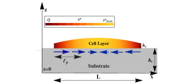
Our model builds on recent work Edwards and Schwarz (2011); Banerjee and Marchetti (2011) describing the cell or cell layer as a contractile elastic medium, with local elastic response of the substrate (as appropriate for micropillar arrays or very thin substrates). In contrast, here we consider substrates of finite thickness where the nonlocality of the elastic response must be included. While previous studies have analyzed the deformations of finite-thickness substrates due to point traction forces on their surface Merkel et al. (2007); Maloney et al. (2008), our work considers the inhomogeneous traction due to an extended contractile cell layer. A central result for our work is the expression for the scaling parameter referred to as the lateral penetration length (Fig. 1). This length scale characterizes the in-plane spatial variations of both adhesion-induced traction stresses on the substrate and cellular stresses within the cell layer in terms of cell and substrate elastic and geometrical properties. Our model also quantifies the experimentally-observed role of substrate thickness in controlling the mechanical response of adhering cell layers Lin et al. (2010). If is small compared to the lateral extent of the cell sheet, the substrate elasticity plays a negligible role in determining the mechanical response of the cell. This may explain why traction forces exerted by cell colonies with appear insensitive to substrate stiffness Trepat et al. (2009). If, in contrast, , then substrate nonlocality controls stress build-up in the cell sheet. This crossover may be observable in large cell colonies on thick substrates. Finally, the importance of long-range substrate elasticity has also been emphasized in recent models of cells as active dipoles on a soft elastic matrix, where it is crucial in controlling cell adhesion Bischofs et al. (2004); De et al. (2007). Long-range interfacial elastic stresses coupled with gel thickness have also been shown to have a profound effect on focal adhesion growth Nicolas and Safran (2006) and to enhance cell polarization Bischofs and Schwarz (2005); Friedrich and Safran (2012). These important effects are not discussed here.
Contractile cell on a soft substrate.
To illustrate the importance of substrate nonlocality, we first analyze a single cell, modeled as a contractile spring of stiffness and rest length , adhering to a continuum substrate (described as an elastic continuum of Young’s modulus and Poisson’s ration ) via two focal adhesion bonds (linear springs of stiffness ) located at and (Fig. 2, top left) Schwarz et al. (2006). This is motivated by the experimental observation that in adhering cells focal adhesions tend to be localized near the cell periphery Wozniak et al. (2004).

For simplicity we consider a one dimensional model, where the cell lies on the axis and the substrate lies in the region of the plane. Contractile acto-myosin fibers connect the focal adhesions and exert active forces of magnitude . Once the cell has fully adhered, the cell-substrate system is in mechanical equilibrium. Force balance at and yields
| (1a) | |||
| (1b) | |||
with the displacements of the contact points from their unstretched positions , and the displacement of the substrate’s surface at . All displacements are defined with respect to an initial state where the cell has length . The net contraction is then . The traction force by the cell on the substrate is localized at and , yielding a traction force density , with . Assuming linear elasticity, the substrate deformation is Landau et al. (1986), , where is the elastic Green’s function at . For a substrate of thickness we use the approximate form 111To enable a direct comparison between the penetration lengths obtained below and experimentally accessible parameters, is the Young modulus of a three dimensional elastic medium.
| (2) |
derived in the Supplemental Material SMp , with the size of adhesion complexes, providing a short-distance cut-off, and denotes the modified Bessel function of the second kind. We obtain , with the effective stiffness of the cell-substrate adhesions. For , is independent of and scales linearly with . Stiffening sets in for , as shown in Fig. 2 (top right), with a crossover controlled by the thickness of the substrate . Using , we solve for both and , shown in Fig. 2 (bottom) as functions of the substrate thickness and stiffness. For very thin () or infinitely rigid substrates, where the substrate elasticity becomes local, , corresponding to a spring in parallel with a series of two focal adhesions springs . In this limit the traction force saturates to . Conversely, for a very soft substrate with , the contraction is maximal and given by , and . The substrate thickness above which both cell contraction and traction force saturate is controlled by the cell size and the substrate elasticity, in qualitative agreement with experiments Lin et al. (2010).
Contractile Cell Layer.
The continuum limit can be obtained by considering a multi-mer of contractile elemental “cells”, connected by springs representing cell-cell interactions. The outcome is a set of coupled equations for a contractile elastic medium. For a cell layer of thickness (Fig. 1), the force balance equation, averaged over the cell thickness, is
| (3) |
where describes the effective strength of the focal adhesions, is the displacement field of the cellular medium at , and is the thickness-averaged cellular stress tensor, , given by , with the longitudinal elastic modulus of the cell layer. The one dimensional model presented here may be relevant to wound healing assays, where the cell layer is a strip with -translational invariance. Although we have neglected components of the cellular displacements and spatial variations along , the cell elastic constants are those of a three-dimensional cellular medium. The active stress arises from acto-myosin contractility Kruse et al. (2005). The substrate deformation at the surface is
| (4) |
with the elastic Green’s function of a substrate of infinite extent in , occupying the region , evaluated at . Eqs. (3)-(4) can be reduced to integro-differential equations for the cellular stress, as
| (5) |
The length scale controls spatial variations of cellular stresses induced by the stiffness of the focal adhesions. It is the size of a region where the areal elastic energy density associated with focal adhesions is of order of the areal elastic energy density of the cell layer. For a cell monolayer with , , , and Balaban et al. (2001), we get , comparable to traction penetration length seen in experiments on stiff microposts Saez et al. (2005, 2007). The second term on the right hand side of Eq. (5) describes spatial variations in the cellular stress due to the (generally nonlocal) coupling to the substrate. In the following we examine solutions to Eq. (5), considering various limiting cases for the substrate thickness and analyze the dependence of traction stresses on cell size, substrate stiffness and substrate depth. The equation governing stress distribution in two dimensional cell layers is derived in the Supplemental material SMp .
Thin substrate.
If the substrate’s elastic response can be approximated as local, as it is the case for or for cells on micropillar arrays, the Green’s function is given by . Eq. (5) can then be written as , where, and describes the combined action of the focal adhesions and the substrate, acting like two linear elastic components in series. Assuming zero external stresses at the boundary, i.e., , the internal stress profile is Edwards and Schwarz (2011); Banerjee and Marchetti (2011); Mertz et al. (2012). The traction stress , is localized within a length from the edge of the cell layer. The penetration length can be written as , with the square root of the ratio of the cell’s elastic energy to the elastic energy density of the substrate. This form highlights the interplay of focal adhesion stiffness and substrate stiffness in controlling spatial variation of stresses in the lateral () direction. The two act like springs in series, where the weaker spring controls the response. If , then and the stiff substrate has no effect. Conversely, if the focal adhesions are stiffer than the substrate, then . For an elastic substrate with , and in the range , lies in the range . This leads to typical values of in the range for a cell layer of length , consistent with experimentally observed traction penetration lengths on thin continuous substrates Mertz et al. (2012) and on micropillar posts Saez et al. (2010).
Infinitely thick substrate.
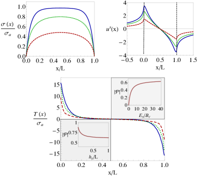
If , the substrate Green’s function can be approximated as that of an elastic half plane, , with the Euler constant Barber (2010). The solution of Eq. (5) with boundary conditions can be obtained by expanding in a Fourier sine series as, and solving the coupled algebraic equations for the Fourier amplitudes given in the Supplementary Material SMp . The effect of the nonlocal elasticity of the substrate is controlled by yet another length scale that can be obtained from the length introduced in the case of thin substrate by the replacement and . This highlights the known fact that cells or cell layers only “feel” the substrate up to a thickness comparable to their lateral size . For parameter values quoted in the preceding paragraphs, takes values between for in the range , indicating that the thin/thick substrate crossover, although not observable in isolated cells, should be seen experimentally in cohesive cell layers where the lateral extent can exceed . The cellular stress and substrate displacement profiles obtained numerically by summing the Fourier series are shown in Fig. 3 (top). The lateral variation of stresses is now controlled by the length scale . One consequence of nonlocal substrate elasticity is that the substrate deformation shown in the top right frame of Fig. 3 extends outside the region occupied by the cell layer, indicated by the two vertical dashed lines. The profile of the local traction stress displayed in Fig. 3 (bottom frame) shows that the traction stress is localized near the edge of the cell layer and its magnitude increases with substrate stiffness. The inset to Fig. 3 (bottom right) shows the magnitude of the net contractile moment defined as . This quantity is negative, as expected for contractile systems. Its magnitude increases with at a rate consistent with experiments, with a rise in upon increasing the substrate stiffness by Wang et al. (2002), and saturates for very stiff substrates.
Substrate of Finite Thickness.
Finally, we consider a substrate of finite thickness, . The calculations are carried out using the approximate Green’s function given in Eq. (2), with the replacement . The variation of the net contractile moment with for is shown in Fig. 3 (bottom left inset). As seen previously in experiments Lin et al. (2010), drops sharply with increasing substrate thickness, quickly reaching the asymptotic value corresponding to infinitely thick substrates. Thinner substrates are effectively stiffer than thick ones, inducing larger contractile moments. Our analysis suggests a general expression for the penetration length that interpolates between the thin and thick substrates limits,
| (6) |
Stress penetration is controlled by a substrate layer of effective thickness given by the geometric mean of the actual substrate thickness and the lateral dimension of the cell or cell layer. If , then and stress penetration is not affected by cell layer size, as in the experiments of Mertz et al. (2012). On the other hand, if , then cells only feel the effect of the substrate down to an effective depth .
Discussion.
In summary, we have examined the dependence of traction stresses in adhering cell layers on the mechanical and geometrical properties of the substrate. Using a generic non-local model, we provide analytical results for the effect of cell and substrate properties on the stress penetration length, that can be tested in experiments. Although the analysis presented here is restricted to one dimensional layers, isotropic planar cell layers with spherical symmetry can also be considered analytically Banerjee and Marchetti (2012), with similar predictions for the dependence of traction fields and their moments on substrate mechanical and geometrical properties. The scaling of traction moments on cell layer size is, however, different in two dimensions Mertz et al. (2012). The model can be extended to incorporate the effects of cell polarization, spatial variations in contractility, heterogeneities in the cell layer or anisotropic elasticity of the substrate.
We thank Eric Dufresne and Aaron Mertz for many useful discussions and the anonymous referees for valuable comments. This work was supported by the National Science Foundation through awards DMR-0806511, DMR-1004789 and DGE-1068780.
References
- Harris et al. (1980) A. Harris, P. Wild, and D. Stopak, Science, 208, 177 (1980).
- Discher et al. (2005) D. Discher, P. Janmey, and Y. Wang, Science, 310, 1139 (2005).
- Balaban et al. (2001) N. Balaban, U. Schwarz, D. Riveline, P. Goichberg, G. Tzur, I. Sabanay, D. Mahalu, S. Safran, A. Bershadsky, L. Addadi, et al., Nature cell biology, 3, 466 (2001).
- Dembo and Wang (1999) M. Dembo and Y. Wang, Biophysical journal, 76, 2307 (1999).
- Butler et al. (2002) J. Butler, I. Tolić-Nørrelykke, B. Fabry, and J. Fredberg, American Journal of Physiology-Cell Physiology, 282, C595 (2002).
- Tan et al. (2003) J. L. Tan, J. Tien, D. M. Pirone, D. S. Gray, K. Bhadriraju, and C. S. Chen, PMNAS, 100, 1484 (2003).
- Polio et al. (2012) S. R. Polio, K. E. Rothenberg, D. Stamenović, and M. L. Smith, Acta Biomaterialia, 8, 82 (2012).
- Ghibaudo et al. (2008) M. Ghibaudo, A. Saez, L. Trichet, A. Xayaphoummine, J. Browaeys, P. Silberzan, A. Buguin, and B. Ladoux, Soft Matter, 4, 1836 (2008).
- Saez et al. (2010) A. Saez, E. A. andM. Ghibaudo, O. du Roure, J.-M. D. Meglio, P. Hersen, P. Silberzan, A. Buguin, and B. Ladoux, J. Phys.: Condens. Matter, 22, 194119 (9pp) (2010).
- Mertz et al. (2012) A. F. Mertz, S. Banerjee, Y. Che, G. K. German, Y. Xu, C. Hyland, M. C. Marchetti, V. Horsley, and E. R. Dufresne, Phys. Rev. Lett., 108, 198101 (2012).
- Trepat et al. (2009) X. Trepat, M. Wasserman, T. Angelini, E. Millet, D. Weitz, J. Butler, and J. Fredberg, Nature physics, 5, 426 (2009).
- Lin et al. (2010) Y. Lin, D. Tambe, C. Park, M. Wasserman, X. Trepat, R. Krishnan, G. Lenormand, J. Fredberg, and J. Butler, Physical Review E, 82, 041918 (2010).
- Sen et al. (2009) S. Sen, A. Engler, and D. Discher, Cellular and molecular bioengineering, 2, 39 (2009).
- Edwards and Schwarz (2011) C. Edwards and U. Schwarz, Physical Review Letters, 107, 128101 (2011).
- Banerjee and Marchetti (2011) S. Banerjee and M. Marchetti, EPL (Europhysics Letters), 96, 28003 (2011).
- Merkel et al. (2007) R. Merkel, N. Kirchgeßner, C. Cesa, and B. Hoffmann, Biophysical journal, 93, 3314 (2007).
- Maloney et al. (2008) J. M. Maloney, E. B. Walton, C. M. Bruce, and K. J. van Vliet, Phys. Rev. E, 78, 041923 (2008).
- Bischofs et al. (2004) I. Bischofs, S. Safran, and U. Schwarz, Physical Review E, 69, 021911 (2004).
- De et al. (2007) R. De, A. Zemel, and S. Safran, Nature Physics, 3, 655 (2007).
- Nicolas and Safran (2006) A. Nicolas and S. Safran, Biophysical journal, 91, 61 (2006).
- Bischofs and Schwarz (2005) L. B. Bischofs and U. S. Schwarz, Phys. Rev. Lett., 95, 068102 (2005).
- Friedrich and Safran (2012) B. M. Friedrich and S. A. Safran, Soft Matter, (2012).
- Schwarz et al. (2006) U. Schwarz, T. Erdmann, and I. Bischofs, Biosystems, 83, 225 (2006).
- Wozniak et al. (2004) M. Wozniak, K. Modzelewska, L. Kwong, and P. Keely, Biochimica et Biophysica Acta (BBA)-Molecular Cell Research, 1692, 103 (2004).
- Landau et al. (1986) L. D. Landau, L. P. Pitaevskii, E. M. Lifshitz, and A. M. Kosevich, Theory of Elasticity, 3rd ed. (Butterworth-Heinemann, 1986) ISBN 075062633X.
- Note (1) To enable a direct comparison between the penetration lengths obtained below and experimentally accessible parameters, is the Young modulus of a three dimensional elastic medium.
- (27) See Supplemental Material at [URL will be inserted by publisher] for derivation of substrate Green’s function, Integro-differential equation governing cellular stress distribution, and corresponding equations for two dimensional cell layers.
- Kruse et al. (2005) K. Kruse, J. Joanny, F. Jülicher, J. Prost, and K. Sekimoto, The European Physical Journal E: Soft Matter and Biological Physics, 16, 5 (2005).
- Saez et al. (2005) A. Saez, A. Buguin, P. Silberzan, and B. Ladoux, Biophysical journal, 89, L52 (2005).
- Saez et al. (2007) A. Saez, M. Ghibaudo, A. Buguin, P. Silberzan, and B. Ladoux, Proceedings of the National Academy of Sciences, 104, 8281 (2007).
- Barber (2010) J. Barber, Elasticity, Vol. 172 (Springer Verlag, 2010).
- Wang et al. (2002) N. Wang, I. Tolić-Nørrelykke, J. Chen, S. Mijailovich, J. Butler, J. Fredberg, and D. Stamenović, American Journal of Physiology-Cell Physiology, 282, C606 (2002).
- Banerjee and Marchetti (2012) S. Banerjee and M. C. Marchetti, “On the role of substrate thickness on traction force distribution of adherent cell layers,” (2012), in preparation.