Magnetic properties of PdAs2O6: a dilute spin system with an unusually high Néel temperature
Abstract
The crystal structure and magnetic ordering pattern of PdAs2O6 were investigated by neutron powder diffraction. While the magnetic structure of PdAs2O6 is identical to the one of its isostructural -homologue NiAs2O6, its Néel temperature (140 K) is much higher than the one of NiAs2O6 (30 K). This is surprising in view of the long distance and indirect exchange path between the magnetic Pd2+ ions. Density functional calculations yield insight into the electronic structure and the geometry of the exchange-bond network of both PdAs2O6 and NiAs2O6, and provide a semi-quantitative explanation of the large amplitude difference between their primary exchange interaction parameters.
pacs:
75.25.-j, 75.50.Ee, 61.05.fm, 71.20.PsI INTRODUCTION
The magnetic properties of transition metal compounds with valence electrons have been one of the central research themes in solid-state physics for the past three decades. In view of the interplay between magnetism and high-temperature superconductivity, particular attention has been focused on oxides and arsenides. Recently, the electronic structure and ordering phenomena of transition metal compounds with and valence electrons (such as ruthenates and iridates) have also captured much attention. The electronic correlations in these materials are generally weaker than those of their counterparts, while the spin-orbit coupling is stronger. The quantitative description of the influence of these parameters on the electronic phase behavior of -electron compounds is an important topic of current research. Here we report a detailed investigation of the magnetic properties of PdAs2O6, a recently synthesized orosel electrically insulating compound with a magnetic lattice of Pd2+ ions in the electron configuration . We compare our results to the isostructural compound NiAs2O6, which is based on Ni2+ ions with the same number of electrons in the -shell.
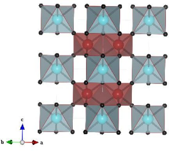
PdAs2O6 crystallizes in the PbSb2O6 structure with Pd2+ and As5+ ions segregated into different layers (Fig. 1). The octahedral coordination of Pd2+ in this structure is unusual, because divalent palladium shows a strong preference for square-planar coordination, which is associated with a diamagnetic ground state. Only a few examples of sixfold-coordinated Pd2+ compounds are known, including the ambient- and high-pressure polymorphs of PdF2 as well fluoro-palladates of composition MPdF4 (M = Ca, Cd, Hg) and CsPd2F5. mueller These compounds are paramagnetic at high temperatures and tend to order antiferromagnetically upon cooling. In accord with this trend, magnetic susceptibility measurements on PdAs2O6 showed paramagnetic behavior at room temperature, and an antiferromagnetic phase transition at the Néel temperature K. orosel This behavior is qualitatively analogous to the one of the isostructural 3d-homologues MnAs2O6, CoAs2O6, and NiAs2O6, which also show antiferromagnetic ordering with , 20, and 30 K, respectively. nakua However, the much higher Néel temperature of PdAs2O6 is surprising, especially because the PdO6-octahedra do not share vertices, edges or faces. The exchange paths connecting neighboring Pd2+ ions are therefore long and involve at least two bridging oxygen sites.
In order to elucidate the microscopic origin of this surprising behavior, we have used neutron diffraction to determine the magnetic structure of PdAs2O6, which turned out to be identical to that of the 3d-homologues (Section II). This implies similar networks of exchange bonds in both sets of compounds. We employed density-functional calculations to obtain insights into the electronic structure of PdAs2O6 and NiAs2O6, and specifically into the origin of the exchange paths (which turned out to be hopping via As dimers) and of the large amplitude difference of the primary exchange interaction parameters (Section III). A model based on these interactions yields an excellent description of the magnetic susceptibilities of both compounds.
II Neutron diffraction
II.1 Experimental details
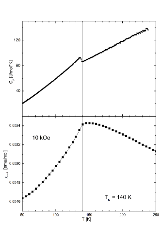
A powder sample of PdAs2O6 of weight g was prepared using the starting materials PdO (99.9 % metals basis, Alfa Aesar) and As2O5 (99.9 %, Alfa Aesar) in the molecular ratio 1 : 1.1 as described earlier. orosel The mixed powder was pressed into pellets and dried in evacuated silica tubes for 12 h at 373 K. Then the evacuated silica tubes were heated up to 973 K with a rate of 100 K/h. The hygroscopic and air sensitive powder of PdAs2O6 was obtained after an annealing process of about 100 hours. Measurements of the magnetic susceptibility and specific heat were carried out in the temperature range between 5 and 300 K (Fig. 2). Both quantities show anomalies indicative of antiferromagnetic ordering of the Pd sublattice at 140 K, in agreement with prior work. orosel
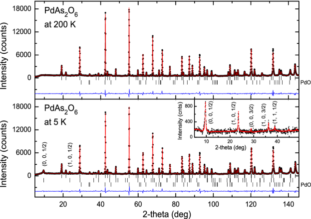
In order to investigate the crystal and magnetic structure of PdAs2O6, a neutron diffraction experiment was carried out at the research reactor FRM-II in Garching. Neutron powder patterns were collected with the instrument SPODI at 5 K and 200 K in the range 4∘–160∘. This instrument uses a germanium monochromator (reflection 551) selecting the neutron wavelength Å. The refinements of the crystal and magnetic structure were carried out with the program FullProf. rodriguez We used the nuclear scattering lengths b(Pd) = 5.91 fm, b(As) = 6.58 fm and b(O) = 5.805 fm. sears The magnetic form factors of the magnetic ions were taken from Ref. brown, .
II.2 Crystal structure of PdAs2O6
The crystal structure of PdAs2O6 was recently refined from x-ray powder diffraction data in the trigonal PbSb2O6-type structure (space group P1m, No. 162), where the Pd, As and O atoms are in the Wyckoff positions 1a(0,0,0), 2d(,,) and 6k(x,0,z), respectively. orosel The same space group was found earlier for the compounds MnAs2O6, CoAs2O6 and NiAs2O6 containing 3d-metal ions. nakua From our neutron powder diffraction data taken at the lower temperatures 5 and 200 K (Fig. 3) the trigonal space group P1m was confirmed. For the Rietveld refinements we used data in the extended range from 4∘ up to 146∘. A total of 14 parameters was refined: an overall scale factor, five profile function parameters, the zero point, two lattice constants, the positional parameters x and z of the oxygen atom as well as three isotropic thermal parameters. The powder sample contained small amounts of the binary oxide PdO, which crystallizes in the tetragonal space group . moore Therefore the overall scale factor of PdO was additionally allowed to vary during the refinements.
In Table I the results of the refinements are compared with those of the x-ray study carried out earlier at room temperature. orosel Here it can be seen that the positional parameters of the oxygen atoms determined at 5 and 200 K are in good agreement, indicating that the structural changes between the magnetically ordered and the paramagnetic states are weak. Only a slight reduction of 0.0036 Å (about 6 ) could be observed for the Pd-O-bond length in the PdO6-octahedra. In contrast, the distances between the As and O-atoms are practically unchanged (Table I). The value d(As-O) = 1.8281(6) Å (at 200 K) found for PdAs2O6 is in very good agreement with the values of other arsenates containing 3d-metal ions: d(As-O) = 1.827(4) Å (NiAs2O6), d(As-O) = 1.830(3) Å, (CoAs2O6), d(As-O) = 1.826(2) Å (MnAs2O6), and d(As-O) = 1.826(1) Å (CdAs2O6). nakua ; weil All of these values are in agreement with d(As-O) = 1.82 Å calculated for an AsO6-octahedron given by Shannon. shannon In Table I it can be seen that the structural parameters obtained at 200 and 300 K show relatively large discrepancies, despite the fact that both data sets were collected in the paramagnetic phase. This can be ascribed to the larger scattering power of the O-atoms in neutron diffraction, with the consequence that the O-positions can be determined more reliably. Furthermore, the shortest oxygen contact d(O-O) = 2.308(3) Å was found to be implausibly short in the x-ray study. orosel From our neutron diffraction study we found the larger values d(O-O) = 2.3757(12) Å at 5 K and d(O-O) = 2.3726(12) Å at 200 K, respectively. The value d(O-O) = 2.410(3) Å found for NiAs2O6 is slightly larger, nakua while the As-O-bond lengths d(As-O) = 1.827(4) Å (NiAs2O6) and d(As-O) = 1.8281(6) Å (PdAs2O6) are practically the same in both compounds. The cell volume of the Ni compound ( Å3) is much smaller than the one of the Pd compound ( Å3). This is due to fact that the ionic radius of Pd2+ is larger than that of Ni2+. In order to keep the As-O-bond lengths almost constant in the AsO6-octahedra, the bond angle (O-As-O) increases from 169.91(3)∘ in PdAs2O6) to 173.23(12)∘ in NiAs2O6.
| 5 K | 200 K | 290 K | |
| a [Å] | 4.81700(4) | 4.81837(5) | 4.8196(1) |
| c [Å] | 4.65618(6) | 4.66014(7) | 4.6646(1) |
| V [Å3] | 93.565(2) | 93.698(2) | 93.835(3) |
| x(O) | 0.37187(15) | 0.37230(16) | 0.3695(7) |
| z(O) | 0.28203(18) | 0.28236(18) | 0.2926(5) |
| B(Pd) [Å2] | 0.46(3) | 0.62(3) | 0.80(3) |
| B(As) [Å2] | 0.49(2) | 0.54(2) | 0.88(3) |
| B(O) [Å2] | 0.62(2) | 0.74(2) | 0.55(6) |
| d(Pd-O) [Å] | 2.2211(6) (6) | 2.2247(6) (6) | 2.2437(20) (6) |
| (O-Pd-O) [∘] | 180 (3) | 180 (3) | 180 (3) |
| 88.61(2) (6) | 88.58(2) (6) | 86.84(10) (6) | |
| 91.39(2) (6) | 91.42(2) (6) | 93.16(10) (6) | |
| d(As-O) [Å] | 1.8288(6) (6) | 1.8281(6) (6) | 1.8076(23) (6) |
| (O-As-O) [∘] | 169.91(3) (3) | 169.79(3) (3) | 170.42(13) (3) |
| 81.01(3) (3) | 80.92(4) (3) | 79.34(15) (3) | |
| 92.18(3) (3) | 92.19(3) (3) | 93.34(15) (3) | |
| 95.49(2) (6) | 95.57(2) (6) | 94.03(10) (6) | |
| d(O-O)min [Å] | 2.3757(12) | 2.3726(12) | 2.308(3) |
| 0.0241 | 0.0298 | 0.0859∗ |
II.3 Magnetic structure of PdAs2O6
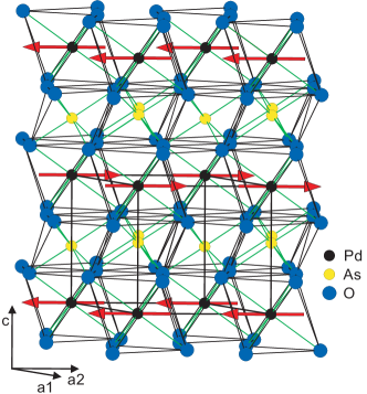
The neutron powder data recorded at 5 K show additional Bragg reflections that can be ascribed to the antiferromagnetic order of the Pd sublattice. The two prominent ones at and can be indexed as (0, 0, )M and (0, 1, )M, respectively. This suggests that the magnetic cell has a doubled c-axis with a propagation vector k = (0, 0, ). All further magnetic reflections were assigned indices according to (hk)M = (hk)N k, where M and N designate the magnetic and nuclear reflections. The same type of magnetic ordering was observed earlier for the isotypic divalent transition metal arsenates CoAs2O6 and NiAs2O6. nakua The presence of the strong magnetic reflection (0, 0, )M indicates that the magnetic moments of the Pd-atoms are aligned ferromagnetically within the hexagonal ab-plane. Due to antiferromagnetic exchange interactions between the palladium moments the ferromagnetic layers form the sequence + - + - along the c-axis (Fig. 4). With this model the magnetic structure of PdAs2O6 could be successfully refined using the magnetic reflections observed in the -range up to 50∘. It has to be noted that the moment direction within the hexagonal plane cannot be determined from the refinements. The wave vector k = (0, 0, ) keeps the full symmetry of the group Gk = G according to magnetic group theory, and it defines the magnetic translation lattice. rossat The existence of three magnetic domains in the hexagonal basis plane prohibits an unambiguous determination of the moment direction.
| (Ni2+) | (Pd+) | |||
|---|---|---|---|---|
| (0, 0, )M | 518 | 445 | 503 | 9.5 |
| (1, 0, )M | 198 | 205 | 178 | 23.5 |
| (0, 0, )M | 9 | 34 | 25 | 28.8 |
| (1, 0, )M | 88 | 87 | 50 | 36.2 |
| (1, 1, )M | 42 | 45 | 23 | 38.8 |
| (2, 0, )M | 30 | 28 | 11 | 44.8 |
| (1, 1, )M | 43 | 28 | 9 | 48.0 |
| (0, 0, )M | 2 | 6 | 2 | 49.1 |
| (Pd2+) () | 1.87(3) | 2.04(3) | ||
| 0.141 | 0.173 |
In order to improve the refinement of the magnetic structure, we used the purely magnetic intensities obtained from the difference between the data sets collected at 5 and 200 K (inset of Fig. 3). Since the magnetic form factor of Pd2+ is not available, we first used the one of the Pd+ ion, brown but Table II shows that the calculated intensities decrease much more strongly with increasing than the observed ones. A considerably better fit was obtained with the form factor of Ni2+, which also shows a -configuration. The relatively large residual [defined as ] reflects the fact that the low intensities of the magnetic reflections at high -values could not be measured with good accuracy, in combination with systematic errors arising from the difference between the form factors of Pd2+ and Ni2+.
The sublattice magnetization resulting from the refinement is per Pd site, similar to the value reported for NiAs2O6 (Ref. nakua, ). While the ordered moment is consistent with the spin-only moment expected for a configuration, a fit to the magnetic susceptibility for yields a -factor larger than 2, which is indicative of an orbital contribution to the Pd moment (see Section III.C below). The difference may in part be due to zero-point fluctuations of the magnetic moment, which reduce the ordered moment of the spin-1 system in the binary oxide NiO by %. hutchings The zero-point reduction is possibly larger in PdAs2O6 because of the low-dimensional exchange-bond network (see Section III.C). While these considerations suggest a small but nonzero orbital contribution to the Pd moment, measurements of the -factor anisotropy by single-crystal neutron diffraction and/or electron spin resonance will be required to quantify this contribution.
III Density functional calculations
III.1 LDA band structure
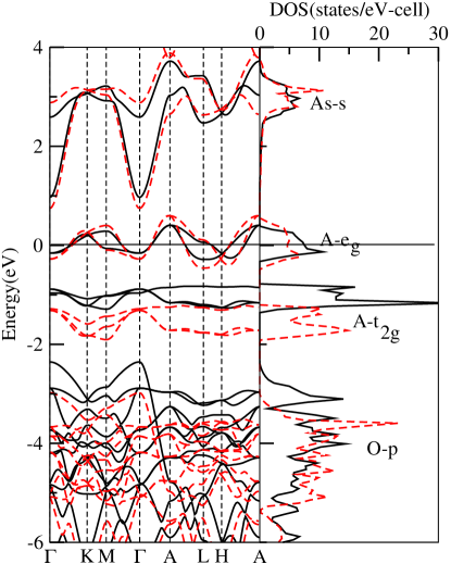
Figure 5 shows the electronic band structure and density of states in the paramagnetic local density-functional approximation (LDALDA1 ) for NiAs2O6 in solid lines and for PdAs2O6 in dashed lines. The self-consistent calculations were performed with the linear muffin-tin orbital (LMTO) method LMTO1 using -points in the Brillouin zone.
The Fermi level falls in the middle of the two narrow transition-metal -bands. These are split above the three narrow -bands, because the -hopping is times stronger than the -hopping to the O orbitals at the corners of the -octahedron. This is in accord with the labeling Similarly, in accord with the labeling O2- and As the oxygen -like bands are below and the As - and As -like bands are above the Fermi level. However, in terms of atomically localized orbitals (LMTOs) rather than Wannier orbitals, the bands denoted as As in the figure have 40% anti-bonding O character, as well as some and O characters. Correspondingly, around 12 eV (below the frame of the figure) there are two bands with As and O bonding characters in about equal amounts.
With the bands lined up at the Fermi level, the and bands lie lower in the Pd than in the Ni compound. This is because orbitals have a larger extent and therefore larger hopping integrals to O than do orbitals. For the same reason, the band is about 1.4 times wider in the Pd than in the Ni compound.
Inclusion of the Coulomb interaction beyond the LDA splits the bands and leads to insulating solutions, as we have checked through LDA+U LDA+U calculations. For the present purpose of calculating and understanding the magnetic properties, it is more convenient to start from the localized description and treat the hopping, , to order
III.2 Pd Wannier orbitals, low-energy tight-binding Hamiltonian and magnetic interactions
We therefore construct a low-energy Hubbard Hamiltonian. Since the LDA band is narrow and well separated from all other bands, we can limit the one-electron Hilbert space to that of the two -centered Wannier orbitals, and describing this band. When using the MTO downfolding method, we thus kept the degrees of freedom as active, and downfolded the rest. The Wannier orbitals are finally obtained by symmetrically orthonormalizing the downfolded, th-order muffin-tin orbitals (MTOs NMTO ). In the representation, of the corresponding one-electron part of the Hamiltonian, is the hopping integral from orbital on site to orbital on site
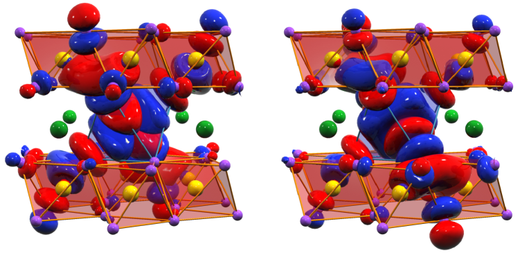
Figure 6 shows the and Wannier orbitals for PdAs2O6 as surfaces with positive lobes colored red and negative blue. The -axis points along the Pd-O bond in the -plane and the - and -axes point to the other oxygens in the Pd-centered octahedron. In the figure, the latter has been given a blue, transparent skin while all As-centered octahedra have been given a red, transparent skin. Pd atoms are green, As atoms yellow, and oxygens violet. Such a Pd-centered Wannier orbital has or character locally, and strong antibonding character on the neighboring oxygens. The unusual feature here is that the back-lobes of the strongest O characters (the four and tails of the Wannier orbital and the two tails of the Wannier orbital) bond to the characters on the closest pair of As atoms and, from there, antibond to the closest oxygen, which now belongs to a neighboring PdO6 octahedron. As an example: The red lobe of the orbital sticking up and out towards the reader, antibonds with the blue lobe of near orbital, whose red back-lobe merges together (bonds) with the red, two-center As bond, giving rise to a “red sausage”. The latter finally antibonds with the O orbital which points upwards towards a near Pd belonging to the upper Pd-sheet (not shown in the figure). Similarly for the orbital on the right-hand side of the figure: Its red lobe, sticking down and out, antibonds with the blue lobe of the near orbital, which merges with the As two-center bond into a red sausage. The latter finally induces strong -like character which points down and out, towards a Pd atom in the lower Pd sheet.
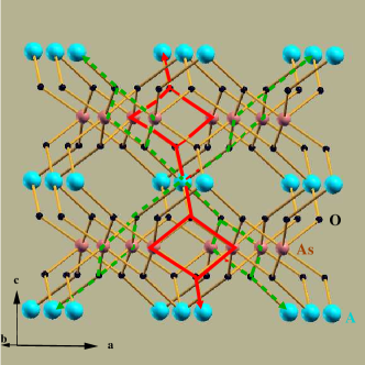
These hopping paths, shown schematically in Fig. 7, are to 3rd-nearest Pd neighbors, but only to six out of the twelve, namely to those at and The integrals for hopping to the 3rd-nearest neighbors at and , which have no bridging oxygen and As pairs, are negligible. The calculated hopping integrals exceeding 10 meV are given in Table III. We see that those to 3rd-nearest neighbors dominate those to 2nd- and 1st-nearest neighbors.
The Ni Wannier orbitals in NiAs2O6 are similar to the Pd Wannier orbitals in PdAs2O6, except that they are more localized. This is consistent with the 1.4 times smaller -band width. Concomitantly, the hopping integrals for NiAs2O6 listed in parentheses in Table III are about 1.4 times larger than those for PdAs2O6.
| Vector from to | ||||||
| . | . | |||||
| . | ||||||
| . | . |
The magnetic exchange interaction, , can be expressed in general as a sum of antiferromagnetic and ferromagnetic contributions. In the limit of large Coulomb correlation, typically valid for late transition metal elements like Pd or Ni, the antiferromagnetic contribution is related to the hopping integral by the second-order perturbation relation , where is the effective on-site Coulomb repulsion defined for the Wannier orbitals. Considering the hopping integrals in Table III, the contribution from the term in the magnetic exchange gives rise to a factor two difference between the Pd and Ni compounds. The estimate of , unlike that of the hopping integral , is a rather delicate issue. In principle, one should compute for the Wannier orbitals shown in Figure 7. However lacking a prescription to do so, we computed the values corresponding to Ni- and Pd- partial waves, which were truncated outside the atomic spheres defined around an Ni or Pd sites. The calculations were carried out within the framework of the constrained DFT scheme DFT ; DFT1 ; DFT2 . The values calculated in this way were eV and eV. But since the Wannier orbitals are far more delocalized than the truncated partial waves, and more so for Pd than for Ni, these values should be significantly reduced, and more so for Pd than for Ni. This could conceivably lead to the factor needed to bring our theoretical estimate for the Néel temperatures of the two compounds into agreement with the measured ratio of 4.7.
III.3 Spin model and susceptibility
Taking into account only the dominant hopping integrals provided by the MTO-downfolding calculation, a spin model can be defined in terms of the six pairs of 3rd-nearest-neighbor magnetic interactions, all of size , obtained by summing over the squares of the hopping integrals between the 3rd-nearest neighbors. Based on this model, we have calculated the magnetic susceptibility by considering the following spin-1 Hamiltonian:
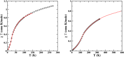
This model was solved by the quantum Monte Carlo method QMC on a lattice. The primary interaction , and the effective Landé -factors were obtained by fitting to the experimental susceptibility. As shown in Fig. 8, the calculated and measured susceptibilities agree very well. The optimal values of the -factor and the exchange parameter , were found to be respectively 2.38 and 62 K for the Pd compound, and 2.48 and 13 K for the Ni compound. We find that the -factors are larger than the spin-only value of 2, in accord with the discussion in Section II.C above. The fit of the susceptibilities yields a value of 5 for the ratio of the predominant magnetic exchange parameters of PdAs2O6 and NiAs2O6, in good agreement with the ratio of the magnetic transition temperatures.
IV Conclusions
Using a combination of susceptibility and neutron diffraction measurements, we have developed a comprehensive experimental description of the magnetic properties of the newly synthesized antiferromagnet PdAs2O6. Density functional theory has provided a detailed understanding of the magnetic bond network of this compound, as well as a semi-quantitative explanation of the large enhancement of the magnitude of its primary exchange interaction parameters compared to its homologue NiAs2O6. This approach may prove useful for research on other Pd compounds including the recently discovered bruns ferromagnet PdS2O7, and for comparative studies of materials with valence electrons and their -electron counterparts in general.
V Acknowledgements
We are grateful to the referee for constructive criticism, to M. Hölzel and A. Senyshyn for help with the neutron diffraction measurements at the FRM-II, and to O. Jepsen for performing the calculations. TSD and OKA acknowledge the MPG-India partner group program and the INDO-EU research network RP7 MONAMI for support.
References
- (1) D. Orosel and M. Jansen, Z. Anorg. Allg. Chem. 632, 1131 (2006).
- (2) For a recent review, see M. Serafin und B.G. Müller, Z. Anorg. Allg. Chem. 633, 2523 (2007).
- (3) A. M. Nakua and J. E. Greedan, J. Solid State Chem. 118, 402 (1995).
- (4) J. Rodriguez-Carvajal, Physica B 192, 55 (1993).
- (5) V. F. Sears, in International Tables of Crystallography, edited by A. J. C. Wilson (Kluwer, Dordrecht, 1992), Vol. C, p. 383.
- (6) P. J. Brown, in International Tables of Crystallography, Vol. C, p. 391.
- (7) W. J. Moore and L. Pauling, J. Amer. Chem. Soc. 63, 1392 (1941).
- (8) M. Weil, Acta. Cryst. E 57, i22 (2001).
- (9) R. D. Shannon, Acta. Cryst. A32, 751 (1976).
- (10) J. Rossat-Mignaud in Methods of Experimental Physics, edited by K. Sköld and D. L. Price (Academic Press, New York, London, 1987), Vol. 23, p. 69.
- (11) M. T. Hutchings and E. J. Samuelsen, Phys. Rev. B 6, 3447 (1972).
- (12) U. von Barth and L. Hedin. J. Phys. C: Solid State Phys. 5, 1629 (1972).
- (13) O. K. Andersen and O. Jepsen, Phys. Rev. Lett. 53, 2571 (1984).
- (14) V. I. Anisimov, I. V. Solovyev, M. A. Korotin, M. T. Czyzyk, and G. A. Sawatzky, Phys. Rev. B 48, 16929 (1993).
- (15) O. K. Andersen and T. Saha-Dasgupta, Phys. Rev. B 62, R16219 (2000).
- (16) P. H. Dederichs, S. Blügel, R. Zeller, and H. Akai, Phys. Rev. Lett. 53, 2512 (1984).
- (17) O. Gunnarsson, O. K. Andersen, O. Jepsen, and J. Zaanen, Phys. Rev. B 39, 1708 (1989).
- (18) V. I. Anisimov, J. Zaanen, and O. K. Andersen, Phys. Rev. B 44, 943 (1991).
- (19) A. W. Sandvik, Phys. Rev. B 59, R14157 (1999).
- (20) J. Bruns, M. Eul, R. Pöttgen, and M. S. Wickleder, Angew. Chem. 124, 2247 (2012).