Electrokinetic-flow-induced viscous drag on a tethered DNA inside a nanopore
Abstract
Recent work has shown that the resistive force arising from viscous effects within the pore region could explain [Ghosal, S. Phys. Rev. E. 71, 051904 (2006) & Phys. Rev. Lett. 98, 238104 (2007)] observed translocation times in certain experiments involving voltage driven translocations of DNA through nanopores. The electrokinetic flow inside the pore and the accompanying viscous effects also play a crucial role in the interpretation of experiments where the DNA is immobilized inside a nanopore [U. Keyser, et al. Nature Physics 2, 473 (2006)]. In this paper the viscous force is explicitly calculated for a nanopore of cylindrical geometry. It is found that the reduction of the tether force due to viscous drag and due to charge reduction by Manning condensation are of similar size. The result is of importance in the interpretation of experimental data on tethered DNA.
pacs:
87.15.Tt, 87.14.GgThe interaction of charged polymers, such as DNA with nanometer sized natural and artificial pores have received considerable attention recently kasianowicz_PNAS96 ; meller_etal_PNAS00 ; ssdna_sequence_nbt ; storm_nature ; storm_physRevE05 ; lubensky_nelson ; ghosal_PRE06 . Such studies are partly motivated by the desire to understand how polymers cross internal membranes of cells Alberts . The possibility of developing devices capable of detecting properties of biopolymers at the single molecule level for applications such as rapid DNA sequencing deamer_trendsinbiotech is also a motivating factor for such studies.
In a recent paper Keyser et al. reported keyser_nature_phys06 experimental measurements in which a single strand of dsDNA was immobilized while threaded inside a nanopore by the application of a pulling force to counteract the electrical force on the DNA. This was achieved by attaching one end of the DNA to a Streptavidin coated polystyrene bead and holding the bead in a laser optical trap. The displacement of the bead from its equilibrium position could be detected and used to measure the pulling force on the DNA. The measured value was found to be about 75 % of the maximum electric force on the DNA within the pore based on its bare charge irrespective of the electrolyte (KCl) concentration. This pulling force is however determined by a complex interplay between electric forces and hydrodynamics, as noted by Keyser et al. keyser_nature_phys06 . The point of this calculation on an idealized physical model is to understand the relative importance of hydrodynamics and the reduction of effective charge on the DNA due to Manning condensation in determining the observed pulling force. Since the DNA as well as the internal walls of the pore are charged, the pore region has a cylindrically symmetric distribution of oppositely charged counter-ions. In the presence of a strong electric field an electroosmotic flow probstein is therefore generated in this region that flows in a direction opposite to the direction in which the DNA would move if it were not immobilized (Figure 1). This flow produces a hydrodynamic drag on the DNA partially balancing the applied electrical force. In this paper, a simplified geometry of the pore region is used to calculate explicitly the viscous drag. It is shown that the drag is a significant fraction of the total force acting on the DNA and needs to be taken into account for a proper interpretation of experimental data on DNA nanopore interactions.

A simplified model is adopted in which the nanopore is regarded as a cylinder of radius (5.0 nm) and length (60 nm). The part of the DNA inside the nanopore is regarded as a uniformly charged cylinder of radius (1.1 nm) along the axis of the pore. The DNA has a linear charge density (2 electron charges every 0.34 nm – the distance between adjacent bases) and a lower “effective” charge density of due to the Oosawa-Manning Oosawa ; Manning phenomenon of counter-ion condensation on its surface. The factor is the Oosawa-Manning factor, it has the value of for an ideal model of an infinite line charge in an unbounded electrolyte. Referring to the system sketched in Figure 1, the fluid velocity in the pore is axially directed and is described by some function where is the distance from the central axis. The electric potential is where the first term is due to the externally applied axial electric field along the pore (the -direction). The functions and are governed by the Stokes equation for viscous flow (with zero pressure gradient and an electric body force term) and the Poisson equation of electrostatics respectively:
| (1) | |||||
| (2) |
where is the permittivity of the electrolyte and is the electric charge density due to ions. The classical boundary conditions of ‘no slip’ are assumed for the velocity:
| (3) |
Eliminating from the pair of equations (1) and (2) and using (3) determines in terms of the potential :
| (4) |
where . The viscous force (along the -axis) on the DNA is then
| (5) |
If is the surface charge density on the channel wall, then by Gauss’ law,
| (6) | |||||
| (7) |
Using the first of these equations to eliminate and noting that the electrical force on the DNA, , equation (5) may be written as
| (8) |
where is the tether force.
In order to calculate the ratio from equation (8), all that remains to be done is to calculate the quantity . If the potential anywhere in the gap does not greatly exceed (about 30 mV at room temperature; is the Boltzmann factor, the absolute temperature and the magnitude of electric charge) then may be computed from the Debye-Hückel model ( is the Debye length):
| (9) |
with the boundary conditions (6) and (7). The solution to that problem is
| (10) |
where the constants and may be compactly expressed in terms of the following dimensionless variables , and . Thus,
| (11) | |||||
| (12) |
where , ( is a non-negative integer) are modified Bessel functions of integral order. The potential drop in equation (8) is now easily found from equation (10)
| (13) |
where and .
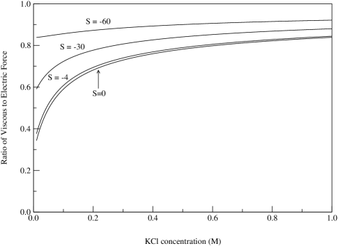
The result of the calculation as described above is shown in Figures 2. The figure shows the magnitude of the viscous to the electric force () evaluated from equation (8) using the value of appropriate for a symmetric binary electrolyte probstein . The effective charge of the DNA is assumed to be the same as the bare charge () of -2e per base pair and the applied Voltage is mV. The surface charge concentration on the substrate, has been assumed independent of the KCl concentration. From measurements of conductance and streaming potentials it has been shown stein_kruithof_dekker_prl04 ; dekker_nano_lett06 that in Si/Si nanopores mC/ for KCl concentrations greater than about M. However, for low concentrations, the surface charge density drops substantially and needs to be calculated from a more elaborate model that takes into account the equilibrium of surface reactions at the interface. For very low concentrations, mC/. Due to the approximate nature of our model it does not seem worthwhile to attempt to incorporate the proper dependence of on KCl concentration. Instead, it suffices to show how varies with salt concentration for several fixed values of between to mC/ as shown in Figure 2. The important feature that these curves illustrate is that is essentially constant for most of the KCl concentration range at a value of around 0.7 – 0.8. Thus, the viscous force is not small, and furthermore, if the viscous force were neglected and the reduction in the electrical pulling force were attributed to counter-ion condensation, it would appear that the DNA effective charge is lowered by about percent (just the right amount to lead one to conclude that charge reduction by the Manning factor of is being validated!). Figure 2 shows an increase in the hydrodynamic drag with increase in the magnitude of , because the surface charge on the pore walls enhance the electroosmotic flow due to the DNA charge. A model that properly accounts for the variation of with KCl concentration is expected to follow the mC/ curve closely for low concentrations (below about M) and asymptote to the mC/ curve at high concentrations. The distribution of counter ions in the calculations presented here was treated by means of the equilibrium Debye-Hückel theory and one may question whether that corresponds to the experimental conditions. Taking the ratio , equation (8) gives mV if for one assumes the DNA bare charge of two electronic charges per base pair. If this is reduced by the Manning factor of , then mV. Though the formal requirement for the Debye-Hückel linearization is mV, in practice the double layer structure calculated from the Debye-Hückel theory does not deviate substantially from the more accurate Poisson-Boltzmann calculation as long as the maximum value of is not substantially larger than mV hunter . Therefore the Debye-Hückel theory certainly suffices for our present purpose. For the cylindrical geometry considered here, the applied potential does not disturb the equilibrium Debye layer structure, since the applied field is always along the iso-concentration surfaces of the ions. However, for the real nanopore, the applied electric field may have a radial component, and one may ask if this is strong enough to distort the equilibrium Debye layer. Since mV and nm, this imposed field is V/m. The radial field within the Debye layer may be estimated as V/m with Manning condensation and V/m with the DNA bare charge. Thus, though the distortion of the double layer can be neglected for the purpose of obtaining a rough estimate, it should be accounted for if one desires an accurate calculation of the viscous force. In order to do so, the cross-sectional shape of the nanopore must be known.
Analysis of this simplified model suggests that a more careful modeling is needed in order to properly interpret the Keyser et al. keyser_nature_phys06 experiments. Such a model should account for hydrodynamic drag while taking into account the proper pore shape, the variation of substrate charge with KCl, possible departures of the equilibrium potential from the Debye-Hückel model and other relevant conditions of the experimental set up. Numerical simulation on a more elaborate model incorporating these details used in conjunction with the experimental data could provide a more complete picture of the effective charge on DNA inside a nanopore. One may be tempted to question the use of classical continuum hydrodynamics to flows on the nanometer scale. However, the classical approach has already been shown to give results in reasonable agreement with experiments on DNA translocation through solid state nanopores ghosal_PRE06 ; ghosal_PRL07 . Molecular dynamic simulations, such as those presented by Aksimentiev et al. aksi_bpj04 could be used to further refine these calculations and to show that the effects described here persist even if continuum hydrodynamics is replaced by a discrete molecular model. In situations where the length of the DNA polymer is much greater than the length of the nanopore, entropic forces due to random coiling of the polymer become significant. Such entropic effects have been considered by Muthukumar muthukumar99 ; muthukumar01 ; muthukumar03 . In the limit , the hydrodynamic friction with the pore walls becomes unimportant and the problem becomes one of determining the electric field that would immobilize a polyelectrolyte acted upon by given non-electrical forces in the presence Brownian fluctuations. This problem has been studied in its general form by Long et al. long_viovy_ajdari_prl96 ; long_viovy_ajdari_biopolymers96 .
References
- (1) J. Kasianowicz, E. Brandin, D. Branton, and D. Deamer, Proc. Natl. Acad. Sci. 93, 13770 (1996).
- (2) A. Meller et al., Proc. Natl. Acad. Sci. 97, 1079 (2000).
- (3) W. Vercoutere et al., Nature Biotechnology 19, 248 (2001).
- (4) A. Storm et al., Nature Materials 2, 537 (2003).
- (5) A. Storm, J. Chen, H. Zandbergen, and C. Dekker, Phys. Rev. E 71, 051903 (2005).
- (6) D. Lubensky and D. Nelson, Biophys. J. 77, 1824 (1999).
- (7) S. Ghosal, Phys. Rev. E 74, 041901 (2006).
- (8) B. Alberts et al., Molecular Biology of the Cell (Garland Publishing, Taylor & Francis Group, New York, U.S.A., 1994).
- (9) D. Deamer and M. Akeson, Trends in Biotech. 18, 147 (2000).
- (10) U. Keyser et al., Nature Physics 2, 473 (2006).
- (11) R. Probstein, Physicochemical Hydrodynamics (John Wiley and Sons, Inc., New York, U.S.A., 1994).
- (12) F. Oosawa, Polyelectrolytes (Marcel Dekker, New York, U.S.A., 1971).
- (13) G. Manning, J. Chem. Phys. 51, 924 (1969).
- (14) D. Stein, M. Kruithof, and C. Dekker, Phys. Rev. Lett. 93, 035901 (2004).
- (15) M. Smeets et al., Nano Letters 6, 89 (2006).
- (16) R. Hunter, Introduction to modern Colloid science (Oxford University Press, Oxford. U.K., 2003).
- (17) S. Ghosal, Phys. Rev. Lett. 98, 238104 (2007).
- (18) A. Aksimentiev, J. Heng, G. Timp, and K. Schulten, Biophys. J. 87, 2086 2097 (2004).
- (19) M. Muthukumar, J. Chem. Phys. 111, 10371 (1999).
- (20) M. Muthukumar, Phys. Rev. Lett. 86, 3188 (2001).
- (21) M. Muthukumar, J. Chem. Phys. 118, 5174 (2003).
- (22) D. Long, J. Viovy, and A. Ajdari, Phys. Rev. Lett. 76, 3858 (1996).
- (23) D. Long, J. Viovy, and A. Ajdari, Biopoloymers 39, 755 (1996).