Human sperm cells swimming in micro-channels
Human sperm cells swimming in micro-channels
Abstract
The migratory abilities of motile human spermatozoa in vivo are essential for natural fertility, but it remains a mystery what properties distinguish the tens of cells which find an egg from the millions of cells ejaculated. To reach the site of fertilization, sperm must traverse narrow and convoluted channels, filled with viscous fluids. To elucidate individual and group behaviors that may occur in the complex three-dimensional female tract environment, we examine the behavior of migrating sperm in assorted micro-channel geometries. Cells rarely swim in the central part of the channel cross-section, instead traveling along the intersection of the channel walls (‘channel corners’). When the channel turns sharply, cells leave the corner, continuing ahead until hitting the opposite wall of the channel, with a distribution of departure angles, the latter being modulated by fluid viscosity. If the channel bend is smooth, cells depart from the inner wall when the curvature radius is less than a threshold value close to 150 m. Specific wall shapes are able to preferentially direct motile cells. As a consequence of swimming along the corners, the domain occupied by cells becomes essentially 1-dimensional. This leads to frequent collisions and needs to be accounted for when modeling the behavior of populations of migratory cells and considering how sperm populate and navigate the female tract. The combined effect of viscosity and three-dimensional architecture should be accounted for in future in vitro studies of sperm chemoattraction.
| 1 | School of Engineering, University of Warwick, Coventry, CV4 7AL, UK. |
|---|---|
| 2 | Department of Applied Mathematics and Theoretical Physics, |
| University of Cambridge, Cambridge, CB3 0WA, UK. | |
| 3 | School of Mathematics, University of Birmingham, Edgbaston, Birmingham, |
| B15 2TT, UK. | |
| 4 | School of Clinical and Experimental Medicine, University of Birmingham, |
| Edgbaston, Birmingham, B15 2TT UK. | |
| 5 | Centre for Human Reproductive Science, Birmingham Women’s NHS |
| Foundation Trust, Mindelsohn Way, Birmingham, B15 2TG, UK. | |
| ∗ | Author for correspondence, p.denissenko@warwick.ac.uk |
Introduction
Sperm motility is influenced by surfaces; this is most simply and strikingly evident in the accumulation of cells on the surfaces of microscope slides and coverslips, a phenomenon known to every andrologist. The effect and its causes have been investigated extensively through a variety approaches, including microscopy [1, 2, 3, 4], computational fluid mechanics, [5, 6, 7, 8, 9], molecular dynamics [10] and mathematical analysis [11]. Principal points addressed by previous studies are the extent to which surface accumulation is a generic feature fluid dynamic effect associated with near-wall swimming, the role of specialized flagellar beat patterns, species-specific morphology, and the relative prevalence of swimming ‘near’ as opposed to ‘against’ walls; discussion of these questions can be found in recent editorials [12, 13]. There has also been a resurgence of interest recently in the fluid mechanics of motile bacteria [14, 15, 16, 17], and generic models for swimming cells [18, 19, 20, 11].
Previous studies have usually focused on the behavior of a cell near a single planar surface or between a pair of planar surfaces, modeling the interior of a haemocytometer or similar device; however, both the female reproductive tract and microfluidic in-vitro fertilization (IVF) devices present sperm with a much more confined and potentially tortuous geometry. The fallopian tubes consist of ciliated epithelium [21], the distance between opposed epithelial surfaces being of the order of m in many regions, particularly cervical crypts and the folds of the ampullary fallopian tube, comparable with the approximate m length of the human sperm flagellum. Microchannel fabrication technology also allows the construction of environments with complex geometries that may be exploited in directing and sorting cells [22]. In this letter we report experimental observations of the motility of populations of human sperm in fabricated microchannel environments, and the effect of fluid viscosity.
Bacterial cell movement in microchannels, particularly those produced with soft lithography, has perhaps received more attention than sperm, and studies have focused more closely on cell tracking and motility characteristics in the channels. Galajda et al. [23] showed that a ‘wall of funnels’ can be used to concentrate bacteria preferentially on one side, producing a non-uniform distribution from an initially uniform one — an apparent example of ‘Maxwell’s Demon’. Hulme et al. [22] showed that a ‘ratchet’ geometry microchannel can be used to direct bacterial movement,and that cells can be sorted by length through their ability to navigate different curvature bends, purely on the basis of cell motility and surface interaction; no external pumping was required. Recently, Binz et al. [24] investigated the effect of channel width and path tortuosity on S. marcescens migration in PDMS microenvironments. These studies lead us to ask — what principles govern sperm motility in microchannel environments, how might they be exploited in IVF technology, and how might they extrapolate to understanding the migration of sperm to the egg?
Results
The first observation is that cells mainly swim along the channel corners as sketched in Fig. 1a. Indeed, contours of the channel appear black (Label 1 in Fig. 2), which indicates that many cells passed during the imaging period, as red, green, and blue stains combine to give black. At the periphery of the frame, due to the short distance between objective and the channel, the vertical channel wall is visible, enabling us to distinguish cells swimming in the ‘top’ and ‘bottom’ corners of the channel: We see two parallel bunches of cell tracks indicated by Label 6. Swimming can be characterized as being almost against rather than simply near walls, similar to chinchilla sperm observations of Woolley [4], and differing from the mixture of near- and against-wall swimming evident from experiment [2] in 400 m capillary tubes, and computation [7]. This disparity may be due to the presence of vertical in addition to horizontal walls, and emphasizes the difference between motility in standard (broad) in vitro environments, where vertical walls are usually not an immediate influencing factor and hence the cells traverse a 2D wall, as opposed to confined spaces of artificial microchannels and female tract physiology where the cell will experience a complex 3D series of surfaces.

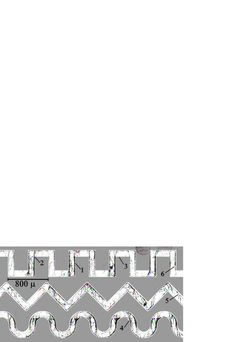
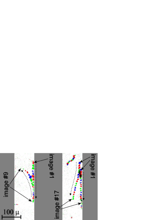
The next clearly observed effect is that cells depart from walls on sharp turns forming ‘fans’ of trajectories, shown by Label 2. After reaching the opposite wall, most cells follow it to the next turn. As a result, few or no cells swim along ‘inner’ segments of channel walls (Label 3). On curved turns, cells may also depart the channel wall (Label 4) though some cells still continue following the wall. Sometimes cells leave the corner in the absence of geometrical features (Label 5) which we attribute to collisions. These collisions may be head-on or on overtaking, as shown in Fig. 3.
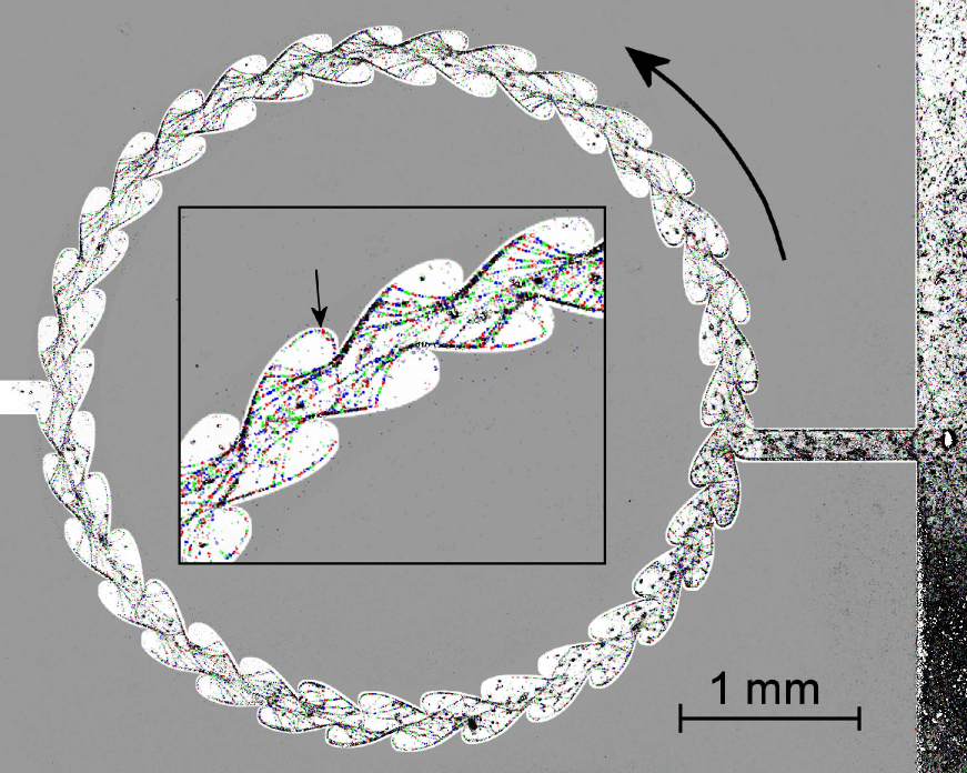
The fact that cells depart from corners can be used to create a channel with ratchet-type walls to force cells to swim in one direction. Cells in a sort of a circular running track are shown in Fig. 4. Certain configurations lead to entrapment of cells for extended times. A defective link in an earlier version of a channel was able to trap cells for as long as 10 minutes before they escape: two crypts on the opposite walls were staggered in such a way that, while following the channel wall, a cell was ejected by one crypt to get into the other and then ejected by the latter to return to the first crypt.
We have studied the influence of medium rheology on the cell near-wall behavior by filling the microchannel with , , and solutions of methylcellulose. The main effects are shown to be robust with respect to medium rheology: spermatozoa swim head-against-the-wall and depart from sharp bends in both pure (Newtonian) medium and in the medium with methylcellulose, which has more than 100 times higher viscosity and complicated rheological properties. A qualitative observation is that at higher concentration of methylcellulose, visibly more cells swim in the middle of the channel. To assess the distribution of cells departing from walls on the channel bends, we analyze the pixel intensity in fans of trajectories starting from channel bends in superposition of image sequences. As the light sensitivity of our CCD camera is linear to a good approximation, pixel intensity is a suitable quantitative parameter to use for reconstruction of the cell distribution by departure angles. The 30 minutes long records have been analyzed and data over four 90∘ channel bends have been analyzed. Typical results are shown in Fig. 5b. Depending on the donor, the mean cell turning angle varies from 10∘ to 20∘ with the width at half maximum at the level of 25∘. Observe that a notable part of cells turn away from the wall (negative angles). No consistent dependence of the departure angle on the concentration of methylcellulose has been detected, with the concentration affecting the sperm from different donors in different ways. This can be attributed to a sophisticated interplay between the flagellum stroke pattern and the medium rheological properties.
Discussion
We can interpret our observations through the following intuitive model, similar to that advanced by Woolley [4]. The amplitude of head oscillations is less that that of the end of the tail, so the head can, on average, be closer to the wall. The conical envelope of the flagellar wave aligns with the surface, resulting in the direction of propulsion being inclined towards the wall (Fig. 1b). Cells therefore are directed towards surfaces, and moreover cells stay against those surfaces. Once a cell reaches a horizontal wall, it is likely to travel along horizontally while translating until it, by chance, reaches a vertical wall (or vice versa). It then remains trapped by both walls, swimming along their intersection, until it finally reaches a sufficiently sharp change to the curvature of a vertical wall (Fig. 1a) to cause departure.
Furthermore, we can use this intuitive model to estimate the minimum turning radius of the cell. Consider similar triangles, the one formed by the cell envelope and the one formed by radii connecting head and tail of the cell with the center of a circle forming the channel wall (Fig. 1c). Equating the ratios of triangle bases and sides, we get that the radius of the channel wall at which a cell of the length is oriented tangent to the surface at the point of the head contact can be estimated as
| (1) |
Substituting m and from microscopic observations, we get 150 m. This value is close to the inner radius of curved (the lowest) channel in Fig. 2. Observe that while most cells depart on the turn, some stay at the wall which is an indication that the wall radius is not far from critical in accordance with the estimate (1).
The effect of viscosity on cell departure angle emphasizes the need to perform laboratory assays and ex vivo sperm-tract interaction studies in medium with viscosity adjusted to the magnitude of physiological fluids. It also suggests a possible role for viscosity in deflecting cells away from crypts in the reproductive tract. Our finding that sperm respond to ratchet geometries in a similar way to bacteria may potentially improve microfluidic IVF devices, through acting to direct high concentrations of motile cells towards the egg. We only have the beginnings of an understanding of how the minute population of sperm reaching the site of fertilization may differ from the vast majority that do not. The existence of this distinguished subpopulation was suggested by in vivo studies in rabbits [25] over 30 years ago, but the determinants of successful migration still remain mysterious; these characteristics may include motility, in addition to immunological markers and morphology. Further experimentation may also enable development of a useful motility-based functional diagnostic or prognostic test for male fecundity. For example, observation of sperm in microchannels may reveal hitherto undiscovered swimming parameters underlying successful tract migration or navigation.
As shown above, sperm cell migration in a micro-channel crucially depends on the channel geometry. Cells swim along boundaries and, if the two flat boundaries intersect, cells follow the corner. This has cardinal consequences for modeling of the cell behavior. Instead of spreading through a 3-dimensional domain, many cells swim along 1-dimensional folds. First, this entails that wall features such as ratchets can prescribe swimming direction. Second, the size of the domain available to the swimmers is drastically reduced, so cells collide more often; this requires special consideration when modeling the spreading of the entire population, either in microchannel environments or the female reproductive tract. The increased likelihood of a sperm-sperm collision may also have a more complex behavioral effect; when cells collide, mechanotransduction may induce cell signaling, altering beat pattern and hence migratory behavior.
The findings now indicate that recent advances in investigating sperm chemoattractants not only need to take account of the rheology of the fluid in which the cells are swimming [26], but also the three-dimensional architecture of the fluid domain. The application of experimental and computational fluid dynamics is beginning to reveal the complexity of the system of sperm-tract interaction, one of the central unsolved problems in reproductive science.
Materials and methods
This study employed channels of a cross-section m to observe trajectories of individual freely migrating human sperm in microchannels of basic geometrical configurations (corners, curves) and more complex features (‘ratchets’). Cell behavior in micro-channels of basic geometrical configurations was studied. Microchannels of m height were produced in elastomer (PDMS) by soft lithography [27] and then bonded to a glass coverslip after oxygen plasma treatment. Swimming cells were observed through the glass wall of the channel using a CCD camera equipped with a standard microscope objective. A green 100 mW diode laser equipped with a condenser was used as the light source. For imaging of the whole channel, we utilized a 160 mm 2x objective attached with an extension tube to a 4 Megapixel Basler avA2300-25gm camera run at 4 fps. Cell swimming was examined in fluid of three different rheologies: 0%, 0.5%, and 1% methylcellulose (M0512, Sigma-Aldrich, Poole, UK, approximate molecular weight 88,000) was added to Earle’s Balanced Salt Solution without phenol red, supplemented with 2.5 mM Na pyruvate and 19 mM Na lactate (06-2010-03-1B Biological Industries, Kibbutz Neuro Haemek, Israel), and 0.3% w/v charcoal delipidated bovine serum albumin (Sigma A7906). Semen samples were obtained by masturbation, at the Centre for Human Reproductive Science, Birmingham Women’s NHS Foundation Trust from normozoospermic research donors giving informed consent, after 2–4 days’ abstinence. Donors provided informed consent under Local Ethical Approval (South Birmingham LREC 2003/239). Experiments were performed between 1 and 3 hours after the semen sample was produced. The raw semen was injected into the wide ‘entry’ branch of the channel from which cells naturally spread to the main section. Results shown are representative of 5 donors.
Acquired images were processed in series of 200 to form superimposition images. Pixels at which the brightness increased from frame to frame above a certain level were stained, so that only moving objects are visible. Additionally, the image sequence was color-coded as order, i.e. cell positions in frames 1 and 2 are stained red, frames 3 and 4 green, frames 5 and 6 blue, frames 7 and 8 red again and so on. Hence, the direction of cell motion can be inferred from superposition images. One such image is shown in Fig. 2. Camera resolution was 2.7 m/pix, too coarse to resolve details of the cell head, but sufficient to determine its position.
Acknowledgments
DJS and JKB acknowledge funding from Birmingham Science City. PD acknowledges the award from Warwick Institute of Advanced Study. The authors thank staff at Birmingham Women’s Hospital and members of the Reproductive Biology and Genetics Group, University of Birmingham, for assistance; the authors also thank Professor Howard Berg for comments on the manuscript.
References
- [1] Rothschild. 1963. Non-random distribution of bull spermatozoa in a drop of sperm suspension. Nature, 198:1221–1222.
- [2] H. Winet, G. S. Bernstein, and J. Head. 1984. Observations on the response of human spermatozoa to gravity, boundaries and fluid shear. J. Reprod. Fert., 70:511–523.
- [3] J. Cosson, P. Huitorel, and C. Gagnon. 2003. How spermatozoa come to be confined to surfaces. Cell Motil. Cytoskel., 54:56–63.
- [4] D. M. Woolley. 2003. Motility of spermatozoa at surfaces. Reproduction, 126:259–270.
- [5] M. Ramia, D. L. Tullock, and N. Phan-Thien. 1993. The role of hydrodynamic interaction in the locomotion of microorganisms. Biophys. J., 65:755–778.
- [6] L. J. Fauci and A. McDonald. 1995. Sperm motility in the presence of boundaries. Bull. Math. Biol., 57:679–699.
- [7] D. J. Smith, E. A. Gaffney, J. R. Blake, and J. C. Kirkman-Brown. 2009. Human sperm accumulation near surfaces: a simulation study. J. Fluid Mech., 621:220–236.
- [8] D. J. Smith and J. R. Blake. 2009. Surface accumulation of spermatozoa: A fluid dynamic phenomenon. The Mathematical Scientist, 465:2417–2439.
- [9] J. R. Blake. 1971. A note on the image system for a Stokeslet in a no-slip boundary. Proc. Camb. Phil. Soc., 70:303–310.
- [10] J. Elgeti, U. B. Kaupp, and G. Gompper. 2010. Hydrodynamics of sperm cells near surfaces. Biophys. J., 99(4):1018–1026.
- [11] D. G. Crowdy and Y. Or. 2010. Two-dimensional point singularity model of a low-Reynolds-number swimmer near a wall. Phys. Rev. E, 81(3 Pt 2):036313.
- [12] D. J. Smith, E. A. Gaffney, H. Shum, H. Gadêlha, and J. Kirkman-Brown. 2011. Comment on the article by J. Elgeti et al. Hydrodynamics of Sperm Cells Near Surfaces. Biophys. J., 100:2318–2320.
- [13] J. Elgeti, U. B. Kaupp, and G. Gompper. 2011. Response to comment on article: Hydrodynamics of sperm cells near surfaces. Biophys. J., 100(9):2321–2324.
- [14] E. Lauga, W. R. DiLuzio, G. M. Whitesides, and H. A. Stone. 2006. Swimming in circles: motion of bacteria near solid boundaries. Biophys. J., 90(2):400–412.
- [15] Guanglai Li and Jay X. Tang. Aug 2009. Accumulation of microswimmers near a surface mediated by collision and rotational brownian motion. Phys. Rev. Lett., 103:078101.
- [16] D. Giacché, T. Ishikawa, and T. Yamaguchi. 2010. Hydrodynamic entrapment of bacteria swimming near a solid surface. Phys. Rev. E, 82(5):56309.
- [17] H. Shum, E. A. Gaffney, and D. J. Smith. 2010. Modelling bacterial behaviour close to a no-slip plane boundary: the influence of bacterial geometry. Proc. R. Soc. Lond. A.
- [18] A. P. Berke, L. Turner, H. C. Berg, and E. Lauga. 2008. Hydrodynamic attraction of swimming microorganisms by surfaces. Phys. Rev. Lett., 101(3):38102.
- [19] Y. Or and R. M. Murray. 2009. Dynamics and stability of a class of low Reynolds number swimmers near a wall. Phys. Rev. E, 79.
- [20] D. Crowdy and O. Samson. 2011. Hydrodynamic bound states of a low-reynolds-number swimmer near a gap in a wall. J. Fluid Mech., 667:309–335.
- [21] S. S. Suarez and A. A. Pacey. 2006. Sperm transport in the female reproductive tract. Hum. Reprod. Update, 12:23–37.
- [22] S. E. Hulme, W. R. DiLuzio, S. S. Shevkoplyas, L. Turner, M. Mayer, H. C. Berg, and G. M. Whitesides. 2008. Using ratchets and sorters to fractionate motile cells of escherichia coli by length. Lab Chip, 8(11):1888.
- [23] P. Galajda, J. Keymer, P. Chaikin, and R. Austin. 2007. A wall of funnels concentrates swimming bacteria. J. Bacteriol., 189(23):8704.
- [24] M. Binz, A.P. Lee, C. Edwards, and D.V. Nicolau. 2010. Motility of bacteria in microfluidic structures. Microelectronic Engineering, 87(5-8):810–813.
- [25] J. Cohen and K.R. Tyler. 1980. Sperm populations in the female genital tract of the rabbit. J. Reprod. Fert., 60(1):213.
- [26] J.C. Kirkman-Brown and D.J. Smith. 2011. Sperm motility: is viscosity fundamental to progress? Mol. Hum. Reprod., 17(8):539–544.
- [27] Y. N. Xia and G. M. Whitesides. 1998. Soft lithography. Annu. Rev. Mater. Sci., 28:153.