Towards a Nano Geometry?
Geometry and Dynamics on Nano Scale
Abstract.
This paper applies I.M. Gelfand’s distinction between adequate and non-adequate use of mathematical language in different contexts to the newly opened window of model-based measurements of intracellular dynamics. The specifics of geometry and dynamics on the mesoscale of cell physiology are elaborated - in contrast to the familiar Newtonian mechanics and the more recent, but by now also rather well established quantum field theories. Examples are given originating from the systems biology of insulin secreting pancreatic beta-cells and the mathematical challenges of an envisioned non-invasive control of magnetic nanoparticles.
Key words and phrases:
Cell physiology, free boundary problems, differential invariants, geometry driven dynamics, non-Newtonian liquids, electro-magnetic fields2000 Mathematics Subject Classification:
Primary 92C37; Secondary 35R35, 53A551. The Challenge of Nano Structures
There are many different geometries around. Do we really need new kinds of geometries? Why and how?
1.1. Nanoparticle Based Transducer for Intracellular Structures
These days, we witness a dramatic progress in various technologies devoted to capturing intracellular dynamics of highly differentiated animal cells like the delicate insulin secreting pancreatic -cell with its thousands of free moving insulin granules, rails of microtubuli, fences of actin filaments, zoos of organelles, proteins, genes, ion channels, electro-static and electrodynamic phenomena. A radically new world of geometry and dynamics is evolving before our eyes. The most decisive technological advances are in the following domains:
-
•
Life imaging, for instance confocal multi-beam laser microscopy, admitting up to 40 frames per second for tracking position and movement of suitably prepared nanoparticles within the cell and without overheating the tissue;
-
•
Magnetic nanoparticle design and coating, admitting electromagnetic manipulation, docking to selected organelles and tracking their movement;
-
•
Computer supported collection and administration of huge databases.
This raises the question: Are we on the verge of a need and, then, of the emergence of radically different new mathematical concepts, as predicted by the late I.M. Gelfand in 2003 at a conference (see [Eti]) on The Unity of Mathematics, held in honor of his 90th birthday. He said:
… we have a “perestroika” in our time. We have computers which can do everything. We are not obliged to be bound by two operations - addition and multiplication. We also have a lot of other tools. I am sure that in 10 to 15 years mathematics will be absolutely different from what it was before. ([Glf, p.xiv])
Then, how reasonable is it to demand and to expect new geometries only in view of the new time and length scales of cell physiology: much larger than the scales underlying particle physics, quantum mechanics, proteomics and genetics with its characteristic operator analysis, geometric foldings and stochastic processes; and much smaller than the scales underlying tissue, organ, patient and population biology and medicine with its characteristic statistics and bifurcations? Can’t we transfer geometric and dynamic concepts all the way up and down the scales? What should be so special for geometers with the mesoscale of a few nanometers and a few seconds and minutes?
1.2. Gelfand’s Dictum
At the mentioned conference, the jubilee surprised by qualifying the general praise of mathematics as adequate language for science. Against the supposed unity and adequacy of mathematics he insisted on the distinction between adequate and inadequate use of mathematical concepts, depending on the context (see [Glf] for the whole talk):
An important side of mathematics is that it is an adequate language for different areas: physics, engineering, biology. Here, the most important word is adequate language. We have adequate and nonadequate languages. I can give you examples of adequate and nonadequate languages. For example, to use quantum mechanics in biology is not an adequate language, but to use mathematics in studying gene sequences is an adequate language.
Clearly, on one side, Gelfand played on the common pride of mathematicians regarding Galilei’s famous dictum of [Gal, Il Saggiatore, cap. 6]:
La filosofia è scritta in questo grandissimo libro che continuamente ci sta aperto innanzi a gli occhi (io dico l’universo), ma non si può intendere se prima non s’impara a intender la lingua, e conoscer i caratteri, ne’ quali è scritto. Egli è scritto in lingua matematica, e i caratteri son triangoli, cerchi, ed altre figure geometriche, senza i quali mezi è impossibile a intenderne umanamente parola; senza questi è un aggirarsi vanamente per un oscuro laberinto.111In English: “Philosophy is written in that great book which ever lies before our eyes I mean the universe but we cannot understand it if we do not first learn the language and grasp the symbols, in which it is written. This book is written in the mathematical language, and the symbols are triangles, circles and other geometrical figures, without whose help it is impossible to comprehend a single word of it; without which one wanders in vain through a dark labyrinth.” The Assayer (1623), as translated by Thomas Salusbury (1661), p. 178, as quoted in The Metaphysical Foundations of Modern Science (2003) by Edwin Arthur Burtt, p. 75.
On the other side, Gelfand warned in the given quote against the misleading playing around with mathematical concepts without due regard to the characteristic lengths, times, data and problems of a concrete context. Is there a contradiction?
1.3. The Common Regard and Disregard of Context
Deep in our heart, we mathematicians believe in the unity and universality of mathematics. We are not topologists, algebraists, pde folk or applied, as little as a music composer is a quartet or a trio composer, as Gelfand also noted in his talk. We are mathematicians, and our belief in the unity and universality of our concepts is based on three solid pillars,
-
(1)
our Emmy Noether and N. Bourbaki belief in the universal meaning of structures;
-
(2)
our semiotic training which assigns to even the most abstract concepts very concrete, worldly, human, mental images (a process intensively studied by the American physicist and philosopher Charles Sanders Peirce); and
-
(3)
our accept of the universality of phenomena, be it the universality of the three conic sections of Apollonius of Perga in the level curves of all binary quadratic forms in two variables , or the universality of René Thom’s seven elementary catastrophes (generic structures for the bifurcation geometries) in all dynamical systems subjected to a potential with two or fewer active variables, and four or fewer active control parameters.
So, on Sundays we are easily seduced to contempt of the context and into belief of universality.
However, from the history of our subject we know that there are no great eternal lines in mathematics. Euclid did not suffice for Newton’s study of planetary motion, and the calculus was created. Classical analysis did not suffice for Bohr’s study of the atom and operator theory in Hilbert space was created. Functional analysis did not suffice for the study of elementary particles and spectral geometry was developed for the sake of quantum field theories. Worst of all, there is no mathematics around or emerging in physics to support a Theory of Everything (TOE) merging all four interactions into one, in spite of the solid mathematical foundations and the high promises of the Grand Unified Theory (GUT) to replace the ad-hoc Standard Model of particle physics. On the contrary, looking through a modern textbook on Quantum Gravity like [Bo10] will support Niels Bohr’s view of the complementarity and - in tendency - the mutually unrelated state of different areas of our investigation. So, we have to study a subject with focused glasses, directed to limited segments, full of surprises. We have grown used to all kinds of confinements, due to peculiar aspects of the chosen level of physical reality or due to fashions, Führers, external impact that can devaluate earlier approaches and demand radically new ideas over night. So, in daily work we have learned to live without universality.
2. Typology of Mathematics Use in Cell Physiology


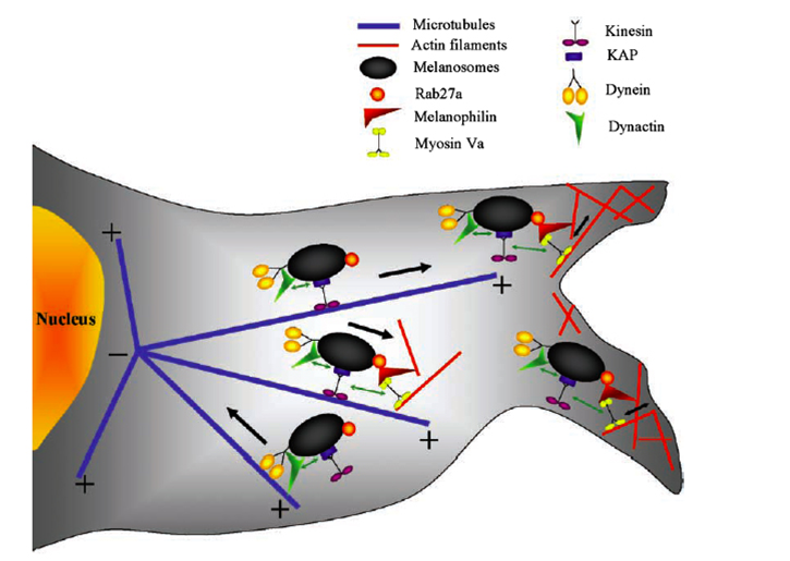
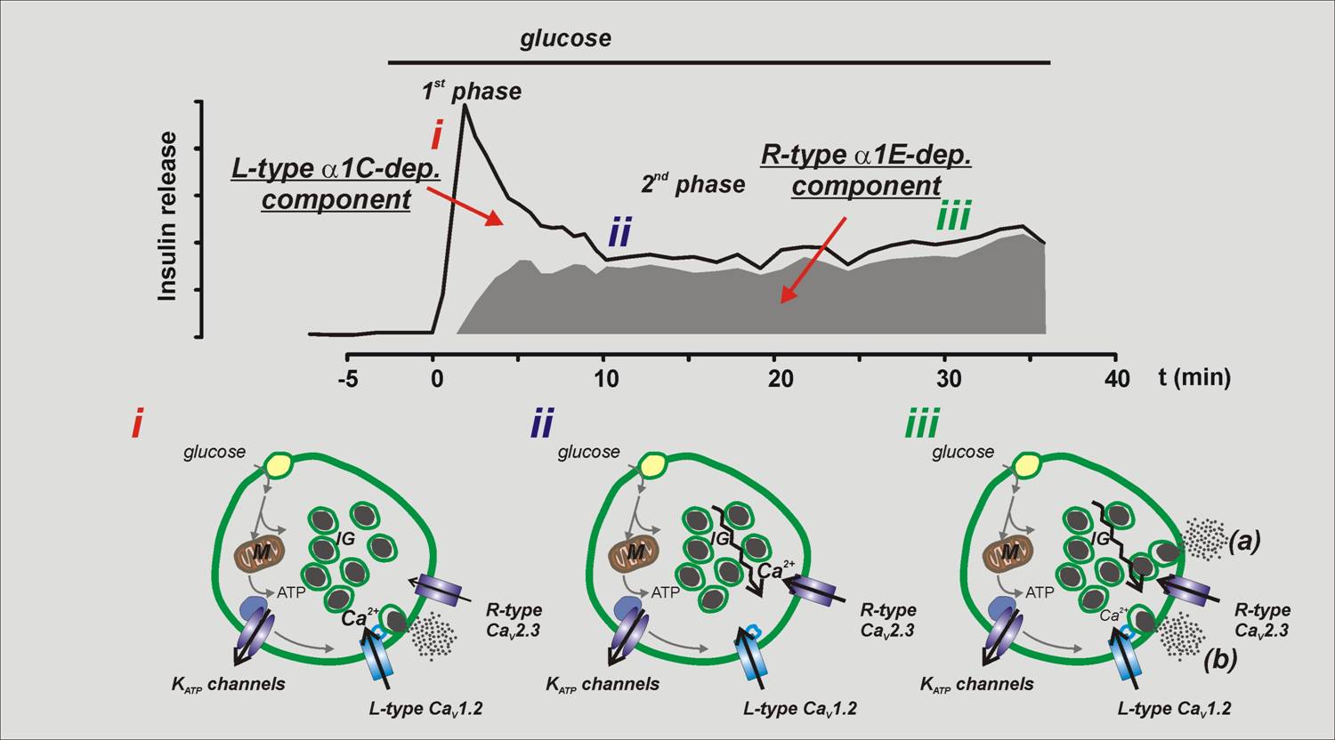
For capturing the geometry and the dynamics of insulin secretion of pancreatic -cells (regulated exocytosis, see Figures 1, 2), it may be helpful to distinguish the following modeling purposes:
2.1. Model Based Capturing of Intracellular Dynamics
With the sudden technology-pushed opening of a window to intracellular positions, shapes and movements, it seems to me that the descriptive role of mathematics will be the most decisive contribution to the progress of medical biology, i.e., supporting model-based measurements in the laboratory. To some extent, the technological progress has given immediate access to machine generated cell data in -cells like
-
•
precise measurements of the quantitative and temporal sequence of glycose-stimulus secretion-response;
-
•
precise determination of changes in the electro-static potential over the plasma membrane and the opening and closing of ion channels across the plasma membrane upon stimulation;
-
•
precise observation of positions of organelles, microfilaments and granules by electron microscopy and electron tomography under rapid freezing, and vaguely by luminescent quantum dots and other fluorescent reporters in living cells;
-
•
identifying proteins, enzymes; and
-
•
determining genes in DNA sequences.
These observations have been around for decades. The drawback with all of them is their static and local character. No matter how valuable they are for some purposes, they don’t give access to the intracellular dynamics. So, the true functioning (or dysfunctioning) of a living -cell is not accessible immediately.
Many biomedical quantities cannot be measured directly. That is due to the subject matter, here the nature of life, partly because most direct measurements will require some type of fixation, freezing and killing of the cells, partly due to the small length scale and the strong interaction between different components of the cell. Just as in physics since Galileo Galilei’s determination of the simple (but at his time not measurable) free vertical fall law by calculating “backwards” from the inclined plan, one must also in cell physiology master the art of model-based experiment design.
Below in Section 3.3 we shall discuss essential parameters for the insulin granule motility in -cells like the visco-elasticity of the cytosol or the magnetic field strength of the pulsating flux of calcium ions between storage organelles (mitochondria and endoplasmic reticulum). For high precision in the critical period of granule preparing, docking and bilayer fusion with the cell membrane, radically new possibilities appear by tracking the movements of labeled magnetic nanoparticles in controlled electro-dynamic fields (see below). In this case, solving mathematical equations from the fields of electro-dynamics and thermo-elasticity becomes mandatory for the design of the experiments and the interpretation of the data. In popular terms, one may speak of a mathematical microscope (a term coined by J. Ottesen [Ott]), in technical terms of a transducer, sensor, actuator that becomes useful as soon as we understand the underlying mathematical equations.
2.2. Simulation and Prediction
Once a model is found and verified and the system’s parameters are estimated for one domain, one has the hope of doing computer “experiments” (i.e., calculations and extrapolations for modified data input) to replace or supplement costly, time-consuming and sometimes even physically impossible experiments. The last happens when we are permitted to change a single parameter or a selected combination of parameters in the calculation contrary to a real experiment, where typically one change induces many accompanying changes. In this way we may predict what we should see in new experiments in new domains (new materials, new temperatures etc). Rightly, one has given that type of calculations a special name of honor, computer simulations: As a rule, it requires to run the process on a computer or a network of computers under quite sophisticated conditions (discussed in [Shi]). Typically, the problem is to bring the small distances and time intervals of well-understood molecular dynamics up to reasonable mesoscopic scales, either by aggregation or by Monte Carlo methods – as demonstrated by Buffon’s needle casting for the numerical approximation of .
One should be aware that the word “simulation” has, for good and bad, a connotation derived from NASA space simulators and Nintendo war games and juke boxes. Animations and other advanced computer simulations can display an impressive beauty and convincing power. That beauty, however, is often their dark side: Simulations can show a deceptive similarity with true observations, so for the lipid bilayer fusion of an insulin vesicle with the plasma membrane and the release of the bulk of hormone molecules. The numerical solution of huge systems of Newton’s equation, i.e., the integration of all the forces between the membrane lipids can be tuned to display a convincing picture of the secretion course in a nanosecond time span whereas that very process in reality takes seconds and minutes. In numerical simulation, like in mathematical statistics, results which fit our expectations too nicely, must awake our vigilance instead of being taken as confirmation.
2.3. Control
The prescriptive power of mathematization deserves a more critical examination. The time will come when the model based understanding of intracellular dynamics in healthy and dysfunctional -cells will lead to new diagnostic approaches, new drugs and new treatments. In physics and engineering we may distinguish between the (a) feasibility, the (b) efficiency, and the (c) safety of a design. A design can be an object like an airplane or a circuit diagram for a chip, an instrument like a digital thermometer, TV set, GPS receiver or pacemaker, or a regulated process like a feed-back regulation of the heat in a building, the control of a power station, the precise steering of a radiation canon in breast cancer therapy - or the design of a new, non-symptomatic diagnostic procedure or therapy.
Mathematics has its firm footing for testing the feasibility of new approaches in thought experiments, estimations of process parameters, simulations and solving equations. For testing efficiency, a huge inventory is available of mathematical quality control and optimization procedures by variation of key parameters. It seems to me, however, that safety questions provide the greatest mathematical challenges. For early diagnosis, say of juvenile diabetes (DT1) and drug design, mathematics does not enter trivially into the certification of the correctness of the design copy and the quality test of the performance. Neither do we come to a situation where it suffices to modify and re-calculate well-established models and procedures. Experienced pharmacologists and medical doctors, we may hope, will not trust mathematical calculations and adaptations. Too many parameters may be unknown and pop up later. Here is a parallel to the early days of traditional railroad construction: A small bridge was easily calculated and built, but then photogrammetrically checked when removing the support constructions. A lowering of more than required re-building. Similarly, even the most carefully calculated and clinically tested diagnoses and therapies will require supplement by the most crazy mathematical imagination of what could go wrong and might show up only after years of treatment and where and how to find or build an emergency exit in the cell.
An additional disturbing aspect of science-integrated medical technology development is the danger of losing transparency. Medical doctors are trained to understand the elements of mechanics and chemical reactions, i.e., purely locally in cell terms. They are not prepared to grasp global cell phenomena like magnetic field density and the geometry and dynamics of long-distance amplification processes within cells. Therefore, it will be very unfortunate when medical doctors shall ordinate a treatment they do not really understand.
2.4. Explain phenomena
The noblest role of mathematical concepts in cell physiology is to explain phenomena. Einstein did it in physics when reducing the heat conduction to molecular diffusion, starting from the formal analogy of Fick’s Law with the cross section of Brownian motion. He did it also when generalizing the Newtonian mechanics into the special relativity of constant light velocity and again when unifying forces and curvature in general relativity.
One may hope that new mathematical models can serve biomedicine by reducing new phenomena to established physical principles; and as heuristic devices for suitable generalizations and extensions.
Physics history has not always attributed the best credentials to explaining phenomena by abstract constructions. It has discarded the concept of a ghost for perfect explanation of midnight noise in old castles; the concept of ether for explaining the finite light velocity; the phlogiston for burning and reduction processes, the Ptolemaic epicycles for planetary motion. It will be interesting to see in the years to come whether some of the common explanations in cell physiology will suffer the same fate.
2.5. Theory development
Finally, what will be the role of mathematical concepts and mathematical beauty for the very theory development in cell physiology? Not every mathematical, theoretical and empirical accumulation leads to theory development. Immediately after discovering the high-speed rotation of the Earth around its own axis, a spindle shape of the Earth was suggested and an infinitesimal tapering towards the North pole confirmed in geodetic measurements around Paris. Afterwards, careful control measurements of the gravitation at the North Cap and at the Equator suggested the opposite, namely an ellipsoid shape with flattened poles. Ingenious mathematical mechanics provided a rigorous reason for that. Gauss and his collaborator Listing, however, found something different in their control. They called the shape gleichsam wellenförmig and dropped the idea of a theoretically satisfactory description. Since then we speak of a Geoid.
Similarly, when analyzing intracellular geometries and dynamics we may meet events which have their own phylogenetic history, dating back to more than 0.6 billion of years in the case of -cells, and have lost their relevance since then. With high probability, many of the phenomena we observe are heritage, meaningless relics of past hundreds of millions of species’ development. Neglecting the probabilistic ruin character of our existence and pressing it into a slick mathematical model may be quite misleading.
3. Non-Invasive Control of Magnetic Nanoparticles
3.1. Emerging Radically New Research Agenda
Addressing the intracellular geometry and dynamics of the cell has many levels and many scales. To give an example, I shall describe an evolving - focussed - systems biology of regulated exocytosis in pancreatic -cells, mostly based on [Bo11]. These cells are responsible for the appropriate insulin secretion. Insufficient mass or function of these cells characterize Type 1 and Type 2 diabetes mellitus (DT1, DT2). Similar secretion processes happen in nerve cells. However, characteristic times for insulin secretion are between 5 and 30 minutes, while the secretion of neurotransmitters is in the millisecond range. Moreover, the length of a -cell is hardly exceeding 4000 nanometres (nm), while nerve cells have characteristic lengths in the cm and meter range. So, processes in -cells are more easy to observe than processes in nerve cells, but they are basically comparable.
It seems that comprehensive research on -cell function and mass has been seriously hampered for 80 years because of the high efficiency of the symptomatic treatment of DT1 and DT2 by insulin injection. Recent advances - and promises - of noninvasive control of nanoparticles suggest the following radically new research agenda, to be executed first on cell lines, then on cell tissue of selected rodents, finally on living human cells:
3.1.1. Optical Tracking of Forced Movement of Magnetic Nanoparticles
Synthesize magneto-luminescent nanoparticles; develop a precisely working electric device, which is able to generate a properly behaving electromagnetic field; measure cytoskeletal viscosity and detect the interaction with organelles and actin filaments by optical tracking of the forced movement of the nanoparticles. Difficulties to overcome: protect against protein adsorption by suitable coating of the particles and determine the field strength necessary to distinguish the forced movement from the underlying Brownian motion.
3.1.2. Optical Tracking of the Intracellular Dynamics of Insulin Granules
Synthesize luminescent nanoparticles with after-glow property (extended duration of luminescence and separation of excitation and light emission); dope the nanoparticles with suitable antigens and attach them to selected organelles to track intra-cellular dynamics of the insulin granules.
3.1.3. Precise Chronical Order of Relevant (Electrical) Secretion Events
Apply a multipurpose sensor chip and measure all electric phenomena, in particular varying potentials over the plasma membrane, the bursts of ion oscillations, and changing impedances on the surface of the plasma membrane for precise chronical order of relevant secretion events.
3.1.4. Geometry and Dynamics of Lipid Bilayer Membrane-Granule Fusion
Describe the details of the bilayer membrane-granule fusion event (with the counter-intuitive inward dimple forming and hard numerical problems of the meso scale, largely exceeding the well-functioning scales of molecular dynamics).
3.1.5. Connecting Dynamics and Geometry with Genetic Data
Connect the preceding dynamic and geometric data with reaction-diffusion data and, finally, with genetic data.
3.1.6. Health Applications
Develop clinical and pharmaceutical applications:
-
•
Quality control of transplants for DT1 patients.
-
•
Testing drug components for -cell repair.
-
•
Testing nanotoxicity and drug components for various cell types.
-
•
Early in-vivo diagnosis by enhanced gastroscopy.
-
•
Develop mild forms of gene therapy for patients with over-expressed major type 2-diabetes gene TCF7L2 by targeting short interfering RNA sequences (siRNAs) to the -cells, leading to degradation of excess mRNA transcript. (This strategy may be difficult to implement, due to the degradation of free RNA in the blood and the risk of off-target effects.)
In the following, we shall not deal with the envisaged true health applications. Only shortly we shall comment upon the mathematical challenges of the non-invasive control of magnetic nanoparticles, the intricacies of the related transport equations, compartment models, electromagnetic field equations, free boundary theory, reaction-diffusion equations, data analysis etc.
3.2. Gentle Insertion by Rolling on Cell Surface
The good news is the newly developed Dynamic Marker technique, see [Ko]: Based on well-established electrical power engineering know-how, arrays of conventional coils are arranged in small engines to generate precisely directed dynamic magnetic field waves of low magnetic field density (mTesla range) and low frequency (1-40 Hz). The use of dynamic and directed magnetic field waves makes the beads roll on the cell surface to rapidly meet willing receptors.
This technology has shown a much better insertion performance in experiments than conventional diffusion or static magnetic fields: With less than 10 minutes characteristic dynamic marking is much faster than waiting for 12-24 hours on diffusion of the nanoparticles across the plasma membrane and reduces dramatically the inflammation risks during waiting. Contrary to the conventional use of static magnetic fields (e.g., by applying MRI machines) for transfection the cell’s nucleus will not be “bombed” and the associated high lysis (= break down) of cells under the process is avoided. Success has only been achieved, however, for magnetic nanoparticles with a diameter . Moreover, the success seems to depend heavily on the correct tuning of magnetic field density and frequency. The guiding transport equations seem not fully understood yet. To generate much higher magnetic field densities of field waves for in-vivo application, portable superconductive coils at relatively high temperature ( K) are expected to be developed in the future.
3.3. Viscosity in Newtonian and Non-Newtonian Cytosol
Like many other biomedical quantities, the viscosity of the cytosol cannot be measured directly. Let us look at the eight to twelve thousand densely packed insulin vesicles in a single -cell. They all must reach the plasma membrane within a maximum of 30 minutes after stimulation, to pour out their contents. Let us ignore the biochemistry of Figure 1 and the many processes taking place simultaneously in the cell and consider only the basic physical parameter for transport in liquids, namely the viscosity of the cell cytosol. From measurements of the tissue (consisting of dead cells) we know the magnitude of viscosity of the protoplasma, namely about 1 milli-pascal-seconds (mPa s), i.e., it is of the same magnitude as water at room temperature. But now we want to measure the viscosity in living cells: before and after stimulation; deep in the cell’s interior and near the plasma membrane; for healthy and stressed cells.
It serves no purpose to kill the cells and then extract their cytosol. We must carry out the investigation in vivo and in loco, by living cells and preferably in the organ where they are located. The medical question is clear. So is the appropriate technological approach with noninvasive control of magnetic nanoparticles, explained in Section 3.2 above. These particles are primed with appropriate antigens and with a selected color protein, so that their movements within the cell can be observed with a confocal multi-beam laser microscope which can produce up to 40 frames per second. The periods of observations can become relatively short, down to 8-10 minutes - before these particles are captured by cell endosomes and delivered to the cells’ lysosomes for destruction and consumption of their color proteins (see also below Section 3.6).
3.3.1. Newtonian Idealization
Assuming (wrongly, see below) that the cytosol is a Newtonian liquid, we get from A. Einstein [Ein] and M. von Smoluchowski [Smo] precise recipes how to determine the viscosity from a few snapshots of the Brownian motion or the forced movement of suspended particles. Roughly speaking, Einstein discovered the scale independence (self-similarity = fractal structure) of the Brownian motion permitting to derive the characteristic diffusion coefficients of the almost continuously happening jumps, from sample geometries of positions at observable, realistic huge time intervals (huge compared to the characteristic time of the process). Smoluchowski worked out a smart observation scheme in the case of the presence of many particles which can no longer be traced individually by then (and now) existing equipment.
More precisely, the simplest mathematical method to determine its viscosity in vivo would be just to pull the magnetized particles with their fairly well-defined radius with constant velocity through the liquid and measure the applied electromagnetic force . Then the viscosity is obtained from Stokes’ Law . The force and the speed must be small so as not to pull the particles out of the cell before the speed is measured and kept constant. Collisions with insulin vesicles and other organelles must be avoided. It can only be realized with a low-frequency alternating field. But then Stokes’ Law must be rewritten for variable speed, and the mathematics begins to be advanced. In addition, at low-velocity we must correct for the spontaneous Brownian motion of particles. Everything can be done mathematically: writing the associated stochastic Langevin equations down and solve them analytically, or approximate the solutions by Monte Carlo simulation, like in [Lea, Schw]. However, we rapidly approach the equipment limitations, both regarding the laser microscope’s resolution and the lowest achievable frequency of the field generator.
So we might as well turn off the field generator and be content with intermittently recording the pure Brownian motion of a single nanoparticle in the cytosol! As shown in the cited two famous 1905/06 papers by Einstein, the motion’s variance (the mean square displacement over a time interval of length ) of a particle dissolved in a liquid of viscosity is given by , where denotes the diffusion coefficient with Boltzmann constant , absolute temperature and particle radius . In statistical mechanics, one expects collisions per second between a single colloid of diameter and the molecules of a liquid. For nanoparticles with a diameter of perhaps only 30 nm, we may expect only about collisions per second, still a figure large enough to preclude registration. There is simply no physical observable quantity at the time scale seconds. But since the Brownian motion is a Wiener process with self-similarity we get approximately the same diffusion coefficient and viscosity estimate, if we, e.g., simply register 40 positions per second. Few measurements per second are enough. Enough is enough, we can explain to the experimentalist, if he/she constantly demands better and more expensive apparatus.
Note that also can be estimated by the corresponding two-dimensional Wiener process of variance , consisting of the 2-dimensional projections of the 3-dimensional orbits, as the experimental equipment also will do.
Now you can hardly bring just a single nanoparticle into a cell. There will always be many simultaneously. Thus it may be difficult or impossible to follow a single particle’s zigzag path in a cloud of particles by intermittent observation. Also here, rigorous mathematical considerations may help, namely the estimation of the viscosity by a periodic counting of all particles in a specified window. As mentioned above, the necessary statistics was done already in [Smo].
3.3.2. Non-Newtonian Reality
Beautiful, but it is still insufficient for laboratory use: There we also must take into account the non-Newtonian character of the cytosol of -cells. These cells are, as mentioned, densely packed with insulin vesicles and various organelles and structures. Since the electric charge of iron oxide particles is neutral, we can as a first approximation assume a purely elastic impact between particles and obstacles. It does not change the variance in special cases, as figured out for strong rejection of particles by reflection at an infinite plane wall in [Smo]. But how to incorporate the discrete geometry and the guiding role of the microtubules into our equations of motion? Here also computer simulations have their place to explore the impact of different repulsion and attraction mechanisms on the variance.
3.4. Sensing Microfilaments, Tracing Insulin Granule Motility, Displaying the Genetic Variety of the Diabetes Umbrella
Up to now, it is not clear what geometry or geometries are underlying the secretion dynamics.
3.4.1. Pressing Need for Geometric Invariants
Soon we may be able to sense and map the extended geometry of the microtubules and their smoothing before secretion; soon we may be able to sense and map the extended geometry of the actin filaments and their dissolution just before secretion. However, we shall need numbers or other mathematical objects to characterize observed geometries and dynamics in order to relate observations of well-functioning secretion and dysfunction to the effect of selected genes. Epidemiological studies in large populations (see [LyGr, PaGl]) have found more than 20 gene deviations which show up in families with high expression of DT1 or DT2. Form that we learned that DT1 and DT2 are not two single diseases (distinguished by simple symptomatic classification) but umbrellas of quite different dysfunctions leading to the same or similar symptoms. To give these patients a cure, we need to know more precisely what is going wrong on the cell level. Proteomic analysis, in particular the disclosure of the proteins a certain gene is coding for, is a very promising approach. However, it will only lead to success if we become able to supplement it by precise description of related deviations in geometry and dynamics.


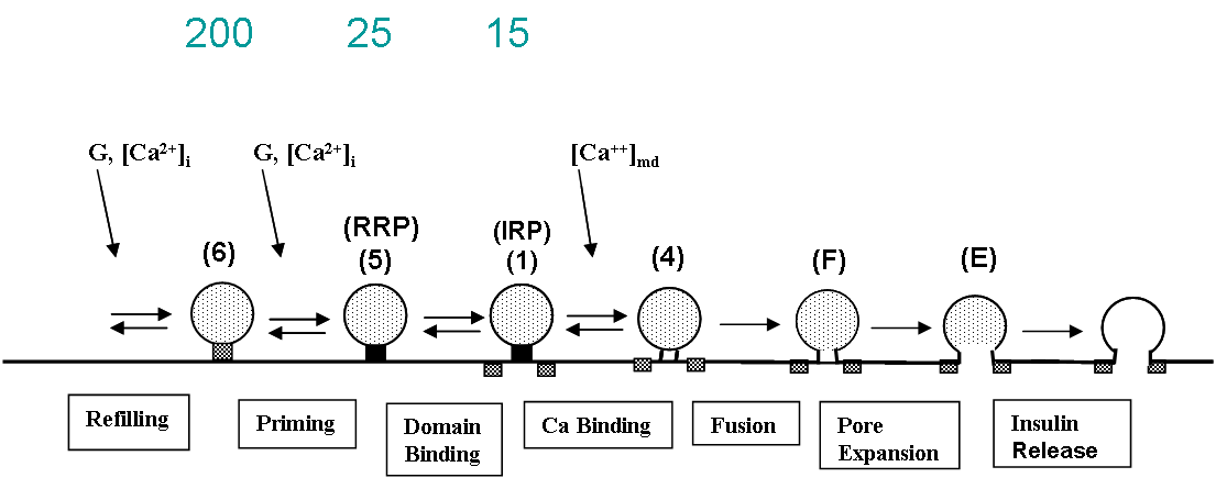
b) Down: Extended six-pool model, incorporating Ca-binding, of Chen, Wang and Sherman
3.4.2. First Steps via Compartment Models and Transition Rates
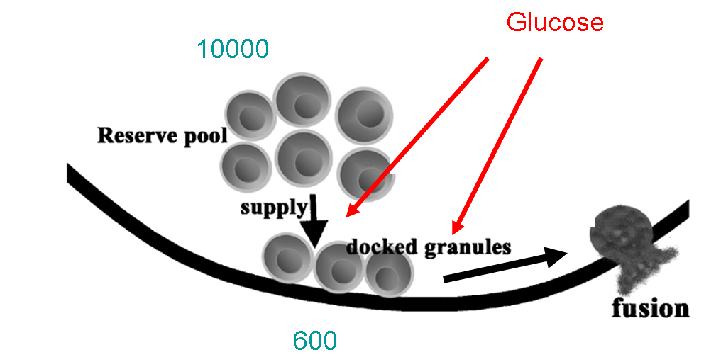
Just as in engineering, economics or anywhere else, also in cell physiology the daily mathematical exercise consists of the estimation of some parameters; testing the significance of some hypotheses; and designing compartment models for the dynamics of coupled quantitative variables. A first step towards integrating spatial geometry and temporal dynamics are the Compartment Models of regulated exocytosis, first introduced by Grodsky [Gro] in 1972. He assumed that there are two compartments (pools) of insulin granules, docked granules ready for secretion and reserve granules, see Figure 3a. By assuming suitable flow rates for outflow from the docked pool and resupply from the reserve pool to the docked pool, the established biphasic secretion process of healthy -cells (depicted in Figure 2) could be modeled qualitatively correct. By extending the number of pools from two to an array of six (Figure 3b) and properly calibrating all flow rates, Chen, Wang and Sherman in [CWS] obtained a striking quantitative coincidence with the observed biphasic process, see also Toffolo, Pedersen, and Cobelli [TPC]. Such compartment models invite the experimentalists (both in imaging and in proteomics) and the theoretician (both in geometry and in mathematical physics) to verify the distinction of all the hypothetical compartments in cell reality and to assign global geometrical and biophysical values to the until now only tuned flow rates. A geometer who is familiar with mathematical physics may, for instance, look for small inhomogeneities in the cell which could drive the global dynamics. One candidate is the viscosity as addressed above in Section 3.3: A slight increase of cytosol viscosity from the cell interior towards the plasma membrane would induce an externally directed vector by diffusion statistics.
A self-imposed limitation is the low spatial resolution of the aggregated compartments, which does not allow to investigate the local geometry and the energy balance of the secretion process.
3.5. Electrodynamic Insulin Secretion “Pacemaker”
In the preceding sections I briefly described the common phenomenological approaches to regulated exocytosis: the focus on the variable discrete geometry of the microfilaments; the numerical treatment of the molecular dynamics visualizing the singularity of lipid bilayer fusion events; the analytic power of compartment models to reproduce biphasic secretion. The phenomenological approaches relate the various data by visible evidence and statistically more or less well supported ad hoc assumptions about the regulation. They focus on the dominant and visible structures (like the filaments) and measurable local states in the neighborhood of the fusion event, neglecting long-distance phenomena like electromagnetic waves across the cell. One may deplore that and also the “enormous gap between the sophistication of the models and the success of the numerical approaches used in practice and, on the other hand, the state of the art of their rigorous understanding” (Le Bris [Leb] in his 2006 report to the International Congress of Mathematicians).
In [Apu], the authors advocate for supplementing the phenomenological approach by a theoretical approach based on first principles, following Y. Manin’s famous parole: The visible must be explained in terms of the invisible, [Ma]. They explain how a combination of rigorous geometrical and stochastic methods and electro-dynamical theory naturally draws the attention to fault-tolerant signalling, self-regulation and amplification (in M. Gromov’s terminology). They consider the making of the fusion pore, anteceding the lipid bilayer membrane vesicle fusion of regulated exocytosis, as a free boundary problem and show that one of the applied forces is generated by glucose stimulated intra-cellular ions oscillations (discussed in [FrPh]) resulting in a low-frequent electromagnetic field wave. It mis suggested that the field wave is effectively closed via the weekly magnetic plasma membrane (containing iron in the channel enzymes). Unfortunately, textbook electrodynamics mostly deals with geometrically simple configurations where the beauty and strength of Maxwell Equations best come out, but is less informative for treating peculiar geometries. This was also noted by Gelfand:
… images play an increasingly important role in modern life, and so geometry should play a bigger role in mathematics and in education. In physics this means that we should go back to the geometrical intuition of Faraday (based on an adequate geometrical language) rather than to the calculus used by Maxwell. People were impressed by Maxwell because he used calculus, the most advanced language of his time. ([Glf, p.xx])
The recent experimental evidence of the bio-compatibility and bio-efficiency of low-frequent electromagnetic fields (addressed above in Section 3.2) gives a hint to the presence of global fields controlling local events in the cell and supports a vision of a possible future electrodynamic pacemaker to stimulate regulated exocytosis in tired, dysfunctional -cells.
3.6. Induced Apoptosis Chain Reaction in Cancer Cells
The continuing almost total lack of understanding of the global aspects of cell physiology can also lead to happy surprises: In the course of the insertion experiments described above in Section 3.2, it was discovered that the inserted iron oxide nanoparticles of diameter nm (with special antibody-conjugated surfaces) were immobilized in less than 10 minutes within the lysosomes (organelles for digestion and destruction). However, continuing the action of the low frequency electromagnetic oscillations tore the membranes of these lysosomes, purely mechanically.
That was bad news for exploring the intracellular geometry and dynamics by nanoparticle transducers, because the observation window is consequently short, only 10 minutes if we come to use the “wrong” antibodies. It was good news for cancer research: Destroying the lysosomes in a probe of cancer cells leads to release of the digestive enzymes and initiates a destructive chain reaction in the neighboring tissue which stops automatically when healthy tissue (with neutral pH) is reached. The range (in time and space) of the obtainable chain reactions has not yet been fully determined. Nanotoxicity for healthy tissue, however, will be excluded conclusively. Testing of the field generator is under preparation for curing skin cancer on model tissue, on model animals and for full proof of concept.
4. Conclusions
Down there, in the nano-world of regulated exocytosis in pancreatic -cells, a heap of geometrical and dynamical information is waiting to be interrelated. Encouragement and, perhaps, inspiration may be gained from the visionary [CaGr] (though restricted to molecular biology). Every mathematician’s conviction is the inseparability of geometry and dynamics. That’s what we teach the students in algebra classes with the concepts of orbits and ideals; in ordinary differential equations classes with the Poincaré-Bendixson Theorem and the significance of the multiplicity and sign of eigenvalues for global behavior and the geometry of bifurcations; and most outspoken in spectral geometry classes with our focus on spectral invariants that characterize both shape and change at the same time.
For the evolving medical biology of highly differentiated cells like the pancreatic -cells it remains to hope that tendencies to futile overspecialization and excessive reductionism can be overcome. Clearly, the base of all future advances must be the precise, controlled single observations. But a real hope for diabetes patients can only come from the integration of the already established local facts into a global geometric and dynamic perception.
References
- [Apu] D. Apushkinskaya, E. Apushkinsky, B. Booß–Bavnbek and M. Koch, Geometric and electromagnetic aspects of fusion pore making, in: [Bo11], pp. 505–538, arXiv:0912.3738 [math.AP].
- [Bo10] B. Booß–Bavnbek, G. Esposito and M. Lesch (eds.), New Paths Towards Quantum Gravity, Springer Lecture Notes in Physics Vol. 807, Springer, Heidelberg-Berlin-New York, 2010, ixx + 359 pages, 47 figures, ISBN: 978-3-642-11896-8, e-ISBN: 978-3-642-11897-5.
- [Bo11] B. Booß–Bavnbek, B. Klösgen, J. Larsen, F. Pociot, E. Renström (eds.), BetaSys - Systems Biology of Regulated Exocytosis in Pancreatic -Cells, Series: Systems Biology, Springer, Berlin-Heidelberg-New York, 2011, XVIII + 558 pages, 104 illustr., 53 in color. With online videos and updates, ISBN: 978-1-4419-6955-2. Comprehensive review in Diabetologia, DOI10.1007/s00125-011-2269-3.
- [CaGr] A. Carbone and M. Gromov, Mathematical slices of molecular biology, Institut des Hautes Études Scientifiques, Bures, 2001, also Gaz. Math. No. 88, suppl. (2001), 80 pp.
- [CWS] Y.D. Chen, S. Wang and A. Sherman, Identifying the targets of the amplifying pathway for insulin secretion in pancreatic beta cells by kinetic modeling of granule exocytosis, Biophys. J. 95/5 (Sept. 2008), 2226–2241.
- [Ein] A. Einstein, Über die von der molekularkinetischen Theorie der Wärme geforderte Bewegung von in ruhenden Flüssigkeiten suspendierten Teilchen, Ann. Phys. 17 (1905) 549–561; Zur Theorie der Brownschen Bewegung, Ann. Phys. 19 (1906) 371–381. Both papers have been reprinted and translated several hundred times.
- [Eti] P. Etingof, V. Retakh and I.M. Singer (eds.), The Unity of Mathematics - In Honor of the Ninetieth Birthday of I.M. Gelfand, Birkhäuser, Boston, 2006, XXII + 631 pages, ISBN-10 0-8176-4076-2, e-IBSN 0-8176-4467-9.
- [FrPh] L.E. Fridlyand and L.H. Philipson, What drives calcium oscillations in -cells? New tasks for cyclic analysis, in: [Bo11], pp. 475–488.
- [Gal] Galileo Galilei, Il Saggiatore, Lettere, Sidereus Nuncius, Trattato di fortificazione, in: “Opere”, a cura di Fernando Flora, Riccardo Ricciardi Editore, 1953.
- [Glf] I.M. Gelfand, Talk given at the dinner at Royal East Restaurant on September 3, 2003 (transcribed by Tatiana Alekseyevskaya), and Mathematics as an adequate language, in: [Eti], pp. xiii–xxii.
- [Gro] G.M. Grodsky, A threshold distribution hypothesis for packet storage of insulin and its mathematical modelling, J. Clin. Invest. 51 (Aug. 1972), 2047–2059.
- [Ko] M. Koch, Transporting (nano): magnetic beads are moved, http://www.feldkraft.de/.
- [Lea] A.R. Leach, Molecular Modelling – Principles and Applications, Prentice Hall, Pearson Education Ltd., Harlow, 2001, 784 pages, ISBN: 0582382106.
- [Leb] C. Le Bris, Mathematical and numerical analysis for molecular simulation: accomplishments and challenges, in: Proc. Int. Cong. Mathematicians, Madrid 2006, eds. M. Sanz-Solé et al. (European Mathematical Society, Zürich, 2006), p. 1506.
- [LyGr] V. Lyssenko and L. Groop, DNA variations, impaired insulin secretion and type 2 diabetes, in: [Bo11], pp. 275–297.
- [Ma] Y. Manin, Mathematics as Metaphor: Selected Essays of Yuri I. Manin with Foreword by Freeman J. Dyson, American Mathematical Society, 2007.
- [Ott] J.T. Ottesen, The mathematical microscope – making the inaccessible accessible, in: [Bo11], pp. 97–118.
- [PaGl] A. Pal and A.L. Gloyn, Genetically programmed defects in -cell function, , in: [Bo11], pp. 299–326.
- [Schw] F. Schwabl, Statistical Mechanics, Springer, Berlin-Heidelberg-New York, 2006, xvi + 577 pages, ISBN-10: 3540323430, ISBN-13: 978-3540323433.
- [Shi] J. Shillcock, Probing cellular dynamics with mesoscopic simulations, in: [Bo11], pp. 459–473.
- [Smo] M. von Smoluchowski, Studien über Molekularstatisktik von Emulsionen und deren Zusammenhang mit der Brownschen Bewegung, Sitzber. Kais. Akad. Wiss. Wien, Mat.-naturw. Klasse 123/IIa (Dec. 1914), 2381-2405, also available on http://matwbn.icm.edu.pl/spis.php?wyd=4&jez=en.
- [TPC] G.M. Toffolo, M.G. Pedersen and C. Cobelli, Whole-body and cellular models of glucose-stimulated insulin secretion, in: [Bo11], pp. 489–503.