Also at ]Department of Bioinformatics, Graduate School of Medicine and Dentistry, Tokyo Medical and Dental University, Tokyo 113-8501, Japan
Phenomenological understanding of
aggregation and dispersion of chemotactic cells
Abstract
We present a simple model that describes the motion of a single chemotactic cell exposed to a traveling wave of the chemoattractant. The model incorporates two types of responses to stimulation by the chemoattractant, i.e., change in polarity and change in motility of the cell. The periodic change in motility is assumed to be induced by the periodic stimulation by the chemoattractant on the basis of previous observations. Consequently, net migration of the cell occurs in a particular direction with respect to wave propagation, which explains the migration of Dictyostelium cells in aggregation processes. The difference between two time delays from the stimulation to the two responses and the wave frequency determined by the frequency of the secretion of the chemoattractant are important parameters that determine the direction of migration and the effective interaction between cells in a population. This result explains the dispersed state of a population of vegetative cells and cells in preaggregation without the assumption of a chemorepellent, and also explains the commencement of the aggregation. The result is extended to a general fact as follows: the temporal oscillation of the magnitude of the random motion for gradient-sensing particles induces spontaneous movement, even when the particles are exposed to a periodic wave of the chemoattractant, which results in the aggregation or dispersion of the particles communicating via their attractant.
pacs:
Valid PACS appear hereI Introduction
Several types of cells can sense the presence of extracellular signals and migrate in the direction of the concentration gradient of these signals. This phenomenon is known as chemotaxis, which plays an important role in a variety of biological systems including mammalian neutrophilis Stephens2008 , fibroblasts Schneider2006 , microglia Rogers2001 , and cancerous cells Kedrin2007 . Dictyostelium discoideum serves as an ideal model for the study of chemotaxis. Each Dictyostelium cell migrates toward the gradient of the chemoattractant, cyclic adenosine monophosphate (cAMP) Song2006 . It is well known that a population of Dictyostelium cell exhibits collective behavior. When bacterial food is available, Dictyostelium cells live as unicellular amoebae. In the absence of food, the developmental phase of the life cycle is induced, that is, the cells aggregate into a multicellular slug, and form a fruiting body whose spores germinate into amoebae. In the aggregation process, the dispersed starved cells periodically emit extracellular pulses of the chemoattractant Tomchik1981 ; Devreotes1983 ; Gregor2010 , a target or rotating wave of the chemoattractant concentration is formed, and the cells migrate toward the center of the wave via chemotaxis Song2006 ; Lee1996 .
In this article, we present a highly simplified phenomenological model that describes the motion of a single chemotactic cell. The analytical results allow us to explain the migration of cells toward the wave source in the aggregation process and to give an explanation for the aggregation of starved cells and dispersed state of vegetative cells and preaggregating cells in a population of Dictyostelium cells.
Simplified and generalized descriptions of chemotactic migration are desired not only for the understanding of natural sciences but also for various industrial applications. Recently, numerous phenomena that emerge through natural self-organization processes have inspired industrial applications such as nanofabrication Whitesides2002 and robotics Ishiguro2006 ; Pfeifer2007 . Dictyostelium is a typical example of gradient-sensing systems that facilitate self-organization of the population; therefore, the understanding of the mechanism as simply as possible would contribute to the development of artificial systems and related technologies. The mechanism of migration presented in this study is sufficiently simple to allow application of the mechanism to other gradient-sensing systems. One such system is presented in the last section.
II Model
Consider a single cell in one dimension, exposed to a traveling wave of the chemoattractant concentration (see Fig. 1(a)) represented by a sinusoidal function:
| (1) |
at position and time . Here, and denote the wave number and the angular frequency, respectively. and are constant parameters which satisfy . Although it has been observed that the typical waveform is not a simple sinusoidal form but a sharply peaked one for a population of Dictyostelium cellsTomchik1981 , as we discuss later, the use of the sinusoid is not essential to the result. The result qualitatively holds as long as the wave is monophasic and periodic.
The model presented in this study is such that the position of a single cell, , is governed by the following equation of motion,
| (2) | |||||
| (3) |
Here , , and are constant parameters which satisfy .
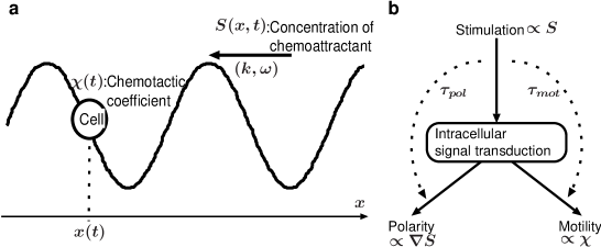
This model equation is constructed as follows (Fig. 1(b)). We use a widely-accepted assumption that the velocity of the cell is proportional to the spatial gradient of the chemoattractant concentration Keller1970 ; Othmer1998 ; Goldstein1995 . The proportional coefficient, , is referred to as chemotactic coefficient in what follows. Recent studies on intracellular signal transduction pathways of chemotactic cells have shown that the stimulation by extracellular chemoattractant triggers three responses which should be separately recognized, namely, directional sensing, polarization, and change in motility FrancaKoh2006 ; Iglesias2008 ; Swaney2010 . In terms of those studies, from the viewpoint of the modeling of the motion of a cell, we assume that the responses induced by the stimulation by a traveling wave consequently can be divided into two responses. One of them is the set of the directional sensing and the polarization, that is, the spatial localization of several signaling proteins along the plasma membrane because of the presence of the spatial gradient of the chemoattractant concentration in the environment, which results in pseudopod extension to a particular direction. We assume that the factor proportional to describes the magnitude of this response. The constant parameter, , is introduced as the time required from the stimulation to this response. Another response is the change in motility, namely, the magnitude and frequency of the extension of pseudopodia. In the observation of cell locomotion, the motility would be recognized as the quantity which indicates how fast a cell translocates on the substrate. Thus, we assume that the chemotactic coefficient, , is the factor which quantify the level of this response; therefore, it is assumed that the chemotactic coefficient oscillates in response to the oscillation of . The constant parameter, , is introduced as the time required from the stimulation to this response.
The assumption of the oscillatory behavior for the chemotactic coefficient is also based on remarkable previous studies on cell locomotion. Soll et al. determined the time series of the instantaneous velocity of a single Dictyostelium cell in detail when the cell was exposed to temporally periodic change in the chemoattractant concentration in the absence of the spatial gradient Soll2002 ; Zhang2002 ; Zhang2003 . It was observed that the magnitude of the instantaneous velocity oscillated at the same frequency as the extracellular chemoattractant concentration. This result implies that the temporal change of the chemoattractant concentration induces the change in the magnitude of the motility, which means here how fast a cell translocates. When a cell is exposed to a periodic traveling wave of the chemoattractant concentration, there is also the oscillation of extracellular chemoattractant concentration around a cell. Therefore, the model assumes that the chemotactic coefficient oscillates. In addition, because a significant constant phase difference was observed between the oscillation of the extracellular chemoattractant concentration and that of the instantaneous velocity in the experiments, the time delay, , corresponding to the phase difference is introduced in the model.
The assumption in this model provided above can be described in another way from the theoretical point of view. The equation of motion of a single cell is generally given by
| (4) |
as long as we neglect the inertial effect of a cell. Here, denotes external forces, denotes forces which induces random locomotion of a cell Li2008 ; Takagi2008 , and the coefficient is referred to as the friction coefficient. The equation reads
| (5) |
In the absence of the spatial gradient of the chemoattractant and other external forces, the governing equation becomes
| (6) |
According to the experiments carried out by Soll et al. Soll2002 ; Zhang2002 ; Zhang2003 , the temporally periodic and spatially uniform stimulation by the chemoattractant induces the periodic change in the instantaneous velocity, , at the same frequency with a constant time delay. Here, we attribute the oscillation of the velocity to the oscillation of . On the other hand, when a cell is exposed to a traveling wave of the chemoattractant concentration , is proportional to , that is,
| (7) |
Omitting the random component since we are interested in the time-averaged quantities in this study, Eq. (4) is reduced to
| (8) |
where is proportional to . Because the magnitude of the extracellular chemoattractant concentration periodically changes when the cell is exposed to a periodic traveling wave of the chemoattractant, we use the results obtained by Soll et al., that is, we assume that the chemotactic coefficient oscillates at the same frequency as the wave. Incorporating the time delays to the responses, we obtain the model equation (2) in consequence. Although the periodic function that precisely describes the chemotactic coefficient has never been determined in the presence of the spatial gradient of the chemoattractant concentration, we choose a sinusoidal function for simplicity. As discussed later, this choice of the sinusoidal function is not essential to the result; the result holds as long as the chemotactic coefficient is periodic function.
In summary, we postulate in the modeling that a response, change in how large or frequently pseudopodia are extended, can be distinguished from a response, determination of the direction of locomotion; the level of the pseudopod extension is affected by the temporal change in the magnitude of the extracellular chemoattractant concentration whereas the direction decision is affected by the spatial gradient of the extracellular chemoattractant concentration; and the composition of these factors determines the instantaneous velocity of a cell exposed to a traveling wave. Explicit incorporation of the two time delays, and , is an essential feature of this model. It should be noted that this model is not another expression of models presented thus far in which the periodic change in motility is considered but an essentially different model which provides another result on cell migration. The significant difference between them is discussed later.
| Abbreviation | Meaning | Used value | Ref. |
| Wave length of the chemoattractant wave | 2000 [] | Muller1998 | |
| Periodic time of the chemoattractant wave | 7 [] | Muller1998 | |
| Averaged velocity of the single cell | 10[] | Steinbock1991 | |
| Amplitude of the oscillation of the velocity of the single cell | 10[] | Steinbock1991 | |
| time delay from the stimulation to the change in motility | 6[] | Soll2002 | |
| time delay from the stimulation to the change in polarity | 1[] | * |
III Result
We immediately see that the propagation of the traveling wave induces net migration of a cell to a particular direction in average. We can safely approximate because the velocity of the traveling wave, Tomchik1981 , is much greater than that of a cell, which varies between and Soll2002 . Thus, the velocity of the single cell is governed by
| (9) | |||||
On the right hand side of this equation, the third term is a constant whereas the first and second terms are periodic with respect to time as long as is treated as a constant during one wave period. Therefore, the third term plays a greater role in the translocation of a cell than the first and second terms. The elimination of these periodic terms yields the averaged velocity of the cell, , as
| (10) |
We see that the cell migrates toward or away from the wave source on average when or , respectively. Thus, the frequency of the wave and the difference between the two time delays, , are important parameters that determine the direction and the magnitude of the net migration.
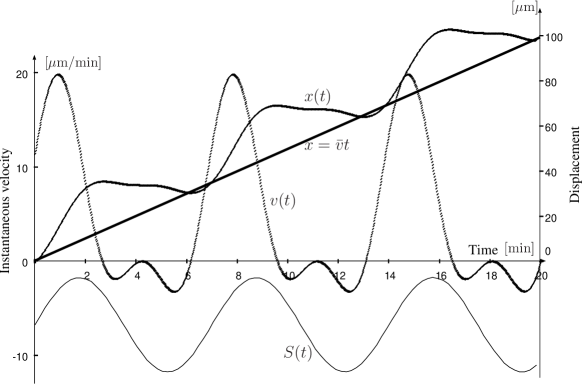
The differential equation (2) is numerically solved without any approximation. The result is illustrated in Fig. 2. Parameter values used here are summarized in Table 1. We see that the averaged velocity well describes the net migration of a cell.
Even if the waveform is not represented by a simple sinusoidal function, and play important roles in determining the direction of the net migration. Consider a more realistic case in which the traveling wave of the chemoattractant concentration is a sharply peaked one represented by
| (11) |
where and are constant parameters, and the chemotactic coefficient is given by Eq. (3). These functional forms of and describe the result of the experiment conducted by Soll et al. more precisely Soll2002 ; Zhang2003 ; Zhang2002 . In the same manner as the case of the sinusoidal traveling wave, it is proved by means of the Fourier expansion that
| (12) |
where . We see that the direction of net the migration of a cell is determined by the sign of ; therefore, the the frequency of the traveling wave and the difference of the two time delays are important parameters again.
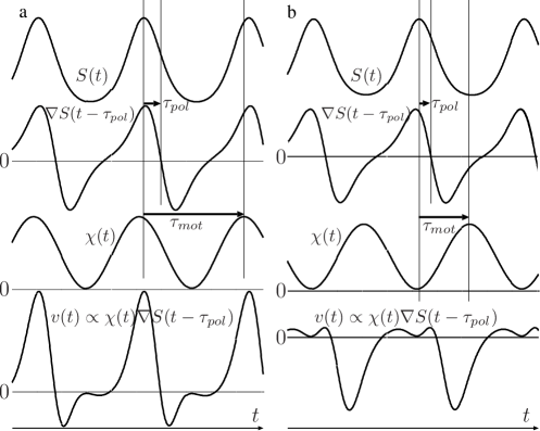
More generally, let us discuss the reason for the emergence of the net migration with referring to Fig. 3. If the shape of the wave of the chemoattractant concentration is monophasic and periodic, its spatial gradient becomes a biphasic wave whose time average is 0. If the time delays are such that the peak response of the oscillating motility appears when is positive, the time average of the product of and , which is proportional to , becomes positive. This implies that the cells migrate toward the source of the traveling wave (Fig. 3a). Conversely, if the time delays are such that the peak response of the motility appears when is negative, the time average of the product of and becomes negative. This implies that cells migrate in the direction of the wave propagation (Fig. 3b). Thus, even if the wave is not represented by a simple sinusoidal function, the difference between the two time delays from the stimulation to the two responses determines the direction of the cell migration of the cells. Moreover, as long as we fix the two time delays, and , Fig. 4a and Fig. 4b illustrate situations in which the frequency of the traveling wave, , is small and large, respectively. This indicates that the direction to which a cell migrates also depends on the wave frequency . Thus, the mechanism of directional migration of a cell is fundamentally the same as long as a wave is monophasic and periodic. The frequency of the traveling wave and the difference of the two time delays are important parameters which determine the direction of the net migration.
Note that, if there are no such time delays, that is, if , the time-average of becomes 0 independently of the functional form of . The directional net migration of cells has been successfully derived using several models in which time dependence of chemotactic coefficient is taken into account Goldstein1995 ; Hofer1994 ; Vasiev1994 ; Oss1996 ; Dolak2005 . On the basis of the discussion provided above, we can conclude that all these models have been constructed such that the time delays are implicitly incorporated in order to introduce an appropriate phase difference between the oscillation of the stimulation and the responses.
IV Discussion
This result (10) and (12) are consistent with the aggregation of Dictyostelium cells in that the each averaged velocity is positive for typical parameter values in the aggregation process shown in Table 1, which indicates that cells migrate toward the center of a target or spiral wave of cAMP concentration.
The numerical result shown in Fig. 2 well duplicates previous observations of the instantaneous velocity of a single Dictyostelium cell during the aggregation processes Soll2002 ; Zhang2002 ; Zhang2003 in that a period in which a cell migrates rapidly alternates with a period in which a cell is virtually stationary. In particular, the result successfully reproduces the observation more precisely than another model Goldstein1995 in that the time courses obtained in the experiments displays complex behavior which cannot be constructed by a simple sinusoid such as the characteristic small peaks in the stationary period.
The result provided above implies that there exists an effective interaction between cells, because, in the case of Dictyostelium, each cell secretes the chemoattractant, the secreted chemoattractant propagates in space, and it serves as a stimulation to nearby cells Gregor2010 . More precisely, if the frequency of the secretion of the chemoattractant, which determines the wave frequency, and the time delays satisfy the condition that a cell migrates toward or away from the wave source, there is effective attraction or repulsion between cells, respectively. This fact indicates that, for systems in which cells communicate with each other using the chemoattractant as an autoinducer, the frequency of the secretion and the time delays determine the effective interaction between the cells. In aggregation process, because the parameter values satisfy , the effective interaction between cells is attraction. This attraction is interpreted as the microscopic reason for the aggregation of cells.
Increase of the frequency of the secretion of the chemoattractant during aggregation has been observed Gregor2010 ; Alcantara1974 . The averaged velocity (10) and (12) indicate that, as the frequency of the secretion of the chemoattractant, , increases, the sign of changes from negative to positive at a critical frequency , which implies that the effective interaction between cells changes from repulsion to attraction. Even if the wave form is not so simple as Eq. (1) or Eq. (11), we can infer that the transition occurs according to the discussion provided above and Fig. 3. Therefore, it is possible that the dispersed state of cells before the commencement of aggregation is due to the effective repulsion between cells induced by the lower frequency of the secretion of the chemoattractant.
In terms of the transition between the dispersion and aggregation in a population of cells, we can estimate for Dictyostelium cells. From Eq. (10) and Eq. (12), the frequency of the secretion of the chemoattractant at which the interaction between cells changes from repulsion to attraction, , satisfies . According to a previous experiment, the time period of the periodic secretion at the commencement of the aggregation was [min] Alcantara1974 . Then, since [min] Soll2002 , we obtain [min]. Although this parameter has not been directly measured in the presence of temporally periodic spatial gradient of the chemoattractant concentration thus far, the result is consistent with an estimation based on the time which requires for the localization of proteins on the plasma membrane Rappel2009 . Inversely, this consistency suggests the existence of the transition at around [min].
Repulsion between Dictyostelium cells in a nutrient environment was reported several decades ago Samuel1961 ; Keating1977 . Although experimental studies on vegetative cells are less than starved cells in the developmental phase, it is certain that vegetative cells are single-celled amoeba and never commence the aggregation. The present model provides us an explanation for this state. It has been observed that right after the nutrient deprivation (within 5 hours precisely), cells pulse chemoattractant randomly every 15 to 30 min Gregor2010 . It can be inferred that vegetative cells secrete the cAMP at the same or lower frequency. According to the discussion above and in terms of Fig. 3, the interaction between cells is repulsion at those frequencies. Note that, even if the wave consists of a single pulse, there arise also repulsion. Thus, it is possible that the effective repulsion mediated by the chemoattractant allows vegetative cells to live as unicellular amoebae and never start to aggregate.
The fact that the interaction between cells may become repulsion indicates the possibility of the non-existence of the chemorepellent. Repulsion between vegetative Dictyostelium cells was originally named negative chemotaxis Keating1977 . Negative chemotaxis was later defined as a variant of chemotaxis in which cells migrate away from higher concentrations of a chemical source. Such a chemical substance is called chemorepellent. Due to this historical background, it was believed that vegetative cells secrete a chemorepellent and repel each other owing to the chemorepellent. A potential candidate for the chemorepellent was reported recently Keizer-Gunnink2007 . However, on the basis of the present model, such a chemorepellent is not required to explain the repulsion between cells. Even if there is no chemorepellent, and cells exhibit only positive chemotaxis, that is, is always positive, attraction and repulsion can be switched depending on the frequency of the secretion of the chemoattractant. We should note that this discussion just gives an possible scenario which explains for behavior of a population of vegetative cells because it has been commonly recognized and accepted that the intracellular molecular dynamics such as signal transduction and gene expression is largely different between vegetative cells and starved cells. The response of vegetative cells to cAMP should be investigated more in the future in order to confirm the discussion here.
It seems that the model presented here is just an another expression of what has been presented thus far in terms of time delay or phase difference of oscillations. However, there is an essential difference between them. Although it has been also assumed that the motility of cells under a traveling wave of the chemoattractant changes, the motility is believed to increase during periods in which the concentration of the external chemoattractant increases, and decrease during periods in which the concentration of the external chemoattractant decreases Soll2002 . This has been considered as the reason why the traveling-wave chemotaxis occurs. However, on the basis of this conventional idea, a cell exposed to a traveling wave always migrate toward the wave source independently of the frequency of the wave; therefore the effective interaction between cells becomes always attraction according to the discussion presented above. This does not explain for the transition from the dispersed state to the aggregation of cells. It should be experimentally confirmed in the future that to which direction cells migrate when cells are exposed to a traveling wave whose frequency is much lower than the wave emerging in aggregation processes. Note that in such an experiment the wave is not necessarily a periodic wave if the durations between peaks are long sufficiently. If those cells migrate away from the wave source, the model presented in this study would be valid. Or this model will be supported if it is observed that cells right before the commencement of aggregation significantly migrate in the direction of the wave propagation.
V Concluding Remarks
A phenomenological model which describes the motion of a single chemotactic cell is presented and analyzed in this study. The result of the analysis implies that systems in which cells communicate with each other using the chemoattractant as an autoinducer exhibit aggregation and dispersion of cells. The frequency of the secretion of the chemoattractant and the time delays from the stimulation by the chemoattractant to the responses of polarity and motility determine the interaction between cells, attraction or repulsion.
It must be controversial whether or not the responses to the stimulation by the chemoattractant can be incorporated as simply as this model because the intracellular signal transduction consists of actually complicated processes. However, the incorporation allows us 1) to understand vegetative cells living as unicellular amoebae and not starting aggregation in terms of repulsion between cells, 2) to understand commencement of the aggregation of starved cells in terms of the transition from the repulsion to attraction, 3) to derive the cell migration toward the source of the target or spiral wave of the chemoattractant in aggregation, 4) to describe the time course of the velocity of a cell in aggregation more precisely that other models presented thus far, and 5) to estimate the time which requires for the polarization.
The model in this study just provides at present a possible mechanism which is used in cell migration and induces dispersion and aggregation of cells. The validity of this model should be confirmed by some experiments in the future. 1) and are regarded as constant in this study. It should be confirmed whether or not values of and depend on the frequency of the stimulation, and taking the dependence into consideration also leads to the same results. Note that, even if those parameters depend on the frequency, the mechanism proposed in this study works as long as there remains the critical frequency at which the transition occurs. 2) We have used the value of that was measured in the absence of spatial gradient of cAMP in this study. Whether or not the change in is the same in the presence of the spatial gradient of cAMP. 3) Because it has been reported that many of responses occur within a few minutes FrancaKoh2006 , why the change in motility requires such a long time, about , should be elucidated in terms of the intracellular dynamics. 4) Net migration of cells exposed to a traveling wave whose frequency is small should be investigated in order to compare with models presented thus far (see the preceding section for detail). As long as we refer to the results obtained in this study, the intriguing question arising next is how cells regulate the frequency of the secretion of chemoattractant depending on the environment, that is, how they choose sufficiently low frequency in order to live as unicellular amoebae when they can survive and grow thanks to enough food, and how they choose sufficiently high frequency in order to start development when they cannot survive alone because of starvation. The answer to this question will elucidate the mechanism of the adaptation of cells to their environment and the strategy taken for the species survival.
From the viewpoint of physics, it should be remarked that the random motion of a cell plays an important role in the generation of directional migration even though the random motion itself is nondirectional, and does not seem to contribute to tactic behavior. In addition, it should be emphasized that the composition of symmetric conditions induces asymmetric behavior, that is, the composition of the periodic stimulation and the nondirectional fluctuation of the position generates directional net migration. This spontaneous symmetry breaking leads to aggregation or dispersion in a population.
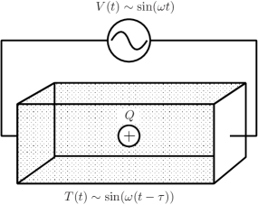
Let us further generalize the results. Consider an isolated element that moves in response to the spatial gradient of a field. When such an element is exposed to the temporally periodic change of spatial gradient of the field and the motility oscillates in the same frequency, spontaneous directional motion occurs on average. The direction of the motion is determined by the frequency of the change in the gradient and the phase difference between the oscillation of the motility and that of the response to the spatial gradient (Fig. 4). Moreover, a group of elements, each of which emits a field with the same frequency (e.g., quantum dots exposed to electromagnetic waves Iida2006 ), is expected to exhibit aggregation or dispersion as seen in Dictyostelium cells. In the future, such a simple mechanism can potentially facilitate the development of self-organizing systems in which elements communicate or interact with each other via a field.
Acknowledgements.
The authors would like to thank Dr. Dan Tanaka for fruitful discussions.References
- (1) Stephens L, Milne L, Hawkins P (2008) Moving towards a better understanding of chemotaxis. Curr Biol 18:R485-R494.
- (2) Schneider IC, Haugh JM (2006) Mechanism of gradient sensing and chemotaxis. Cell Cycle 5:1130-1134.
- (3) Rogers J, Lue L-F (2001) Microglial chemotaxis, activation, and phagocytosis of amyloid -peptide as linked phenomena in Alzheimer’s disease. Neurochem Int 39:333-340.
- (4) Kedrin D, van Rheenen J, Hernandez L, Condeelis J, Segall JE (2007) Cell motility and cytoskeletal regulation in invasion and metastasis. J Mammary Gland Biol Neoplasia 12:143-152.
- (5) Song L, Nadkarni SM, Bdeker HU, Beta C, Bae A, Franck C, Rappel W-J, Loomis WF, Bodenschatz E (2006) Dictyostelium discoideum chemotaxis: Threshold for directed motion. Euro J Cell Biol 85:981-989.
- (6) Tomchik KJ, Devreotes PN (1981) Adenosine 3’, 5’-monophosphate waves in Dictyostelium discoideum: A demonstration by isotope dilution-fluorography. Science 212:443-446.
- (7) Devreotes PN, Potel MJ, MacKay SA (1983) Quantitative analysis of cyclic AMP waves mediating aggregation in Dictyostelium dicoideum. Dev Biol 96:405-415.
- (8) Gregor T, Fujimoto K, Masaki N, Sawai S (2010) The onset of collective behavior in social amoebae. Science 328:1021-1025.
- (9) Lee KJ, Cox EC, Goldstein RE (1996) Competing patterns of signaling activity in Dictyostelium discoideum. Phys Rev Lett 76:1174-1177.
- (10) Whitesides GM, Grzybowski B (2002) Self-assembly at all scales. Science 295:2418-2422.
- (11) Ishiguro A, Shimizu M. Kawakatsu T (2006) A modular robot that exhibits amoebic locomotion. Robot Autom Syst 54:641-650.
- (12) Pfeifer R, Lungarella M, Iida F (2007) Self-organization, embodiment, and biologically inspired robotics. Science 318:1088-1093.
- (13) Keller EF and Segel LA (1970) Initiation of Slime Mold Aggregation Viewed as an Instability. J. Theor. Biol. 26:399-415.
- (14) Othmer HG and Schaap P (1998) Oscillatory cAMP Signaling in the Development of Dictyostelium discoideum. Comments Theor. Biol. 5:175-282.
- (15) Goldstein RE (1995) Traveling-wave chemotaxis. Phys Rev Lett 77:775-778.
- (16) Franca-Koh J, Kamimura Y, and Devreotes P (2006) Navigating signaling networks: chemotaxis in Dictyosteilum discoideum Curr Opin Gen Dev 16: 333-338.
- (17) Igresias PA, Devreotes PN (2008) Navigating through models of chemotaxis. Cur Opin Cell Biol 20:35-40.
- (18) Swaney KF, Huang C-H, Devreotes PN (2010) Eukaryotic chemotaxis: A network of signaling pathways controls motility, directional sensing, and polarity. Annu Rev Biophys 39:265-289.
- (19) Soll DR, Wessels D, Heid PJ, Zhang H (2002) A contextual framework for characterizing motility and chemotaxis mutants in Dictyosteium discoideum. J Muscle Res Cell Motil 23:659-672.
- (20) Zhang H, Wessels D, Petra F. Daniels K, Chisholm RL, Soll DR (2002) Phosphorylation of the myosin regulatory light chain plays a role in motility and polarity during Dictyostelium chemotaxis. J Cell Sci 115:1733-1747.
- (21) Zhang H, Heid PJ, Wessels D, Daniels KJ, Pham T, Loomis WF, Soll DR (2003) Constitutively active protein kinase A disrupts motility and chemotaxis in Dictyostelium discoideum. Eukaryotec Cell 2:62-75 (2003).
- (22) Li L, Nrrelykke SF, Cox EC (2008) Persistent cell motion in the absence of external signals: A search strategy for eukaryotic cells. PLoS One 3:e2093.
- (23) Takagi H, Sato MJ, Yanagida T, Ueda M (2008) Functional analysis of spontaneous cell movement under different physiological conditions. PLoS One 3:e2648.
- (24) Muller SC, Mair T, and Steinbock O (1998) Traveling waves in yeast extract and in cultures of Dictyosteium discoideum Bio Chem 72: 37-47.
- (25) Steinbock O, Hashimoto H, Muller SC (1991) Quantitative analysis of periodic chemotaxis in aggregation patterns of Dictyostelium discoideum. Physica D 49:233-239.
- (26) Hfer T, Maini PK, Sheratt JA, Chaplain MAJ, Chauvet P, Metevier D, Montes PC, Murray JD (1994) A resolution of the chemotactic wave paradox. Appl Math Lett 7:1-5.
- (27) Vasiev BN, Hogeweg P, Panfilov AV (1994) Simulation of Dictyostelium discoideum aggregation via reaction-diffusion model. Phys Rev Lett 73:3173-3176.
- (28) van Oss C, Panfilov AV, Hogeweg P, Siegert F, and Weijer CJ. (1996) Spatial Pattern Formation During Aggregation of the Slime Mould Dictyostelium discoideum. J. Theor. Biol. 181:203-213.
- (29) Dolak Y, Schmeiser C (2005) Kinetic models for chemotaxis: Hydrodynamic limits and spatio-temporal mechanism. J Math Biol 51:595-615.
- (30) Alcantara E, Monk M (1974) Signal propagation during aggregation in the slime mould Dictyostelium discoideum J Gen Microbiol 85:321-334.
- (31) Rappel W-J, Loomis WF (2009) Eukaryotic chemotaxis. WIREs Syst Biol Med 1:141-149.
- (32) Samuel EW (1961) Orientation and rate of locomotion of individual amebas in the life cycle of the cellular slime mold Dictyosteium mucoroides. Dev Biol 3:317-335.
- (33) Keating ME, Bonner JT (1977) Negative chemotaxis in cellular slime molds. J Bacteriology 130:144-147.
- (34) Keizer-Gunnink I, Kortholt A, Van Haastert JM (2007), Chemoattractants and chemorepellents act by inducing opposite polarity in phospholipase C and PI3-kinase signaling. J Cell Biol 177:579-585.
- (35) Iida T, Ishihara H (2006) Force control between quantum dots by light in polaritonic molecule states. Phys Rev Lett 97:117402.