Origin of the large polarization in multiferroic YMnO3 thin films revealed by soft and hard x-ray diffraction
Abstract
We investigated the magnetic structure of an orthorhombic YMnO3 thin film by resonant soft x-ray and hard x-ray diffraction. We observed a temperature-dependent incommensurate magnetic reflection below 45 K and a commensurate lattice-distortion reflection below 35 K. These results demonstrate that the ground state is composed of coexisting E-type and cycloidal states. Their different ordering temperatures clarify the origin of the large polarization to be caused by the E-type antiferromagnetic states in the orthorhombic YMnO3 thin film.
pacs:
75.80.+q, 78.70.Ck, 75.25.-j, 73.61.-rpacs:
71.30.+h, 71.28.+d, 79.60.Dp, 73.61.-rRecently, there has been a lot of interest in multiferroic materials, which display both ferroelectric and magnetic orders with giant magnetoelectric coupling tokuramulti ; cheong ; Sekitokura . It is of particular importance to control magnetization (electric polarization) by electric (magnetic) field for novel device applications. Orthorhombic (-) MnO3 (: rare-earth) with perovskite structure are prototype multiferroic materials. For example, in TbMnO3, ferroelectricity occurs below 28 K, concomitant with the onset of cycloidal spin ordering kimuratmo ; kimuratmoprb ; Kenzelmann . The ferroelectricity in the cycloidal states is realized by the shifts of the oxygen ions through the inverse Dzyaloshinskii-Moriya interaction katsura ; Mostovoy . This is in contrast to E-type antiferromagnetic structures ( type), where ferroelectricity is caused by symmetric exchange striction sergienko . E-type magnetic structures occur in -MnO3 with smaller ions. It is predicted that the E-type structure leads to a larger polarization, which has been experimentally confirmed in the -MnO3 systems pomja ; ishiwatamulti .
The fabrication of the -MnO3 thin films has been especially important for device application of the multiferroic materials. Moreover, bulk -MnO3 samples with smaller ions ( Y, Ho - Lu) can only be synthesized under high oxygen pressure ishiwatamulti , which strongly limits studies on the most interesting materials due to the absence of significantly large high-quality single crystals. Recently, Nakamura et al. reported the fabrication of -YMnO3 thin films onto the YAlO3 (010) substrate YMOnakamura . Their thin film showed a ferroelectric transition at 40 K with a large saturation polarization of 0.8 C/cm2. The ferroelectric polarization could be controlled by magnetic fields, demonstrating magnetoelectric behaviors.
Therefore it is interesting and important to clarify the exact magnetic structure of YMnO3 thin films. In this study we use the technique of resonant soft x-ray diffraction at Mn edges to obtain the information of magnetic ordering in YMnO3 thin films. Resonant soft x-ray diffraction has recently been used to study the magnetic ordering in multiferroic TbMnO3 and Eu3/4Y1/4MnO3 forest ; tmowilkins ; RSXS2011 using single crystals for the larger -ion orthorhombic MnO3 series. This technique is especially suitable for studying magnetism in thin films (as demonstrated on NiO3 valerio ) because even small sample volume of thin films can be used due to the large resonant enhancement of magnetic scattering at the transition-metal edges. We detect (0 0) () magnetic peak, and observed temperature-dependent incommensurabilities. From hard x-ray diffraction we found a commensurate superlattice reflection (0 1 0) that reflects the lattice distortion caused by the E-type magnetic structure. These results reveal that the ground state of the YMnO3 can be described by the coexistence of E-type and cycloidal states, while the E-type state is a dominant source for the large electric polarization of 0.8 C/cm2 by the symmetric exchange striction.
The thin film (40 nm) of YMnO3 was grown on a YAlO3 (010) substrate by pulsed-laser deposition. The details of the sample fabrication were described elsewhere YMOnakamura . Resonant soft x-ray diffraction experiments were performed on the RESOXS endstation SLS at the surfaces/interfaces microscopy (SIM) beamline of the Swiss Light Source of the Paul Scherrer Institut, Switzerland. For the azimuthal scans (rotation around the Bragg scattering wave vector), the sample transfer line was used to rotate the sample holder. With pins attached in a threefold symmetry on the sample holder, an accuracy of approximately 5 deg was obtained. A continuous helium-flow cryostat allows measurements between 10 and 300 K. Hard x-ray diffraction experiments were performed on beamlines 3A and 4C at the Photon Factory, KEK, Japan. The photon energy of the incident x-ray was 12 keV.
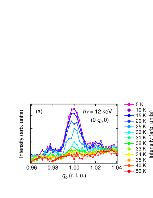
Figure 1 shows the temperature dependence of the (0 0) () peak with (a) and (b) incident x-ray polarizations. The experimental geometry is shown in Fig. 1 (c), together with the definition of the azimuthal angle . Here the diffraction data were taken with at eV (Mn absorption edge). We measured in both cooling and heating cycles, and observed no hysteresis behavior. This peak, which is indicated by vertical bars, appears at 45 K, which coincides with the antiferromagnetic transition temperature determined from magnetization measurements YMOnakamura . Weaker peaks are observed on both sides of the reflection. These are antiferromagnetic Kiessig fringes, and describe the limited thickness of the magnetic contrast of the film. There is almost no difference between (a) and (b) polarizations. The intensity of the peaks increases monotonically with cooling. The peak position deviates from the commensurate position for all temperatures. The peak position shifts to higher angle for decreasing temperatures; the temperature variation of the corresponding wave vector and intensity is summarized in Fig. 2. The intensity increases monotonically and smoothly with decreasing temperatures from K. The peak position, e.g. at 44 K and at 11 K, is temperature-dependent and always incommensurate () in the temperature range of 11 - 44 K. In TbMnO3 the peak position is also incommensurate, but lock to the value of at the ferroelectric transition temperature K forest ; tmowilkins . Such a behavior is not observed in this YMnO3 film; there is no locking of the peak position at K, which was determined from electric polarization measurements YMOnakamura .
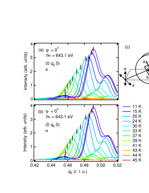
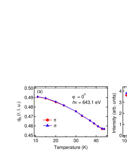
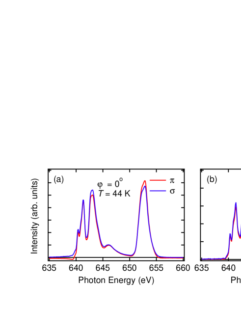
Figure 3 shows the intensity of the (0 0) () peak as a function of photon energies at the Mn absorption edge at 44 K (a) and 11 K (b). There is no polarization dependence at this scattering geometry of at both temperatures. In addition, the spectral shape is identical at these two temperatures and very similar to the one observed for TbMnO3 and Eu3/4Y1/4MnO3 forest ; tmowilkins ; RSXS2011 . This shows that the line shape of the spectrum does not depend on the values of but is rather common in multiferroic -MnO3.
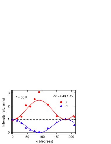
To gain more information on the spin structure, it is important to study the magnetic reflection with linear polarized incident radiation for different azimuthal angles. The (azimuthal angle) dependence of the intensity of the magnetic (0 0) reflection is shown in Fig. 4. For , the intensities are identical for and polarizations within experimental uncertainty. When increases from to , the intensity increases with incident polarization and decreases with incident polarization. The azimuthal-angle dependence allows us to gain information on the directions of the Mn spins. In the electric-dipole transition, the magnetic contribution to the structure factor is given as
where and are unit vectors of the incident and scattered polarization, respectively, and is a unit vector in the direction of the magnetic moment of the ion Hannon ; jphill . We use the notations in Fig. 1 in Ref. jphill which lead to the following expression,
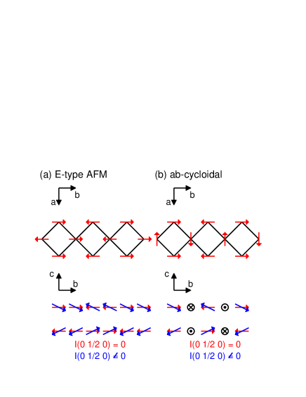
Here is the Bragg angle for the (0 0) reflection. When the magnetic Fourier components contribute only along the axis, , , and . Then the intensity for and incident polarizations are given with at 30 K.
The values of these equations are shown as solid lines in Fig. 4, and are in good agreement with our experimental observations. This reflects an ab cycloid with a spin canting along the -axis as shown in Fig. 5, and indicates that the experiment is only sensitive to its magnetic sinusoidal axis component.
In order to investigate the lattice distortions associated with magnetic order and electric polarization, we additionally performed hard x-ray diffraction measurements of the YMnO3 thin film. The commensurate (0 1 0) reflection appears below 35 K as shown in Fig. 6. This reflection is a structurally forbidden in the chemical high-temperature structure (Pbnm) and caused by the lattice distortion accompanying ferroelectricity. Interestingly, no incommensurability of this reflection is observed by hard x-ray diffraction, in clear contrast to the observed magnetic reflection. Moreover, this reflection does appear below 35 K, at lower temperatures than the onset of the magnetic reflection, in accord with the step onset of the spontaneous electric polarization [12], as can be seen from the temperature-dependent integrated intensity shown in Fig. 6 (b).
We can obtain a full picture of the magnetic states of the epitaxial YMnO3 thin film by combining the above results with the macroscopic measurements of magnetization and electric polarization YMOnakamura . From the macroscopic measurements, three transitions were observed: antiferromagnetic transition at K, ferroelectric transition at K, and an increase of electric polarization at 35 K. The incommensurate magnetic peak was observed at all temperature below 45 K. It reflects spin moments solely along the axis as indicated by its x-ray polarization and azimuthal dependence. This supports the scenario that in the temperature range of 40 - 45 K a sinusoidal state with a spin canting along the axis is realized. Note that the in-plane magnetic moment components cancel for this magnetic wave vector in the structure factor. This state is also consistent with the absence of observed electric polarization in this temperature regime (see Fig. 6 (b)). By cooling through 40 K, the sinusoidal magnetic phase transforms into a cycloidal magnetic structure with significant magnetic moment contributions along the axis. Below 35 K, we can observe both the incommensurate magnetic reflection and the commensurate lattice-distortion reflection. This state can be therefore explained by the coexistence of the cycloidal and the E-type states as theoretically predicted in Ref. mochi1 . In this coexistence region, magnetic reflection is incommensurate as shown in Ref. mochi1 and lattice peaks are commensurate because the E-type phase has a much larger lattice distortion than the cycloidal phase. The existence of the E-type phase causes the large electric polarization of 0.8 C/cm2 due to the symmetric exchange striction YMOnakamura . In other words, the weak polarization emerging at 40 K from the cycloidal magnetic structure causes also weak lattice distortion, which is too weak to be observed in our experiment. On the other hand, the large induced electric polarization below 35 K caused by the E-type structure induces a significant lattice distortion, as observed by the x-ray diffraction experiments on a YMnO3 single crystal okuYMO . However, spin canting in its magnetic structure is so small that no additional magnetic contribution is observed in our experiment. It is difficult to distinguish between the occurrence of - and -cycloids based on our experimental data. However, electric polarization is parallel to the axis YMOnakamura , which clearly indicates the -cycloid. The -cycloids can easily adopt a spin canting along the axis, whereas -cycloids would get anisotropically distorted.
In summary, we investigated the magnetic structures of the YMnO3 thin film by resonant magnetic soft x-ray and hard x-ray diffraction. We observed temperature-dependent incommensurate magnetic peaks below 45 K and commensurate lattice-distortion peaks below 35 K, indicating that E-type and cycloidal states coexist below 35 K. This shows that the occurrence of the large electric polarization below 35 K is directly related to E-type magnetic ordering component in the epitaxial YMnO3 films.
Informative discussions with S. Ishiwata are greatly acknowledged. The authors thank the experimental support of the X11MA beamline staff. Financial support of the Swiss National Science Foundation and its NCCR MaNEP is gratefully acknowledged. This work is also supported by the Japan Society for the Promotion of Science (JSPS) through its Funding Program for World-Leading Innovative R&D on Science and Technology (FIRST Program). Hard x-ray diffraction measurements were performed under the approval of the Photon Factory Program Advisory Committee (Proposals Nos. 2009S2-008 and 2010G678) at the Institute of Material Structure Science, KEK.
References
- (1) Y. Tokura, Science 312, 1481 (2006).
- (2) S.-W. Cheong and M. Mostovoy, Nature Mater. 6, 13 (2007).
- (3) Y. Tokura and S. Seki, Adv. Mater. 22, 1554 (2010).
- (4) T. Kimura, T. Goto, H. Shintani, K. Ishizaka, T. Arima, and Y. Tokura, Nature 426, 55 (2003).
- (5) T. Kimura, G. Lawes, T. Goto, Y. Tokura, and A. P. Ramirez, Phys. Rev. B 71, 224425 (2005).
- (6) M. Kenzelmann, A. B. Harris, S. Jonas, C. Broholm, J. Schefer, S. B. Kim, C. L. Zhang, S.-W. Cheong, O. P. Vajk, and J. Lynn, Phys. Rev. Lett. 95, 087206 (2005).
- (7) H. Katsura, N. Nagaosa, and A. V. Balatsky, Phys. Rev. Lett. 95, 057205 (2005).
- (8) M. Mostovoy, Phys. Rev. Lett. 96, 067601 (2006).
- (9) I. A. Sergienko and E. Dagotto, Phys. Rev. B 73, 094434 (2006).
- (10) V. Y. Pomjakushin, M. Kenzelmann, A. Donni, A. B. Harris, T. Nakajima, S. Mitsuda, M. Tachibana, L. Keller, J. Mesot, H. Kitazawa, and E. Takayama-Muromachi, New J. Phys. 11, 043019 (2009).
- (11) S. Ishiwata, Y. Kaneko, Y. Tokunaga, Y. Taguchi, T. Arima, and Y. Tokura, Phys. Rev. B 81, 100411(R) (2010).
- (12) M. Nakamura, Y. Tokunaga, M. Kawasaki, and Y. Tokura, Appl. Phys. Lett. 98, 082902 (2011).
- (13) T. R. Forrest, S. R. Bland, S. B. Wilkins, H. C. Walker, T. A. W. Beale, P. D. Hatton, D. Prabhakaran, A. T. Boothroyd, D. Mannix, F. Yakhou, and D. F. McMorrow, J. Phys. Condens. Matter 20, 422205 (2008).
- (14) S. B. Wilkins, T. R. Forrest, T. A. W. Beale, S. R. Bland, H. C. Walker, D. Mannix, F. Yakhou, D. Prabhakaran, A. T. Boothroyd, J. P. Hill, P. D. Hatton, and D. F. McMorrow, Phys. Rev. Lett. 103, 207602 (2009).
- (15) H. Jang, J.-S. Lee, K.-T. Ko, W.-S. Noh, T. Y. Koo, J.-Y. Kim, K.-B. Lee, J.-H. Park, C. L. Zhang, S. B. Kim, and S.-W. Cheong, Phys. Rev. Lett. 106, 047203 (2011).
- (16) V. Scagnoli, U. Staub, A. M. Mulders, M. Janousch, G. I. Meijer, G. Hammerl, J. M. Tonnerre, and N. Stojic, Phys. Rev. B 73, 100409(R) (2006).
- (17) U. Staub, V. Scagnoli, Y. Bodenthin, M. Garcia-Fernandez, R. Wetter, A. M. Mulders, H. Grimmer, and M. Horisberger, J. Synchroton Radiat. 15, 469 (2008).
- (18) J. P. Hannon, G. T. Trammell, M. Blume, and D. Gibbs, Phys. Rev. Lett. 61, 1245 (1988).
- (19) J. P. Hill and D. F. McMorrow, Acta Crystallogr. Sect. A 52, 236 (1996).
- (20) M. Mochizuki, N. Furukawa, and N. Nagaosa, Phys. Rev. Lett. 105, 037205 (2010).
- (21) D. Okuyama, S. Ishiwata, Y. Takahashi, K. Yamauchi, S. Picozzi, K. Sugimoto, H. Sakai, M. Takata, R. Shimano, Y. Taguchi, T. Arima, and Y. Tokura, Phys. Rev. B 84, 054440 (2011).