Protein Regge Trajectories, Phase Coexistence and Physics of Alzheimer’s Disease
Abstract
Alzheimer’s disease causes severe neurodegeneration in the brain that leads to a certain death. The defining factor is the formation of extracellular senile amyloid plaques in the brain. However, therapeutic approaches to remove them have not been effective in humans, and so our understanding of the cause of Alzheimer’s disease remains incomplete. Here we investigate physical processes that might relate to its onset. Instead of the extracellular amyloid, we scrutinize the intracellular domain of its precursor protein. We argue for a phenomenon that has never before been discussed in the context of polymer physics: Like ice and water together, the intracellular domain of the amyloid precursor protein forms a state of phase coexistence with another protein. This leads to an inherent instability that could well be among the missing pieces in the puzzle of Alzheimer’s disease.
pacs:
05.45.Yv 87.15.Cc 36.20.EyThe neurological origin of Alzheimer’s disease involves both genetic and environmental factors gene1 -gene5 . Its hallmark is the accumulation of the amyloid isoform A42 into senile plaques abeta1 -abeta2 . This has motivated several immunotherapic approaches to either clear or prevent the cerebral A deposits immu1 -immu3 . Unfortunately there are serious side-effects such as the development of aseptic meningoencephalitis meni1 -meni3 . As a consequence we do not know whether targeting of A42 will cure or even curb the disease in humans. The excess production of A42 might just be an indication that something else has gone wrong greek , critic1 .
The A42 is a derivative of the transmembrane amyloid precursor protein (APP) by proteolytic cleavages abeta1 , swede , appbeyond . APP comes in several isoforms, it is naturally present in many organs. Its physiological function remains under a debate and the understanding of its proteolytic processing is also incomplete greek , swede , appbeyond . Both the dominant, non-amyloidogenic pathway and the disease related, A generating amyloidogenic pathway produce isoforms of the APP intracellular domain (AICD) swede , cleave . We have scrutinized the physical properties of various AICD complexes, searching for an intracellular agent that might correlate with the onset of the anomalous A42 production. We identify an inherently unstable physical phenomenon that has never before been discussed in the context of polymer research.
After the cleavage of APP the AICD may form a transcriptionally active state with the Fe65 family of nuclear multidomain adaptor proteins swede , FEreview -adaptor . Even though the relation between AICD and Alzheimer’s disease is not yet understood, we know that AICD is a product and Fe65 is a participant in the proteolytic cleavage processing of APP into A42. Not surprisingly Fe65 already appears among the potential therapeutic targets FEreview , AICD , immu3 .
In isolation AICD is presumed to be an intrinsically unstructured protein unst . However, upon binding to Fe65, AICD can assume a regular form that can be analyzed with x-ray crystallography. Unfortunately, the high precision data remains limited. Here we shall investigate the structure with PDB code 3DXC (chain B) exp . It describes a complex of a 28 residue segment of AICD with the larger, 65 residue host Fe65. There are also the closely related 3DXD and 3DXE, these can be analyzed similarly and with identical conclusions. We find that the complex appears to have physical properties that seems to set it apart from all but a very few oligomers. It is an example of an apparently previously unrecorded but seemingly systematic phenomenon of protein (polymer) phase coexistence: Like ice with water the two proteins are in two different phases. As such, an oligomer that displays the rare and inherently unstable phenomenon of phase coexistence is for sure an interesting object for future research. But the delicate balance of the AICD/Fe65 complex has the supplementary potential of being an important piece in the puzzle to find a cure for Alzheimer’s disease.
The phases of a protein and more generally those of a polymer, are characterized by their fractal (Hausdorff) dimension. This is an order parameter that can be computed by inspecting the scaling properties of the radius of gyration . Asymptotically, in the limit where the number of monomers becomes very large degennes
| (1) |
Here are the coordinates of the backbone , the pre-factor is an effective inter-monomer distance that is independent of , and is the compactness index that equals the inverse fractal dimension of the backbone. The remarkable property of (1) is that is a universal quantity degennes , schafer . Different values of correspond to different phases, and once we know we can unanimously compute the radius of gyration by simply counting the number of monomers. All the effects of temperature and chemical microstructure and all atomary level details of a polymer are contained in the value of the a priori non-universal and in principle computable pre-factor .
The relation (1) becomes truly precious only in those exceptional circumstances where assumes no more than a small number of different values. We argue that this is indeed the case in proteins: When increases, proteins become increasingly uniform in their chemical composition. Consequently it makes sense to employ (1) to study their phase structure. Different, clearly identifiable trajectories (1) that are labelled by the different well defined values of are then the protein analogs of the Regge trajectories in high energy physics regge .
Proteins and other polymers degennes schafer have four major phases: Under physiological conditions and in other bad solvents a protein collapses into a space filling conformation with . For a fully flexible chain we have the -point value while in the self-avoiding random walk phase we have the Flory value . Finally, when the protein looses its inherently fractal structure and becomes like a one dimensional rigid rod. Examples of this phase are monotonous -helices and -strands that have no additional twists, turns or loops.
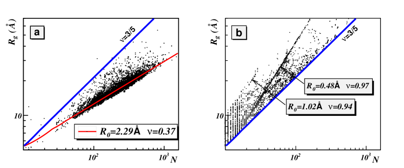
In Figure 1a we plot all individual single chain proteins in PDB that have resolution less than 2.0 Ȧ and homology equivalence which is less than 30. With a few exceptions they assemble around a Regge trajectory with . We also plot the dominant mostly--helical trajectory with and ; The numerical values are slightly different for the different protein subclasses like mostly--helical, mostly--sheets etc. and this fine structure has been discussed in add1 , add2 .
When we extend our analysis to individual chains within oligomers we find two previously unobserved clearly visible Regge trajectories (Figure 1b). These two trajectories are
| (2) |
These trajectories both have very close to one. Thus they must be in the same universality class, and the difference is a finite size effect. This is the universality class of one dimensional rods and sticks. Unlike the other three polymer phases, it has no fractal structure.
The trajectory includes several membrane proteins and viral capsomers, an example of the latter is 1AIK in PDB. The trajectory is mainly populated by collagen proteins such as for example 2CUO in PDB. In both trajectories the oligomers commonly consist of several individual chains that are each located on the same Regge trajectory. Their mutual interactions provides a supportive lattice structure that protects the individual chains against a collapse into the phase.
Remarkably, there are also protein complexes in the Regge trajectories of Figure 1b that do not follow the structural pattern of collagens, membrane proteins or viral capsomers. In particular, we have found that there is a small number of oligomers that are composed of proteins on different Regge trajectories. These complexes consist of (host) sub-chains that are in a -point trajectory and (guest) sub-chains on a similarly uncollapsed trajectory of Figure 1b. These oligomers are examples of a previously unrecorded physical phenomenon of protein phase coexistence: The interaction between the host and the guest provides a support that maintains each of them in an inherently unstable uncollapsed conformation.
We have found two different classes of such phase coexistent oligomers in PDB. The first class consists of an apparently single protein but with multiple sub-chains that are in different phases. The present PDB codes for these proteins are 1WDC, 1G72, 1GOT, 1HTR, 1LTS, 2FP7, 2RIV, 3ABK, 3ARC, 3CX5, 3DBO. Here we concentrate on the second class, formed by complexes with two or more a priori different proteins. The present PDB codes are 1L2W, 1JDH, 1TH1, 2F8X, 2EPV, 2PRR, 2BFX, 2D7C, 2VGO, 2K8F, 2QKH, 3EGG, 3HTU, 3HPW, 3IXS and 3DXC (3DXD, 3DXE). In Figure 2 we display the distribution of the individual sub-chains of the second class in the () plane, they clearly gather around a Regge trajectory and the trajectory in (2).
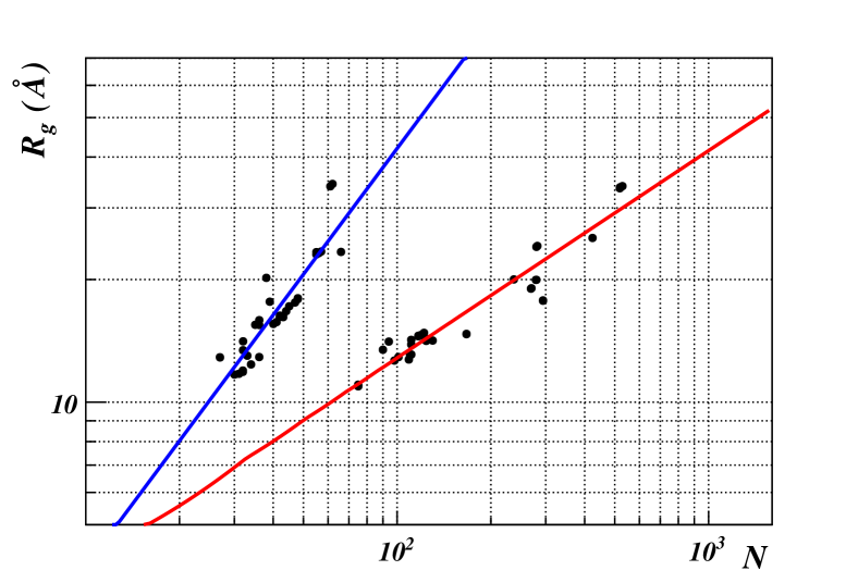
Biologically, the two most notable are 2K8F and 3DXC (3DXD, 3DXE). The former is a bound state of the ”molecular interpreter” p300 p300 , unst in the Regge trajectory , with the tumor suppressing protein p53 in the trajectory. The second is the one of interest here, the Alzheimer related AICD/Fe65 complex with AICD in the trajectory and Fe65 in the trajectory. We now proceed to analyze the peculiar physical properties of the Alzheimer related AICD of the second complex.
In Figure 3a we display the Cα backbone Frenet frame bond and torsion angles of the AICD protein in 3DXC.
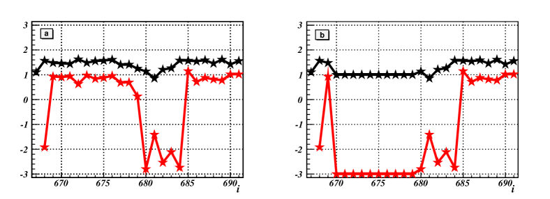
This Figure reveals that the AICD consists of two very closely located loops that are separated from each other by a very short -strand. We can describe the profile of each of these angles using the soliton solution of nonlinear Schrödinger equation, for the backbone bond angles we have maxim , peng
| (3) |
while the backbone torsion angles are computed in terms of the bond angles from
| (4) |
and the parameter values are listed in Table I: The corresponding () profiles (3), (4) describe the first loop at sites 676-683 (we use PDB indexing) with RMSD accuracy of 0.29 Ȧ and the second loop at sites 681-688 with RMSD accuracy of 0.17 Ȧ. Both accuracies are substantially better than the experimental B-factor accuracies.
| loop | match | |||||
|---|---|---|---|---|---|---|
| 676-683 | 51.517 | 51.766 | 2.984 | 2.983 | 679.909 | 177 |
| 681-688 | 39.274 | 38.617 | 3.327 | 3.347 | 682.174 | 896 |
In Table I we also list the number of times each of these loops appear in PDB with RMSD accuracy 0.5 Ȧ or better. Both are abundant in the Regge trajectory of collapsed proteins. In fact, at the outset there is nothing in the secondary structures of this AICD fold that appears unusual for a protein in the collapsed phase. Nevertheless it is very accurately, almost exactly, located on the Regge trajectory .
Since the compactness index is universal and can only have definite discrete values, any continuous and local deformation of the protein shape can never cause any kind of discontinuous transition such as a jump between the two phases and . This makes the present combination of the two loops in AICD highly unusual. Even if we continuously translate the two loops apart from each other along the backbone by shifting the value in (3) that determines the position of the center of the loop, we can never reach a collapsed Regge trajectory but will always remain in the phase.
A scrutiny of the amino acid structure reveals that AICD has a proline at site 669. Since proline often acts as an anchor of a loop in a protein in isolation, we propose that the presence of Fe65 prevents the first loop from sliding towards its natural position, where it becomes attached with Pro(669). Note that there is another proline at the site 685 that appears to stabilize the position of the second loop. Using the explicit profile (3), (4) we investigate what might happen if the first soliton starts sliding towards Pro(669) along the backbone. For this we shift the value of the parameter accordingly. In Figure 4 we show how the radius of gyration of the AICD depends on the position of the first loop as we slide it towards Pro(669) while keeping the second loop anchored by Pro(685); the final () profile is displayed in Figure 3b.
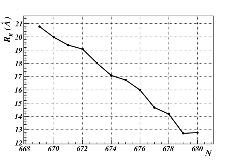
We find that increases monotonically when the two loops drift apart. When the first loop reaches the position where it becomes locked by Pro(669), the ensuing AICD configuration has relocated itself from the Regge trajectory to the Regge trajectory . Since it should be highly natural for a protein to always try and locate itself on a Regge trajectory, we propose that these are the two likely configurations of AICD in the complex with Fe65. The presence of two natural alternatives is an indicative of a genetic switching mechanism. It would be highly interesting to find out how the biological function of the AICD/Fe65 complex differs between these two unfolded conformations of AICD, when the complex becomes translocated to the nucleus and participates in gene transcription. Is there a correlation with the onset of Alzheimer’s disease? Moreover, the genetic switch could even operate solely around Pro(669), the first soliton could conceivably be on either side of this proline. Suppose it is located on the other side of Pro(669) when the cleavage takes place. It could then become part of A. Could this cause the formation of senile amyloid plaques?
In isolation, the phase of AICD must be extremely unstable under in vivo conditions, we have not found any single strand protein above our Flory line. Since its two solitons are very common among proteins, we predict that an isolated AICD becomes subject to a phase transition that takes it into the collapsed trajectory. Thus we propose that when the two proteins are disengaged, AICD collapses either in a process where the two loops first pair-annihilate each other and a new loop structure is formed to bring about the phase transition, or alternatively there could be the formation of a new loop near Pro(669). Alternatively, AICD enters a highly unstructured and dynamic state where the first loop bounces back and forth between the two prolines, causing AICD to oscillate between the two Regge trajectories. Other alternatives also exist. For example the first loop could become locked by Pro(669), and the relatively long -strand could then buckle to form a new loop. We propose xperiments are designed to find out the properties of AICD under various bad solvent conditions.
We conclude that some of the proteins that are involved in the onset of Alzheimer’s disease can be set apart by their rare physical properties. In particular the presence of a protein oligomer, and more generally a polymer complex, with a phase co-existence should be a challenge for future investigations. In particular, if it turns out that the origin of Alzheimer’s disease is due to the ensuing instabilities.
References
- (1) M.C. Chartier-Harlin et.al. Nature 353 (1991) 844
- (2) R. Sherrington et.al. Nature 375 (1995) 754
- (3) E. Levy-Lahad et.al. Science 269 (1995) 973
- (4) M. Gatz et.al. Arch. Gen. Psych. 63 (2006) 168
- (5) W.B. Grant, A. Campbell, R.F. Itzhaki and J. Savory, Journ. Alz. Dis. 4 (2002) 179
- (6) G.G. Glenner, C.W. Wong, V. Quaranta and E.D. Eanes, Appl. Pathol. 2 (1984) 357
- (7) J. Hardy and D. Allsop, Trends Pharmacol. Sci. 12 (1991) 383
- (8) J. Hardy and D.J. Selkoe, Science 297 (2002) 353
- (9) H.J. Fu, B. Liu, J.L. Frost and C.A. Lemere, CNS Neurol. Disord. Drug Targets 9 (2010) 197
- (10) K. Rezai-Zadeh, D. Gate, G. Gowing and T. Town, Curr. Alzheimer Res. 8 (2011) 156
- (11) E.D. Roberson and L. Mucke, Science 314 (2006) 781
- (12) J.-M. Orgogozo et.al. Neurology 61 (2003) 47
- (13) C. Holmes et.al. Lancet 372 (2008) 216
- (14) N.F. Schor, Ann. Neurol. 69 (2011) 237
- (15) N.K. Robakis, Neurobiol. Aging 32 (2011) 372
- (16) A. Mudher and S. Lovestone, Trends Neurosci. 25 (2002) 22
- (17) K.T. Jacobsen and .K. Iverfeldt, Cell. Mol. Life Sci. 66 (2009) 2299
- (18) H. Zheng and E.H. Koo, Molec. Neurodeg. (online) 1 (2006) 1
- (19) S.B. Roberts, J.A. Ripellino, K.M. Ingalls, N.K. Robakis and K.M. Felsenstein, J. Biol. Chem. 269 (1994) 3111
- (20) D.M. McLoughlin, C.J. Christopher and C.C.J. Miller, Journ. Neurosci. Res. 86 (2008) 744
- (21) T. Müllera, H.E. Meyera, R. Egenspergerb and K. Marcusa, Prog. Neurobiol. 85 (2008) 393
- (22) S.L. Sabo et.al. J. Biol. Chem. 274 91999) 7952
- (23) H.J. Dyson and P.E. Wright, Nature Rev. Molec. Cell Biol. 6 (2005) 197
- (24) J. Radzimanowski et.al. Embo Rep. 9 (2008) 1134
- (25) P.G. De Gennes, Scaling Concepts in Polymer Physics (Cornell University Press, Ithaca, 1979)
- (26) L. Schäfer, Excluded Volume Effects in Polymer Solutions, as Explained by the Renormalization Group (Springer Verlag, Berlin, 1999)
- (27) P.D.B. Collins, An Introduction to Regge Theory and High-Energy Physics. (Cambridge University Press, Cambridge, 1977)
- (28) T.G. Dewey, Journ. Chem. Phys. 98 (1993) 2250
- (29) L. Hong and J. Lei, Polym. Sci. B47 (2009) 207
- (30) J.L. Smith et.al. Proc. Natl Acad. Sci. USA 101 (2004) 11554
- (31) M. Chernodub, S. Hu and A.J. Niemi, Phys. Rev. E82 (2010) 011916
- (32) S. Hu, A. Krokhotin, A.J. Niemi and X. Peng, Phys. Rev. E83 (2011) 041907