Undulation instability of epithelial tissues
Abstract
Treating the epithelium as an incompressible fluid adjacent to a viscoelastic stroma, we find a novel hydrodynamic instability that leads to the formation of protrusions of the epithelium into the stroma. This instability is a candidate for epithelial fingering observed in vivo. It occurs for sufficiently large viscosity, cell-division rate and thickness of the dividing region in the epithelium. Our work provides physical insight into a potential mechanism by which interfaces between epithelia and stromas undulate, and potentially by which tissue dysplasia leads to cancerous invasion.
pacs:
87.19.R-,47.20.Gv,87.19.xjInterfaces between epithelial tissues and stromas often present different degrees of undulations. In pre-cancerous abnormalities of epithelial tissues—called dysplasia—such undulations are often especially pronounced and can evolve into long fingers that extend into the stroma Tavassoli et al. (2003). In a stratified epithelium, an important indicator of tissue dysplasia is the thickness of the region in which cells divide. While in healthy epithelia only the cells directly at the basement membrane divide, cell division in dysplastic tissues takes place in a larger domain, and in severe cases throughout the entire epithelium.
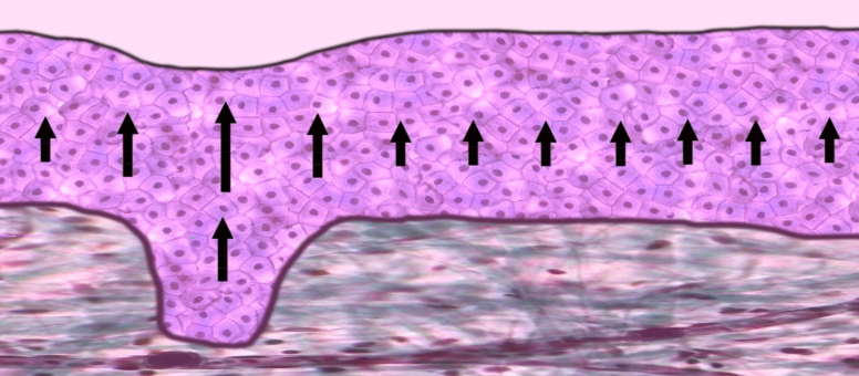
The instability of monolayered epithelia has been modeled as the result of a buckling phenomenon Drasdo (2000). Other studies have used the framework of nonlinear elasticity to describe the instabilities in growing tissues Goriely and Ben Amar (2005). As motivated in earlier work Basan et al. (2009) and shown experimentally Gordon et al. (1972); Marmottant et al. (2009), tissues behave as viscous liquids on long time scales. This is illustrated for example by the existence of surface tension at tissue boundaries Foty (1996); Lecuit and Lenne (2007); Guevorkian et al. (2010); Schötz et al. (2008). Theoretically, viscous descriptions have already been applied in other contexts of tissue growth Bittig et al. (2008). Here, we propose that the fingering of a stratified epithelium originates from viscous friction effects driven by cell division. We treat the epithelium as a viscous fluid lying on top of a viscoelastic stroma (Fig. 1).
As the epithelium consists mostly of cells, the stroma is made of a network of collagen and elastin fibers, constantly remodeled by fibroblasts present at low densities Alberts et al. (2008). On short time scales, this network responds elastically to deformations, but its constant remodeling by fibroblasts allows the tissue to flow on long time scales. A qualitative understanding of the full viscoelastic picture can be gained by interpolating between the results of the elastic and viscous regimes. In this letter, we present these two limit cases.
For the sake of simplicity, the epithelium and the stroma are each considered incompressible. In this case, the continuity equation for the epithelium reads , where is the global production rate of cells, taking into account cell division and apoptosis. The associated constitutive relation is that of an incompressible fluid with shear viscosity 111We assume a ratio of between bulk and shear viscosities.: . Here the total stress tensor has been split into a dynamic part and a velocity-independent part , where is the tissue pressure. The system of equations describing the epithelium is completed by the force-balance condition , which leads to:
| (1) |
Similarly, for an elastic stroma, we obtain:
| (2) |
together with , where is the displacement field, the shear modulus and the pressure.
Boundary conditions are as follows. The stress vanishes at the apical surface of the epithelium, taking into account the Laplace pressure due to the epithelium apical surface tension . At the bottom of the stroma, the displacement vanishes. At the epithelium-stroma interface, the normal component of the velocity is continuous and the normal component of the displacement of the stroma is equal to the variation of the interface location. The discontinuity of the normal component of the stress is given by Laplace’s law with interfacial tension . Finally, the tangential components of the stress are continuous and equal to a finite surface-friction term with coefficient .
The physical origin of the instability discussed in this work can be qualitatively understood as follows. Consider a fingering protrusion of the epithelium into the stroma and assume for simplicity that cell division occurs over the entire height of the protrusion (Fig. 1). The dividing cells create a flow in the epithelium. Since the cells above the finger have more dividing layers underneath them than their neighbors, they flow toward the apical surface faster than the cells in the adjacent regions. This results in a shear flow of cells within the epithelium. The associated shear stress builds up pressure at the bottom of the finger, favoring the development of the protrusion.
Let us now discuss the solution of our model for the flat, unperturbed epithelium-stroma interface. Here and in the following, we make the assumption that, due to the lack of nutrients and growth factors away from the stroma, the overall cell production decreases exponentially over a length scale with increasing distance from the epithelium-stroma interface: 222Note that we do not expect our results to crucially depend on the detailed form of the cell-production function.. When the interface is flat, the cell velocity and pressure in the epithelium read:
| (3) | |||||
| (4) |
where the origin of the coordinate is at the bottom of the stroma, and is the stroma thickness. The height of the epithelium is determined from the condition that the cell velocity vanishes at its apical surface. Together with Eq. (3), this condition reads . The deformation of the stroma vanishes everywhere ().
We now address the question of the stability of the system under a small perturbation. Since we do not expect the origin of the instability to depend on dimensionality, we consider the case of a system translationally invariant in the -direction, with a perturbation of the epithelium-stroma interface of the form . In the linearized system of equations, the solutions for the perturbations all take this form. Eq. (1) then reads:
| (5) |
together with the continuity equation . The bulk equations for the stroma keep their previous forms.
Stress balance at the apical surface of the epithelium reads:
| (6) | |||||
| (7) |
The perturbation of the apical surface is determined by the boundary condition , which takes the form to linear order. Stress balance at the epithelium-stroma interface reads:
| (8) | |||||
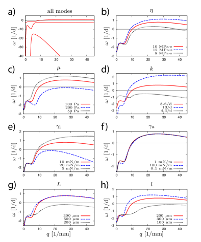
Also at this interface, continuity of velocity and displacement yields and . Finally, the displacement vanishes at the bottom of the stroma: . The growth rate is obtained by imposing the existence of a non-trivial solution to this set of linear equations. From this condition, we obtain three relaxation modes for the system (Fig. 2).
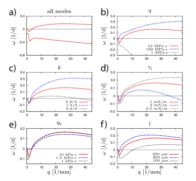
In the case where the stroma is treated as a viscous fluid, the previous equations need to be modified by replacing the displacement by a velocity () and the shear modulus by a viscosity (). In addition, the following boundary conditions are altered: the friction term in Eq. (Undulation instability of epithelial tissues) and the condition at the epithelium-stroma interface are replaced by and , respectively. This results into two relaxation modes (Fig. 3).
The number of modes that we get can be understood as follows. For the elastic stroma, the set of boundary conditions generates three modes because it contains the inverse relaxation rate three times: in the velocity-continuity conditions at both interfaces and in the tangential stress-balance equation at the epithelium-stroma interface. In the case of a fluid stroma, we loose the mode associated with the latter equality.
It is instructive to look at the analytic expansions of these different modes in the limit of large wave numbers . In this regime, the modes associated with respectively the epithelium-stroma interface and the apical surface decouple, since their characteristic decay lengths are of the order , which is much smaller than . For an elastic stroma, their expansions to constant order read:
| (9) |
Among these expressions, only the one related to can be positive, indicative of an unstable mode. It results at the epithelium-stroma interface from a balance of the stabilizing surface tension and stroma resistance to deformations on the one hand, and the overall positive cell-production on the other hand. This expression gives a necessary condition for the existence of an unstable regime (). The condition also yields a leading-order expression for the upper crossover wavenumber from the unstable to the stable regime, provided that this crossover occurs in the large- domain. The expression for results from a balance of surface tension and cell production at the apical surface, and the one for from a balance of tangential stress and surface friction at the epithelium-stroma interface. Both expressions correspond to modes that are always stable in their region of validity.
In the case of a viscous stroma, the potentially unstable mode reads:
| (10) |
The second mode has an identical expansion to that of the elastic case, and the third mode is lost.
Similar expansions can be obtained in the small- regime, but the expressions to next-to-leading order are complicated and mix the different physical origins described above. In the case of an elastic stroma, two of the three relaxation rates diverge to minus infinity, indicative of the elastic resistance of the stroma to a uniform compression. To leading order, they read:
| (11) | |||||
| (12) |
The third mode however has a finite small- limit, which reads . We can retrieve this expression by integrating the continuity equation at over the height of the perturbed epithelium and to leading order in the perturbations. In the case of a fluid stroma, one of the modes has the same finite limit, which is consistent with the argument presented above. However, the other relaxation rate approaches zero as rather than infinity:
| (13) |
Therefore, as the system is also always stable at sufficiently small , the relaxation time diverges in this case. This is because the relaxation here is associated with lubrication-like viscous flows over large distances in the -direction rather than elastic relaxation over short distances in the -direction.
These results show that the instability always occurs at finite wave vector. In Figs. 2 and 3, we analyze the behavior of the most unstable mode as a function of the parameters. We see that the interface is destabilized when either the epithelium viscosity , the cell-division rate , or the thickness of the dividing region is increased, because of a higher resulting shear stress 333Note that, since there is a relation between and , in the case where (resp. ) is varied, (resp. ) is varied in proportion in order to keep the geometry (resp. the amount of cell production) constant.. This is also true for the thickness of the stroma in the elastic case, since a thicker stroma resists less to a given deformation. Increasing the other parameters has a stabilizing effect. This is intuitive for the elastic shear modulus of the stroma in the elastic case and the stroma viscosity in the viscous case, as well as for the surface tension in both cases. The parameter in both cases as well as in the fluid case have little influence on the dispersion curves (not shown for the fluid case).
For a viscoelastic material with relaxation time , we do not expect anything qualitatively different from the fluid or elastic cases to occur at large and intermediate wave vectors. In the small- regime, as the relaxation rate goes toward a finite negative value in the case of an elastic stroma, it vanishes when the latter is fluid. Getting the correct behavior in the generic viscoelastic case would require a complete study. As a general fact, we expect the curves presented in Fig. 2 (resp. Fig. 3) to be valid when (resp. ).
In this work, we have shown the existence of a hydrodynamic instability of an interface between a viscous fluid with production terms and a viscoelastic material. The instability stems from the generation of viscous shear stress in the fluid due to material production. As such, this mechanism constitutes a new hydrodynamic instability that has not yet been described. We propose that this effect provides a potential mechanism for the undulations at epithelium-stroma interfaces in vivo. Our analysis might explain why such undulations are more pronounced in neoplastic tissues Tavassoli et al. (2003). Indeed, tumorous epithelial cells divide faster than healthy cells and in a larger domain away from the basement membrane Weinberg (2007). The large-deformation regime of the instability might correspond to such fingering phenomena. It is commonly accepted that cancerous invasion requires the production of proteases that can degrade the basement membrane and remodel the extracellular matrix Weinberg (2007). Such a digestion could decrease the interfacial tension between the tissues as well as the elastic modulus of the stroma, thereby triggering the present instability. The digestion of the extracellular matrix is thus not an alternative to the mechanism proposed here, but one of its determinants. While proteases enhance the instability and allow the growth of protrusions to proceed deeper into the stroma, we expect the physical forces driving this process to originate from the mechanism presented here.
The undulation instability investigated in this work is potentially relevant for many biological systems in which interfaces of growing cell populations are present. For example, at interfaces between many tumors and healthy tissues, similar effects are observed Tavassoli et al. (2003). More generally, we expect this type of instability to occur in all sufficiently viscous fluids with source terms. It would therefore be interesting to conceive other systems that show the same type of instability, while being easier to characterize experimentally than living tissues.
Acknowledgements.
We thank A. Callan-Jones, M. Lenz and X. Sastre-Garau for many useful discussions.References
- Tavassoli et al. (2003) F. Tavassoli, P. Devilee, I. A. for Research on Cancer, and W. H. Organization, Pathology and genetics of tumours of the breast and female genital organs (International Agency for Research on Cancer, 2003).
- Drasdo (2000) D. Drasdo, Phys. Rev. Lett. 84, 4244 (2000).
- Goriely and Ben Amar (2005) A. Goriely and M. Ben Amar, Phys. Rev. Lett. 94, 198103 (2005); M. M. Müller, J. Guven and M. Ben Amar, Phys. Rev. Lett. 101, 156104 (2008); J. Dervaux and M. Ben Amar, Phys. Rev. Lett. 101, 068101 (2008).
- Basan et al. (2009) M. Basan et al., HFSP J. 3, 265 (2009); J. Ranft et al., Proc. Natl. Acad. Sci. U.S.A. 107, 20863 (2010).
- Gordon et al. (1972) R. Gordon et al., J. Theor. Biol. 37, 43 (1972); R. A. Foty et al., Phys. Rev. Lett. 72, 2298 (1994); G. Forgacs et al., Biophys. J. 74, 2227 (1998).
- Marmottant et al. (2009) P. Marmottant et al., Proc. Natl. Acad. Sci. U.S.A. 106, 17271 (2009).
- Foty (1996) R. A. Foty et al., Development 122, 1611 (1996).
- Lecuit and Lenne (2007) T. Lecuit and P. F. Lenne, Nat. Rev. Mol. Cell. Biol. 8, 633 (2007).
- Guevorkian et al. (2010) K. Guevorkian et al., Phys. Rev. Lett. 104, 218101 (2010).
- Schötz et al. (2008) E. M. Schötz et al., HFSP J. 2, 42 (2008).
- Bittig et al. (2008) T. Bittig et al., New J. Phys. 10, 063001 (2008); T. Bittig et al., Eur. Phys. J. E Soft Matter 30, 93 (2009).
- Alberts et al. (2008) B. Alberts et al., Molecular Biology of the Cell (Garland Science, 2008), 5th ed.
- Fung (1993) Y. Fung, Biomechanics: mechanical properties of living tissues (Springer, 1993).
- Weinberg (2007) R. A. Weinberg, The biology of cancer (Garland Science, New York, 2007).
- Oliver et al. (1999) T. Oliver, M. Dembo, and K. Jacobson, J. Cell Biol. 145, 589 (1999).