Shear unzipping of double stranded DNA
Abstract
We use a simple nonlinear scaler displacement model to calculate the distribution of effect created by a shear stress on a double stranded DNA (dsDNA) molecule and the value of shear force which is required to separate the two strands of a molecule at a given temperature. It is shown that for molecules of base pairs less than 21, the entire single strand moves in the direction of applied force whereas for molecules having base pairs more than 21, part of the strand moves in the opposite direction under the influence of force acting on the other strand. This result as well as the calculated values of as a function of length of dsDNA molecules are in very good agreement with the experimental values of Hatch et al. (Phys. Rev. E , 011920 (2008)).
pacs:
87.15.-v,64.70.qd,05.90.+m,82.37.RsI. INTRODUCTION
A double stranded DNA (dsDNA) molecule consists of two polynucleotide strands connected loosely by hydrogen bonds through the base pairs and base-stacking between nearest neighbour pairs of base pairs and wound around each other to make a helix. The constraints of this helical structure require that the two base sequences on opposite strands must be complementary, with Adenine (A) always binding to Thymine (T) and Guanine (G) binding to Cytosine (C) saenger . The force that holds the complementary strands of DNA together is an important regulator of life’s processes because the binding of regulatory proteins to DNA often involves the procedure of mechanical separation of its strands. The intermolecular forces of DNA have been studied extensively using a variety of techniques, which cover a broad range of forces from a few piconewton(pN) up to several hundred piconewtons (pNs).
These techniques use either atomic force microscopy (AFM) or laser optical traps and magnetic tweezers strick ; bockel ; smith ; reif ; conroy ; kumar . The experiments fall into two general categories; those conducted on short DNA and those on long DNA. Experiments on long DNA focussed on the overall properties of the molecule bockel ; zhang and resulted in the discovery of S-DNA reif ; bloom ; clausen ; busta ; the occurance of this so called B-S transition has also been found in a short DNA chain of 30 base pairs morfill . Those on short duplexes focussed on the reaction pathway of melting, and contributed to the understanding of the local unbinding of DNA lee ; kerall ; pope ; strunz ; kuhner . In the single molecule experiments, the result may depend on choice of which variables are fixed and which can fluctuate. In some of the experiments (like AFM) extension is fixed and force is allowed to fluctuate (constant extension ensemble) while in others (like magnetic tweezers) the force is fixed and extension is allowed to fluctuate (constant force ensemble).
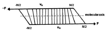
When a force is applied to pull apart the two strands from one end of a dsDNA molecule in a direction perpendicular to the helical axis, bases are sequentially stretched as the duplex is unzipped. On the other hand, in shear unzipping in which the applied force pulls the two strands from opposite ends as shown in Fig.1, the stretching is spread out over many base pairs. The study of distribution of shearing force along the length of a DNA molecule may lead to valuable informations about the force distribution across the phosphate backbone of a single strand of DNA versus the force distribution across the paired bases of complementory DNA. Such informations are of relevance not only to understanding the biological processes but also in material science application such as determining the strength of DNA/gold nanoparticle assemblies chak .
Following the work of Lee et al. lee many workers pope ; strunz ; sattin ; grange ; schu ; neurt ; milam ; wal ; noy ; hatch measured the value of shear force which separates the two strands of a dsDNA molecule. It was found that the force increases linearly with the length. Using a simple ladder model of DNA and expressing the backbone bond energy as well as the interaction energy among complementary bases in the form of harmonic springs, de Gennes gennes in 2001 predicted that the critical force for shear unzipping which for short chains shows linear dependence would saturate to a finite value in the limit where the number of base pairs approaches infinity and that the shear stress relaxes over a distance (number of base pairs) (=, where is the spring constant characteristic of stretching the backbone, and R is the spring constant characterstic of stretching the hydrogen bonds between base pairs) on either side of the chain. The critical force for shear unzipping as a function of number of bound base pairs was given gennes as
| (1) |
where is the rupture force of a single bond and (N+1) is the number of base pairs in the molecule.
Chakrabarti and Nelson chak have generalized the simple harmonic model of deGennes gennes by representing the interaction among complimentary bases by the Lennard-Jones (12-6) potential model and found that the strain is indeed localized over a narrow range of on either side of the chain and the chain unzips when the force exceeds a critical value of for short chains and for long ones as predicted by deGennes gennes . It may, however, be noted that the calculations of deGennes gennes and also of Chakrabarti and Nelson chak correspond to zero temperature. The Langevin dynamics simulation has recently been used to study shear unzipping of dsDNA at finite temperature mishra . The results found for are in agreement with the results of deGennes gennes and Chakrabarti and Nelson chak .
Hatch et al. hatch have measured the value of for several dsDNA molecules of length ranging from 12 to 50 base pairs at the room temperature and found that is linear function of molecular length only up to 20 base pairs and approaches to an asymptotic value as the number of base pairs increases. In fact they found that for a molecule of 32 base pairs has already reached within of the asymptotic value which was found to be 61.4 pN. But when Hatch et al. hatch tried to fit their data to Eq.(1) they found that this can be done only by assuming 7 base pairs of all chains to be in the open state, i.e. the effective length of a molecule of (N+1) base pairs is (N-6) irrespective of the value of N. They attributed this to temperature effect as deGennes calculation did not include temperature.
In this paper we calculate the value of as a function of the number of base pairs at room temperature and compare our results with the experimental data of Hatch et al. hatch and show that the need to adjust the length of a molecule is not as much due to temperature as due to use of the values of and in Eq.(1) and the nature of the curve of N. We derive another form of Eq.(1) and show that with reasonable choice of values of and one gets values of which are in better agreement with experiment without adjusting the length of molecules than that found from Eq.(1). The model which we describe in Sec. II is similar to the nonlinear scaler displacement model of Chakarbarti and Nelson chak . We calculate the force in both the constant force and constant extension ensembles. The paper is organized as follows. In Sec II we describe the model and calculational procedures. In Sec III we give results found for distribution of effects created by shear force along the length of a molecule and the value of as a function of number of base pairs. In Sec IV we compare our results with those of deGennes gennes and with the experimental values hatch . The paper concludes with a brief comment given at the end of Sec IV.
II. MODEL
We consider a dsDNA molecule of length (N+1) base pairs which both 5′-ends (or both 3′-ends) are pulled along the helical (molecular) axis by a force as shown in Fig.1. The displacements of nucleotide from its equilibrium position are denoted by for one (the lower one in Fig.1) single stranded DNA (ssDNA) chain and by for the other (the upper one) chain. The effective Hamiltonian of the system can be written as chak
| (2) | |||||
The first term of this equation represents the stretching energy of nucleotides, excecuting a simple harmonic motion with a spring constant along each chains in dsDNA. In the second term, represents the potential energy of interaction between bases in the pair and the last term of Eq.(2) represents the energy contribution due to the shear stress. In writing Eq.(2) we did not include the contribution arising due to the helicity of DNA as this effect was found to be negligible by Hatch et al. hatch and also by Lavery and Lebrun lavery .
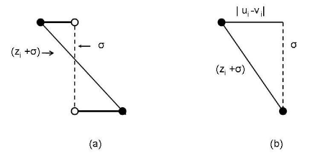
For the potential we use a simple model which has a hard-core repulsion and a long range attraction,
| (3) |
Here is the magnitude of increase in length of hydrogen bonds connecting bases in the pair from its equilibrium value, is the depth of potential at the equilibrium separation and is the diameter of dsDNA (see Fig.2). The repulsion represents the steric hindrance which forbids the molecule from getting compressed along the bond linking the bases with respect to its equilibrium value. From Fig.2 one finds ,
| (4) |
We take the value of equal to which is the diameter of the Canonical DNA (B-DNA) at room temperature.
Since in shear unzipping, a molecule is stretched along its axis we consider the longitudinal displacements of nucleotides and neglect the transverse displacements; the transverse displacements have been found in ref chak an order of magnitude smaller than those in the direction of shear. We define new variables,
| (5) |
where, and now represent the longitudinal displacements of respective strands. In the notations used here and are positive when the nucleotide of upper and lower strands move to r.h.s. and negative when they move to l.h.s.. For the experimental situation shown in Fig. 1 it is clear that for , and magnitute of is greater than that of and for , and the magnitute of is greater than that of . This leads to following relation for variables and ;
| (6) |
When we substitute these variables in Eq.(2) it decouples into two independent components;
| (7) |
where
| (8) |
and
| (9) |
Here the potential defined in Eq.(3) is expressed in terms of variable . Using Eqs. (4) and (5), one can rewrite Eq.(3) as
| (10) |
Note that the expression of does not contain the on-site potential and simply corresponds to a harmonic chain which is being pulled at the two ends by a force whereas the expression of contains the on-site potential as well as the force term.
In view of the relations given by Eq.(6) the average value of displacement of base pair can be calculated from the relation,
| (11) |
where , being the Boltzmann constant and is the temperature. Since for given by Eq.(7) the integrals in Eq.(11) are Gaussians, one can solve them analytically to give
| (12) |
This result agrees with the one found by deGennes gennes .
The average value of displacement of base pair can be found from the relation,
| (13) |
The integrals appearing in this expression cannot be evaluated analytically because of the form of on-site potential . However, for the expression of given by Eq.(9) the integral appearing in Eq.(13) reduces to multiplication of () matrices. The discretization of the coordinate variable and introduction of a proper cut-off on the maximum values of determine the size of the matrices. We have taken -40 and 40 as the lower and upper limit of integration for each co-ordinate variable and discretized space using the Gaussian-Legendre method with the number of grid points equal to 900. By changing the limits of integration as well as the number of grid points we made it sure that the values of are independent of the limit of integration and the number of grid points chosen to discretize coordinate variable.
III. RESULTS
When a shear force is applied on a dsDNA molecule, its two ssDNA strands get pulled in opposite directions as shown in Fig.1. The bonds in the backbone of DNA as well as bonds connecting bases in a pair are stretched. In a case of being infinitely large the two strands will move like a rigid body pulling all base pairs in the sequence in parallel. The effect of the shear force will then be uniformly distributed across all of the base pairs. However, if is finite, then both the backbone and the base pairs will stretch when a shear force is applied. The effect of the shear force may then be confined to limited lengths on both ends of the molecule. To see how the effect caused by shearing of a dsDNA molecule is distributed along the length of a molecule and how this depends on the energies associated with the stretching of backbone and base pairs we calculate the value of from Eq.(13) for different values of when the two end base pairs are stretched to a given length by the shear force.
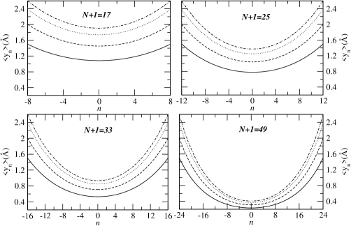
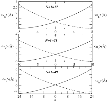
In Fig.3 we plot our results for four dsDNA molecules of length 17, 25, 33 and 49 base pairs and for , , and . The values shown in the figure correspond to and . When and are allowed to be free (i.e.), then is found to be zero for all . From Fig.3 it is clear that for short chains the effect created by shear force is distributed along the entire length of a molecule affecting all base pairs, whereas, for relatively larger molecules the base pairs in the central part of molecules are only marginally affected. For example, for a molecule of length 17 base pairs the value of is 1.76 when , whereas, for the similar situation (i.e. ) the value of for a molecule of length 49 base pairs is only 0.27. The qualitative nature of these results are in agreement with the results found in refs chak and mishra . The other point to be noted from the figure is that the qualitative nature of the distribution of the effect of shear stretching along length of a molecule is same for all values of plotted in the figure.
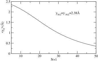
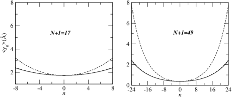
In Fig.4 we plot the value of when = 2.38 as a function of length of dsDNA molecules. As discussed below, the value of = 2.38 is assumed to be the critical value of stretching in the sense that initiation of separation of two strands starts at this value of and the shear force which creates this value of stretching of the end base pairs is equal to , the minimum ( or critical) force required to separate the two strands of a dsDNA molecule. From Figs. 3 and 4 it is clear that as one moves from either ends the differential force across the base pairs decreases and becomes very small at the centre for larger molecules but has not become zero at the centre even for a molecule of length 49 base pairs. The extension of the curve of Fig.4 shows that it would be zero at the centre only for molecules of length larger than 80 base pairs.
The average value of displacements and of base pair can be found from the known values of and . From Eq.(5) one gets
| (14) |
In Fig.5 we plot the values of and as a function of for molecules of length 17, 21 and 49 base pairs. The values plotted in this figure correspond to = 2.38 and F=40.9, 47.4 and 60.6 pN , respectively for molecules of length 17, 21 and 49 base pairs. These values of force, as is shown below, are the critical force of shearing of the respective molecules. From the figure we note that while for a molecule of 17 base pairs the entire ssDNA strand moves in the direction of applied force whereas in the case of a molecule of 49 base pairs nearly half of the strand moves in the opposite direction. While for a molecule of 17 base pairs, , and , , for a molecule of 49 base pairs , and , .
As long as the entire ssDNA strand moves in the direction of applied force the shear force, as explained in ref chak , depends linearly on molecular length. The departure from the linear dependence of the shear force on molecular length is expected to take place when and become zero i.e. the displacement of nucleotides on the opposite side of a ssDNA strand remains unaffected by the applied force. Indeed, we find that and =0 for a molecule of length 21 base pairs whcih is in very good agreement with the experimental value hatch . Since for molecules of base pairs larger than 21, part of a ssDNA moves in the opposite direction, a region develops inbetween on each strand which remains unaffected by the force. This region moves towards the centre on increasing the molecular length; the force gets saturated as soon as the region reaches the centre of the chain and stays there on further increasing the molecular length.
The value of shear force needed to separate the two strands of a dsDNA molecule can be calculated in two different ways. In one, we follow a method proposed by deGennes gennes and which from hereon is referred to as a method of constant force ensemble. The other method is based on the constant extension ensemble.
In the method of constant force ensemble one first defines a critical distance for the rupture of a base pair (i.e. when of base pair becomes larger than the bases of the pair become free) and calculate the value of force which can stretch a base pair to the critical distance . This force can be found from the on-site potential . Thus
| (15) |
If we take the value of ,we find for the value of and given above. This value of is close to the value used by Hatch et al hatch to fit their experimental data and the value estimated by Chakrabarti and Nelson chak . To rupture the end base pairs of a given molecule they must be stretched by the shear force to distance . The balance of force at one ends of one of the ssDNA gives gennes ;
| (16) |
Using the relations of Eq.(5) and Eq.(12) we get
| (17) |
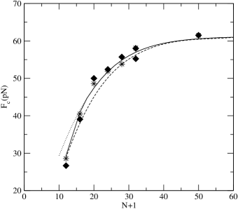
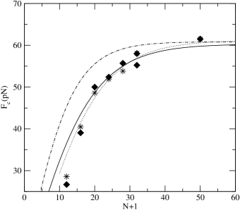
Taking the value of =2.38 we calculate the values of from Eq.(13) for several molecules of length 10-60 base pairs. The value of force found from Eq.(17) is shown by dotted line in Fig.6.
In the constant extension ensemble one first calculate the work done in stretching the two end base pairs of a given molecule to distance . This work is found from the relation
| (18) |
where
| (19) |
is the constrained partition function and
| (20) |
is the partition function. is the Dirac function.
We used the matrix multipliction method to evaluate from the above equations for , and . The derivative of with respect to gives the average force that is needed to keep the extension of end base pairs of a given molecule equal to . Thus
| (21) |
To get the value of critical force we have to chose a value of which corresponds to rupturing of base pairs. The values shown in Fig.6 by dashed line are found when was taken equal to ; this value is slightly lower than the one taken for the constant force ensemble. The difference in the value of critical stretching in the two ensembles may be due to difference in the path of unzipping.
We note that the values of found by methods of the constant force and the constant extension ensembles are close but not identical. The difference between the two as expected swigon is large for molecules of smaller lengths but they becomes close as molecular length increases. Both methods give the same asymptotic value, equal to 61.2pN, which is in very good agreement with the experimental value 61.4pN hatch . The experimental values shown in the figure are of Hatch et al hatch . In view of large spread in experimental data we find good agreement between experimental values of and the theoretical values found using the constant force ensemble as well as the constant extension ensemble.
IV DISCUSSIONS
(a) Comparison with the experiment
Since the experimental values hatch of given in Fig.6 are found using the fixed force ensemble, we concentrate our discussion with the theoretical values found using the same ensemble and shown in Fig.6 by dotted line. The agreement between theory and experiment is excellent except for a molecule of length 12 base pairs. We know that at a given temperature shorter dsDNA molecules are less stable compared to longer molecules. It is quite possible that at room temperature due to surface effects few of base pairs at the two ends of a molecule of length 12 base pairs are nearly in open state. The curve shown in Fig.6 by solid line is found when the effective length of a molecule of length 12 base pairs is taken to be equal to 10 base pairs and that of a molecule of length 16 base pairs equal to 15 base pairs. The values of force shown by solid line is in excellent agreement with the experimental values for the entire range of molecular length investigated by Hatch et al. hatch . The saturation value of found to be 61.2 pN is also in very good agreement with the experimental value of 61.4 pN hatch .
(b) Comparison with the results of deGennes gennes
In order to compare our results with those of deGennes gennes we first estimate the value of defined as , where R is the spring constant of a simple harmonic potential between the bases of a pair. Expanding in ascending powers of one gets
| (22) |
| (23) |
Substituting the values of , and given above we find which corresponds to . This value of is in good agreement with the predicted value of 77 based on calculation of the spring constants for base pairs and backbones value .
The expression for the displacement found by deGennes gennes can be written as
| (24) |
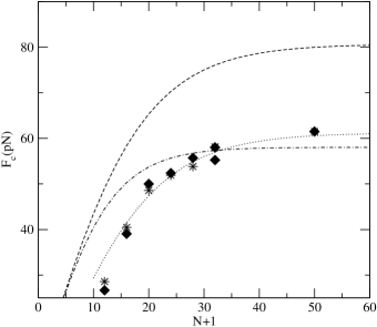
Taking the value of =6.44 and the values of determined above (shown in Fig 4) we calculate the value of from Eq.(24) for molecules of length 23 and 49 base pairs and compare them in Fig 7 with the values of found from Eq.(13). The two values do not agree; the difference increases with the length of molecules. A good agreement is, however, found when =9.4 is taken. This value of is about larger than the value found above. We calculate from the deGennes equation given by Eq.(1) taking =6.44 and 9.4 and =4.1 pN. The results are plotted in Fig 8 in which we also plot experimental values and the values found from our approach and shown in Fig 6 by dotted line. The values found from Eq.(1) are very different from both experimental values and values shown by dotted line. In ref mishra the value of as a function of N were found to be in good agreement with the values found from Eq.(1) for .
Since Eq.(1) involves the rupture force for a base pair and , the value of depends on the values of these quantities. Most often quoted value of is in the range of 4-5 pN chak ; hatch and that of chak ; hatch ; gennes ; mishra . Hatch et al. hatch took =3.9 pN and =6.8 to fit their data to Eq.(1) and found that they have to adjust the length of molecules which amounted to shifting the curve of to the right direction by 7 unit of base pairs in order to get good agreement with the experimental values. We now examine whether such an adjustment of length is essential or there is some other reason for not getting good agreement.
In the appendix we derive Eq.(24) from Eq.(9) and show that which appears in Eq.(24) should be defined as =. Substituting the values of and R we find =9.2 which is close to 9.4 found by using the values found from Eq.(13). With this definition of we find the following expression for (see Eq.(A8)).
| (25) |
The appearance of instead of (in Eq.(1)) is due to difference in the definition of . The definition seems more appropriate in the sense that it is ratio of the two harmonic spring constants than the definition of ref gennes in which one spring constant is multiplied by two.
The value of found from Eq.(25) when =9.4 and =4.1 pN are compared in Fig 9 with the values using the method of Sec. III and shown in Fig 6 by dotted line and values which are found from Eq.(1) when =6.8 and =3.9 pN hatch . From the figure we first note that Eq.(25) gives values of as a function of molecular length which are close to the values found in Sec. III (dotted line); the difference between the two is due to the combined effect of nonlinearity and the temperature. Thus the effect of temperature is not as large as has been suggested in ref hatch . The necessity for adjusting the length of molecules is due to the values of and used in Eq.(1) by Hatch et al. hatch . If one takes =9.4 and =4.1/ pN and calculate using Eq.(1) one gets values shown by full line in Fig 9 which are found from Eq.(25) with =9.4 and =4.1 pN.
In conclusion, we developed a method to calculate the distribution of shear force along the length of a dsDNA molecule at a finite temeprature. The value of shear force which is required to separate the two strands of a molecule has been calculated in both the constant force and the constant extension ensembles. The values of found by these methods differ for molecules of shorter length but approach to each other as length increases. Both methods gave the saturation value equal to 61.2 pN which is in very good agreement with the experimental value 61.4pN. The value of is found to increase linearly with length up to 21 base pairs in agreement with experimental results. The plots of and given in Fig.5 show that as long as the applied force pulls the entire strand in its direction of application, depends linearly on length of molecules. The departure from linear behaviour takes place when part of a strand moves in opposite direction under the influence of force pulling the other strand. The saturation value is achieved when half of a strand moves in the direction of force applied on it and the other half in the opposite direction. It is shown that the value of quickly attains its saturation value on increasing length of DNA molecule; for a molecule of 32 base pairs = 57.0pN as compared to saturation value 61.2pN.
The agreement between theorectical and experimental values of shown in Fig.6 indicates that the model proposed in this paper is capable of describing the responses of dsDNA molecules to shear stress. The model, however, neglects the effect of helicity of DNA and has assumed the molecule to be homogeneous. As far as the effect of helicity is concerned, it has already been shown to be negligible by Hatch et al. hatch and by Lavery and Lebrun lavery . The effect of heterogenity arising due to random distribution of A-T and C-G base pairs in a sequence is also expected to be small as long as the shear unzipping from the two ends is symmetric. This is corroborated by the fact that although the molecules investigated by Hatch et al hatch are heterogeneous with half G-C and half A-T base pairs, yet their response to shear stress is described very well by a model which assumes molecules to be homogeneous. The values of parameters and will, however, depend on the percentage of A-T and C-G base pairs in a given sequence and on the temperature. But, in the absence of symmetry in the unzipping from the two ends a qualitative new features, as argued in ref chak , may arise.
ACKNOWLEDGEMENTS
We are grateful to Sanjay Kumar for drawing our attention to the paper of P. G. deGennes and to B. P. Mandal for useful discussions. One of us (SP) acknowledges the financial support provided by the University Grants Commission, India.
Appendix
In this appendix we derive an expression for from Eq.(9). Writing as (see Eqs.(22) and (23))
where = and differentiating with for we get the following equilibrium conditions gennes from Eq.(9)
For , one gets
From these equations we find
where =, and
Let the force on the last hydrogen bonds (=) is when we reach the threshold . ( being the rupture force for a base pair). Thus
From Eq.(A3) we get
This equation differs from Eq.(1) in the definition of and the mutiplying factor which is now instead of 2.
References
- (1) W. Saenger, Principle of Nucleic Acid Structure (Springer-Verlag, Berlin, 1984).
- (2) T. Strick, J.-F. Allemand, V. Croquette and D. Bensimon , Phys. Today 54, 46 (2001).
- (3) U. Bockelmann, Curr Opin. Struc. Biol. 14, 368 (2004); U. Bockelmann, B. Essevaz-Roulet, and F. Heslot, Phys. Rev. Lett. 79, 4489 (1997).
- (4) S. B. Smith, Y. Cui and C. Bustamante, Science 271, 795 (1996).
- (5) M. Reif, H. Clausen-Schaumann and H. E. Gaub, Nature Struct. Biol. 6, 346 (1999).
- (6) R. S. Convory and C. Danilowicz, Contemp Phys. 45, 277 (2004).
- (7) S. Kumar and M. S. Li, Phys. Rep. 486, 1 (2010).
- (8) Y. Zhang, H. Zhou and Z. -C. Ou-Yang, Biophys. J. 81, 1133 (2001).
- (9) I. Rouzina and V. A. Bloomfield, Biophys. J. 80, 882 (2001).
- (10) H. Clausen-Schaumann, M. Reif, C. Tolksdorf and H. E. Gaub, Biophys. J. 78, 1997 (2000).
- (11) C. Bustamante, S. B. Smith, J. Liphardt and D. Smith, Curr. Opin. Struct. Biol. 10, 279 (2000).
- (12) J. Morfill , F. Khner, K. Blank, R. A. Lugmaier, J. Sedlmair and H. E. Gaub, Biophys. J. 93, 2400 (2007).
- (13) G. U. Lee, L. A. Chrisey, and R. J. Colton, Science 266, 771 (1994).
- (14) A. D. Mc Kerall and G. U. Lee, Eur. Biophys. J. 28, 415 (1999).
- (15) L. H. Pope, M. L. Davies, C. A. Laughton, C. J. Roberts, S. J. B. Tendler and P. M. Williams, Eur. Biophys. J. 30, 53 (2000).
- (16) T. Strunz, K. Oroszlan, R. Schafer and H.-J. Guntherodt, Proc. Natl. Acad. Sci. U.S.A. 96, 11277 (1999).
- (17) F. Khner, J. Morfill, R. A. Neher, K. Blank and H. E. Gaub, Biophys. J. 92, 2491 (2007).
- (18) B. Chakrabarti and D. R. Nelson, J. Phys. Chem. B 113, 3831 (2009).
- (19) B. D. Sattin, A. E. Pelling, and M. C. Goh, Nucleic Acids Res. 32, 4876 (2004).
- (20) W. Grange, T. Strunz, I. Schumakovitch, H.-J. Guntherodt, and M. Hegner, Single Mol. 2, 75 (2001).
- (21) I. Schumakovitch, W. Grange, T. Strunz, P. Bertoncini, H.-J. Guntherodt and M. Hegner, Biophys. J. 82, 517 (2002).
- (22) G. Neuert, C. H. Albrecht and H. E. Gaub, Biophys. J. 93, 1215 (2007).
- (23) Y. Zhang, V. T. Milam, D. J. Graves and D. A. Hammer, Biophys. J. 90, 4128 (2006).
- (24) M. V. Wal, S. Kamper, J. Headley and K. Sinniah, Langmuir 22, 882 (2006).
- (25) A. Noy, D. V. Vezenov, J. F. Kayyem, T. J. Meade and C. M. Leiber, Chem. Biol. 4, 519 (1997).
- (26) K. Hatch, D. Danilowicz, V. Coljee and M. Prentiss, Phys. Rev. E 78, 011920 (2008).
- (27) P. G. de Gennes, Soryushiron Kenkyu 2, 1505 (2001).
- (28) R. K. Mishra, G. Mishra, M. S. Li and S. Kumar, arXiv:1104.3059 (Cond-mat.soft) (2011).
- (29) R. Lavery and A. Lebrun, Genetica 96, 75 (1999).
- (30) D. Keller, D. Swigon and C. Bustamante, Biophys. J. 84, 733 (2003).
- (31) This value quoted in Ref.hatch was found from M. Fytas and E. Kaxiras through private communication by the authors of hatch .