Abnormal effective connectivity in migraine with aura under photic stimulation
Abstract
Migraine patients with aura show a peculiar pattern of visual reactivity compared with those of migraine patients without aura: an increased effective connectivity, connected to a reduced synchronization among EEG channels, for frequencies in the beta band. The effective connectivity is evaluated in terms of the Granger causality. This anomalous response to visual stimuli may play a crucial role in the progression of spreading depression and clinical evidences of aura symptoms.
pacs:
87.19.L-, 05.45.Tp, 05.45.Xt, 42.66.LcImportant information on the structure of complex systems can be obtained by measuring to what extent the individual components exchange information among each other. Transfer entropy schreiber ; lehnertz and Granger causality granger ; bli ; ding ; kc have emerged in recent years as leading statistical techniques to detect cause-effect relationships between time series; they are equivalent in the case of Gaussian stochastic variables seth . These approaches provide further insights on the architecture of complex systems, in addition to those from correlation and synchronization analysis boccaletti ; rosenblum ; quiroga . In neuroscience, interdependencies estimated by correlation or spectral coherence are referred to as functional connectivity, whilst effective connectivity is a notion related to Granger causality or transfer entropy friston ; the relationship between functional and effective connectivity in the cortex represents a significant challenge to present-day neuroscience sporns ; noineuroimage ; neuro ; fr .
Migraine, an incapacitating disorder of neurovascular origin, consists of attacks of headache, accompanied by autonomic and possibly neurological symptoms class . This pathology affects a relevant fraction of the general population and represents a social problem. The study of phase synchronization in EEG rhythms showed a pattern of alpha rhythm (8-12.5 Hz) hyper-synchronization under repetitive flash stimulation in migraine without aura patients, opposite to a de-synchronization trend in non-migraine subjects noiprl ; this nonlinear EEG pattern was found to be modulated by anti-epileptic drugs drugs . Approximately one-third of people who suffer migraine headaches perceive an aura (visual abnormality lasting 10-30 minutes) as a sign that the migraine will soon occur aureview . There are evidences sp of relationships between migraine aura and the spreading depression (SD) phenomenon (a wave of electrophysiological hyperactivity followed by a wave of inhibition leao ) but the link between SD and headache is far to be explained in humans zhang . SD in clinically less conspicuous or extended brain regions may be the trigger of migraine attacks ostensibly without a ”perceived” aura ayata , so it may be plausible that, migraine with and without aura may differ for those neuronal factors favoring the clinical expression of SD phenomenon.
The question we address here is the following: how does the response of aura migraine patients, to external stimuli, differ from those of patients without aura? We show that, in presence of visual stimulations, migraineurs with aura show a pattern of increased effective connectivity between EEG channels in the beta band, and correspondingly a decrease of the synchronization between channels. This variation of connectivity, due to stimuli, is statistically significant for migraine with aura: we remark that in our knowledge neurophysiological patterns separating migraine with and without aura have been rarely detected conte , nor relevant differences in regard to visual reactivity genco .
Our data are as follows. EEG is recorded from 19 patients (7 males, 20-44 age) affected by migraine with aura, 19 (4 males, 21-45 age) patients affected by migraine without aura, and from 11 healthy subjects (control group, 3 males, 20-46 age). All patients are in the interictal state, the time from the end of the last attack being at least 72 h. No patient was under preventive treatment nor had assumed symptomatic drugs in the 72 hours preceding the recording session. During the acquisition, flash stimuli are presented at a rate of 9-18-21-24-27 Hz; also EEG in the absence of stimuli (base) is recorded. Each frequency of stimulation is delivered by a flash with 0.2 J luminance for about 20 sec. EEG data are recorded by six scalp electrodes: two occipital channels (O1 and O2), two parietal ones (P3 and P4), a central electrode (Cz) and a frontal one (Fz); the sampling rate is 256 Hz, and the EEG is digitally filtered off line by a filter with a band-pass 0.3-30 Hz.
Next, we describe our findings. Firstly we investigate the alpha band hyper-synchronization phenomenon noiprl in presence of flash stimuli. The pattern of noiprl is confirmed for migraineurs without aura, whilst patients with aura do not show alpha band hyper-synchronization in presence of light stimuli: in this range of frequencies, aura patients seem to behave as controls.
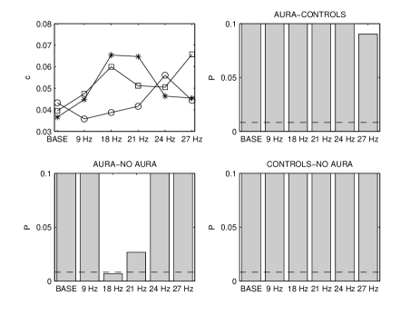
Moving to higher frequencies (beta band, 12.5-30 Hz), instead, we find a peculiar pattern of visual reactivity for aura patients. We set the order of the regression model equal to bic and evaluate the linear Granger causality in the beta band seth1 : linear causalities are weak and show significant differences among classes only at 18 Hz stimulations, see figure (1). On the other hand, evaluating the nonlinear Granger causality among the filtered EEG signals with the kernel approach described in kc and a Gaussian kernel, we find that aura migraine patients exhibit increased values of causality in presence of stimuli, whilst controls and no aura patients do not show significant variation w.r.t. basal conditions. In particular the discrimination between aura and no-aura patients is excellent in presence of flash stimuli. Figure (2) refers to as the width of the Gaussian kernel, however the results are robust to variations of .
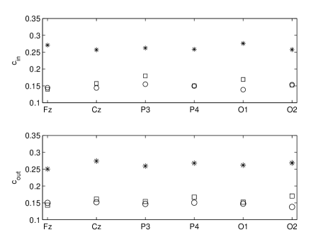
A topographic analysis is also performed: for each electrode, we evaluate the total incoming causality (the sum of the causalities from the other electrodes to the electrode under consideration) as well as the total outgoing causality (the sum of the causalities from the electrode under consideration to the other electrodes). In figure (3) we depict these quantities for the three classes at 18 Hz stimulations in the beta band: it shows that the phenomenon is diffuse over the scalp.
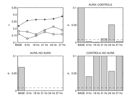
Use of the Gaussian kernel ensures that all orders of nonlinearities are taken into account. If one considers only nonlinearities up to the second order, weaker causalities are detected and less discriminating power is obtained, see figure (4) where we describe the application of nonlinear Granger causality with a polynomial kernel of degree two. We conclude that a relevant amount of nonlinear information transmission characterizes this phenomenon.
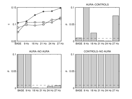
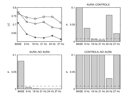
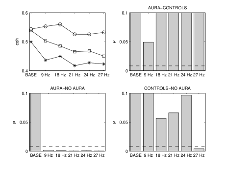
Turning to synchronization, we consider the Pearson linear correlation between channels and find that the increased flow of information, due to flash stimuli, is connected to weakening of the correlations between them. In figure (5) we depict the linear correlation between signals filtered in the beta band, averaged over all pairs of channels; in presence of light stimulations the strength of correlations decreases for aura patients. We find similar results also in terms of the coherence function averaged in the beta band, see figure (6), as well as for the beta band phase synchronization tass .
We also quantify the separation among classes in terms of the ROC area roc , which is directly related to the separation of two conditional distributions, and measures the discrimination ability of the forecast. For any frequency of stimulation, using the Gaussian kernel Granger causality we obtain a roc area equal to 0.87 for aura vs no-aura patients. A similar result is found using the linear correlation, roc area equal to 0.82 for aura vs no-aura at all frequencies.
Summarizing, we have described for the first time a neurophysiological pattern which seems peculiar of migraine patients perceiving visual aura, where the loss of synchronization between channels, due to light, induces stronger statistical causal connections among them, diffuse over the scalp. This pattern is characterized by nonlinear transfer of information among channels and provides excellent discrimination between aura and no-aura patients. The biological implications of this complex phenomenon in facilitating SD progression and aura symptoms perception is the challenge for a better understanding of migraine pathophysiology.
References
- (1) T. Schreiber, Phys. Rev. Lett. 85, 461 (2000).
- (2) M. Staniek, K. Lehnertz, Phys. Rev. Lett. 100, 158101 (2008).
- (3) C.W.J. Granger, Econometrica 37, 424 (1969).
- (4) K. J. Blinowska, R.Kus, and M. Kaminski, Phys. Rev. E 70, 050902 (2004).
- (5) M. Dhamala, G. Rangarajan, M. Ding, Phys.Rev.Lett. 100, 18701 (2008).
- (6) D. Marinazzo, M. Pellicoro, S. Stramaglia, Phys. Rev. Lett. 100, 144103 (2008).
- (7) L. Barnett, A.B. Barrett, A. K. Seth, Phys. Rev. Lett. 103, 238701 (2009).
- (8) S. Boccaletti, J. Kurths, G. Osipov, D.L. Valladares and C. Zhou, Physics Reports 366, 1 (2002).
- (9) M.G. Rosenblum et al., Phys. Rev. Lett. 76, 1804 (1996).
- (10) R. Q. Quiroga, A. Kraskov, T. Kreuz, and P. Grassberger, Phys. Rev. E 65, 041903 (2002).
- (11) K.J. Friston, Human Brain Mapping 2, 56 (1994).
- (12) O. Sporns, G. Tononi, and R. Kotter, PLoS Computational Biology 1, 245 (2005).
- (13) D. Marinazzo, W. Liao, H. Chen and S. Stramaglia, Nonlinear connectivity by Granger causality, in press in Neuroimage, doi:10.1016/j.neuroimage.2010.01.099.
- (14) P.A. Valdes-Sosa, J. Daunizeauc and K. Friston, Effective connectivity: Influence, causality and biophysical modeling, in press in Neuroimage, doi:10.1016/j.neuroimage.2011.03.058.
- (15) K. Friston, Functional and effective connectivity: a review, in press in Brain Connectivity, doi:10.1089/brain.2011.0008.
- (16) Headache Classification Committee. The International Classification of Headache Disorders II. Cephalalgia 24, 24 (2004).
- (17) L. Angelini et al., Phys. Rev. Lett. 93, 038103 (2004).
- (18) M. de Tommaso et al., Clin. Neurophysiol. 118, 2297(2007).
- (19) F.M. Cutrer, K. Huerter, Neurologist. 13, 118 (2007).
- (20) M. Lauritzen, Brain 117, 199 (1994).
- (21) A.A.P. Leao, J. Neurophysiol. 7, 359 (1944).
- (22) X. Zhang et al., Activation of Meningeal Nociceptor by Cortical Spreading Depression. Proceedings of the Society for Neuroscience Annual Meeting, Vol. 339.5. Chicago, IL: Society for Neuroscience (2009).
- (23) C. Ayata, Headache 50, 725 (2010).
- (24) A. Conte et al., PAIN 148, 43 (2010).
- (25) S. Genco et al., CEPHALALGIA 14, 41 (1994).
- (26) G.E. Schwarz, Annals of Statistics 6, 461 (1978).
- (27) A.K. Seth, Journal of Neuroscience Methods 186, 262 (2010).
- (28) M. Bland, An introduction to medical statistics, Oxford Medical Publications, Oxford, 2009.
- (29) P. Tass et al., Phys. Rev. Lett. 81, 3291 (1998).
- (30) M. H. Zweig, G. Campbell, Clin. Chem. 39, 561 (1993).