Controlling the self-assembly of binary copolymer mixtures in solution through molecular architecture
Abstract
We present a combined experimental and theoretical study on the role of copolymer architecture in the self-assembly of binary PEO-PCL mixtures in water-THF, and show that altering the chain geometry and composition of the copolymers can control the form of the self-assembled structures and lead to the formation of novel aggregates. First, using transmission electron microscopy and turbidity measurements, we study a mixture of sphere-forming and lamella-forming PEO-PCL copolymers, and show that increasing the molecular weight of the lamella-former at a constant ratio of its hydrophilic and hydrophobic components leads to the formation of highly-curved structures even at low sphere-former concentrations. This result is explained using a simple argument based on the effective volumes of the two sections of the diblock and is reproduced in a coarse-grained mean-field model: self-consistent field theory (SCFT). Using further SCFT calculations, we study the distribution of the two copolymer species within the individual aggregates and discuss how this affects the self-assembled structures. We also investigate a binary mixture of lamella-formers of different molecular weights, and find that this system forms vesicles with a wall thickness intermediate to those of the vesicles formed by the two copolymers individually. This result is also reproduced using SCFT. Finally, a mixture of sphere-former and a copolymer with a large hydrophobic block is shown to form a range of structures, including novel elongated vesicles.
I Introduction
Amphiphiles such as block copolymers and lipids can self-assemble into many different structures when dissolved in solution Jain and Bates (2003, 2004). The case of block copolymers has proved especially interesting to researchers in recent years, for a variety of reasons Smart et al. (2008). First, the study of block copolymers is a promising route to a fundamental model of the self-assembly of amphiphiles in solution, since the theoretical understanding of the constituent polymer molecules is on a firm footing. Well-established methods such as self-consistent field theory (SCFT) Edwards (1965); Schmid (1998); Matsen (2006) have provided considerable insight into the self-assembly of polymers, especially in melts Maniadis et al. (2007); Drolet and Fredrickson (1999), whilst using simple models of the individual polymer molecules. Second, vesicles formed from block copolymers show more promise as vehicles for drug delivery Kim et al. (2005) than similar structures formed from lipids, as the thickness and low solubility of their membranes means that they can be longer-lived and less permeable Discher et al. (1999); Discher and Eisenberg (2002).
For solutions of a single type of diblock copolymer, it is often relatively straightforward to understand why a given type of aggregate forms in a given system. The main factor that determines the shape of the structures is the architecture of the copolymer; that is, the size of its hydrophilic and hydrophobic blocks Kinning et al. (1988) (although other factors, such as the overall size of the copolymer, may also play a role Kaya et al. (2002)). If the hydrophilic component is large compared to the hydrophobic component, then curved aggregates such as spherical or cylindrical micelles form. Conversely, if the hydrophobic component is large, lamellar structures such as vesicles are observed Kinning et al. (1988). Recently, we demonstrated this behavior in a study of PCL--PEO block-copolymers with various volume fractions of the hydrophobic block (PCL) Adams et al. (2009). Here, large volume fractions of the hydrophilic block (PEO) resulted in micelles (), lower favored wormlike micelles () and still lower fractions () led to the formation of vesicles.
We can gain increased control over the self-assembly by mixing two types of amphiphile that individually form aggregates of different curvatures. Such mixtures are well known in cell biology, where different lipids can be sorted by segregation to regions of high and low curvature Sorre et al. (2009); Zidovska et al. (2009), and have also been studied in the context of lipid-detergent systems Vinson et al. (1989); Oberdisse et al. (1998). More recently, they have been investigated in block copolymer solutions. For example, Jain and Bates Jain and Bates (2004) have studied mixtures of polyethylene oxide-polybutadiene (PEO-PB), and have found that blending ratio can be used to control self-assembly, and furthermore that novel structures such as undulating cylinders form. They also found that different aggregates form depending on whether the two polymer species are mixed before or after their individual self-assembly Jain and Bates (2004).
In a recent study of a mixture of sphere-forming and lamella-forming polycaprolactone-co-polyethylene oxide in water-THF mixed solvents Schuetz et al. (2011), we have built on this work by controlling the quantities of water and THF to mix sphere- and lamella-forming copolymers not only before and after but also during their individual self-assembly. Those copolymers mixed before self-assembly (pre-mixed) formed a sequence of aggregates of increasing curvature as the amount of sphere-former was increased, forming vesicles, then a mixtures of vesicles, rings and worms, and finally spherical micelles. This series of shape transitions has also been observed in lipid-detergent mixtures Vinson et al. (1989); Oberdisse et al. (1998), and has been studied theoretically using self-consistent field theory Schuetz et al. (2011); Li et al. (2009); Greenall and Gompper (2011) and models of chain packing Fattal et al. (1995) and membrane curvature Andelman et al. (1994). When mixed after self-assembly (post-mixed), the two species remained locally in the equilibrium states of the pure components, and a mixture of vesicles and spherical micelles was observed. The structures found when the two species were allowed partially to self-assemble before mixing (intermediate mixing) were more unusual, and included metastable paddle- and horseshoe-shaped aggregates. Using self-consistent field theory, we reproduced the transitions between morphologies observed in the pre-mixed system and also details of the aggregates such as the bulbous ends of the rods Schuetz et al. (2011). We also gained insight into the complex metastable structures seen at intermediate mixing, by showing in SCFT calculations that the segregation of the two types of copolymer can stabilize regions of different curvature within a single aggregate.
In the current paper, we extend this study by varying the architectures of the copolymer species. We consider three specific cases: varying the length of the lamella-former in a blend of sphere- and lamella-formers, blending two lamella-formers of different lengths, and blending sphere-former with a polymer that has such a large hydrophobic block that it precipitates in solution if not mixed with more hydrophilic molecules. In all cases, we observe the quantitative and qualitative changes in the self-assembly as the copolymer architectures are changed. As in our previous work Schuetz et al. (2011), we perform self-consistent field theory calculations in tandem with our experiments and discuss how the distribution of the two copolymer species within the self-assembled structures leads to the formation of the structures seen in the experiments.
The article is organised as follows. In the following section, we give details of our experimental and theoretical methods. The Results section is divided into three subsections, one for each of the mixtures introduced above. We then present our conclusions.
II Methods
II.1 Materials
The PEO-PCL block copolymers were purchased from Advanced Polymer Materials Inc., Montreal and used as received. GPC analysis was also provided by Advanced Polymer Materials Inc. and was referenced against PEO standards. Degrees of polymerization for the PCL block were calculated by NMR in by comparison to the PEO block (the degrees of polymerization for the monomethoxypoly(ethylene oxides) used in these polymerizations are known). The molecular weight and molecular weight distributions are given in Table 1. All other reagents with the exception of NMR solvents were purchased from Sigma Aldrich Company Ltd., Gillingham. Standard solvents were of spectrophotometric grade and inhibitor free. Deuterated NMR solvents were purchased from Euriso-top S.A., Saint-Aubin. All solvents were filtered before use through Pall Acrodisc PSF GHP filters. For all experiments, distilled and de-ionized Millipore water (resistivity = ) was additionally filtered through Sartorius Ministart filters directly before use.
| Commercial sample code | Sample formula | Morphology c | ||||
| PCLPEO | PEOPCL | 17300 | 1.36 | 0.15 | V | 220 |
| PCLPEO | PEOPCL | 7100 | 1.15 | 0.17 | V | 170 |
| PCLPEO | PEOPCL | 7800 | 1.16 | 0.3 | C,S | 30 |
| PCLPEO | PEOPCL | 6380 | 1.15 | 0.08 | P | n/a |
II.2 Preparation of Solutions
Aqueous dispersions of block-copolymer aggregates were prepared by dissolving the polymer in THF to a concentration of . These solutions were then mixed in the volume ratio noted for the experiments. All the mixing ratios are thus ratios of the masses of the respective polymers as opposed to molar ratios. Due to the close molecular weights (Table 1) of the two copolymers PCLPEO and PCLPEO these ratios are not very different in this case, while for the other combinations the conversion is easily calculated. Millipore water was added either manually or by an Eppendorf EDOS 5222 Electronic Dispensing System. 125 aliquots of of water were added at one-minute intervals.
II.3 Turbidity Measurements
We performed turbidity measurements during the preparation of the samples using an adapted Perkin Elmer UV/Vis Lambda 40 Spectrometer. A wavelength of was used with a slit width of . Stirring was performed using a standard magnetic stirrer/hotplate placed under the spectrometer. The polymer was dissolved in THF (, ) and a zero reading was taken (transmittance, ). Millipore water was then added either in aliquots every using an Eppendorf EDOS 5222 Electronic Dispensing System and a turbidity reading was taken after each addition.
II.4 Cryo-TEM
Samples for thin-film cryo-TEM were loaded onto plasma-treated (30 seconds) holey-carbon grids and prepared using a GATAN cryo-plunge into liquid ethane and then transferred using a GATAN 626 cryo-transfer system. Samples were examined using a JEOL 2100 TEM operating at . Images were obtained using a Bioscan or a GATAN Ultrascan 4k camera and analyzed by GATAN Digital Micrograph version 1.71.38. During our previous investigations Adams et al. (2009) we observed that imaging of the self-assembled structures (especially vesicles) is greatly improved in samples containing ca. THF compared to samples in pure aqueous solution. Similar structures were observed in both solvent conditions, which indicates that at THF fractions of and below, the mobility of the block-copolymers is too restricted to allow for further growth of the aggregates. However, we found that, over a timescale of a few months, internal rearrangements occurred that evened out local variations in surface curvature transforming the more complex metastable aggregates into vesicles or nested onion-like structures. In order to focus on the initial metastable structures that are formed (prior to any slow internal rearrangement processes) and to obtain as high quality imaging as possible, we prepared the solutions for cryo-TEM in aqueous solutions containing THF and imaged these samples within a maximum time of two weeks.
II.5 Self-consistent field theory
To further our understanding of the role of polymer architecture on self-assembly, we performed self-consistent field theory (SCFT) calculations Edwards (1965) on a model system of two species of AB diblock copolymers (lamella- and sphere-forming respectively) blended with A homopolymer ‘solvent’. SCFT is a coarse-grained mean-field theory in which the individual polymer molecules are modeled by random walks and composition fluctuations are neglected. For sufficiently long polymers Müller (2006), these approximations prove extremely effective and the predictions of the theory are very accurate for a wide range of systems Matsen (2006). We use a simple implementation of the theory where the interactions of the polymers are included by imposing incompressibility and introducing a contact potential between the A and B monomers Matsen (2006). The fine details of the polymer molecules are not taken into account, so that monomers of all species are taken to have the same length and volume .
From a technical point of view, a self-consistent field theory calculation consists of solving a series of differential (diffusion) equations to calculate the density profiles of the various polymer species. An initial guess for the profiles is made, which has the approximate form of the structure that we wish to study. The density profiles are then recalculated until a set of equations reflecting the physical properties of the system (such as its incompressibility) Matsen (2006, 2004) is satisfied. The SCFT differential equations are solved using a finite-difference method Press et al. (1992) with a spatial step size of , where is the number of monomers in the sphere-forming species, and reflecting boundary conditions are imposed at the origin and edges of the box. Full technical details of our calculation can be found in a recent publication Greenall and Gompper (2011).
In the current paper, we use SCFT calculations in two slightly different ways. First, we perform effectively one-dimensional calculations on spherical micelles and infinite cylinders and bilayers to calculate simple phase diagrams as a function of sphere-former volume fraction and reproduce the basic phenomenology observed in the experiments. These calculations proceed as follows Greenall et al. (2009a, b). To begin, we calculate the free-energy density of a box containing a single spherical, cylindrical or planar aggregate surrounded by solvent. The shape of this box is set by the symmetry of the aggregate; for example, a spherical micelle is formed at the center of a spherical box. The calculation is therefore effectively one-dimensional. The volume of this simulation box is then varied, keeping the volume fraction of copolymer constant, until the box size with the minimum free-energy density is found. Provided the system is dilute, so that each aggregate is surrounded by a large volume of solvent and the aggregates do not interact with each other, this provides a simple model of a larger system (of fixed volume and fixed copolymer volume fraction) containing many aggregates. The reason for this is that such a system minimizes its free energy by varying the number of aggregates and hence the volume (‘box size’) occupied by each. Although computationally inexpensive, this approach yields accurate information on micelle shape transitions and its results agree well with experiment Greenall et al. (2009b).
We also carry out more detailed calculations on the rod and ring structures seen in the experiment. We have two aims here. First, we wish to show that these more complex structures can be reproduced in detail in our calculations. Second, we will study the distribution of the sphere-forming and lamella-forming copolymers within the aggregates. Since both rods and rings have cylindrical symmetry, we will perform our (effectively two-dimensional) calculations in a cylindrical box. We note that it is not possible to include information on the distribution of the sphere- and lamella-formers within the micelles in the initial guess for the SCFT calculations. Any segregation of the two species will therefore arise naturally from the theory and need not be artificially introduced Greenall and Gompper (2011).
Although this model is relatively simple, we found in our previous work on the self-assembly of binary PEO-PCL mixtures Schuetz et al. (2011) that it contains enough detail to yield information on the structures formed in such systems and on the distribution of the two polymer species within these aggregates. In this earlier paper, we focused on a mixture of lamella-forming PCLPEO and sphere-forming PCLPEO. We modeled the lamella-former by a symmetric AB diblock with equal numbers of monomers and in its hydrophilic (A) and hydrophobic (B) sections. For simplicity, the homopolymer was taken to have the same length as the lamella-former. In line with the experiments, the sphere-former contained the same number of hydrophobic monomers as the lamella-former but a larger number of hydrophilic monomers, so that . The parameter setting the strength of the interaction between the A and B species was set to , where is the total number of monomers in the sphere-forming species. Our aim here was not to match the experimental polymer parameters exactly, but to reproduce the basic phenomenology of the system (sphere- and lamella-forming species, matched hydrophobic blocks) as simply as possible. We take a similar approach in the current paper. To study the effect of lamella-former length on the blend of sphere- and lamella-former, and to observe the result of blending two different lamella-formers, we introduce the larger lamella-forming copolymer PCLPEO. We model this new molecule by an SCFT polymer with monomers, while keeping . The number of monomers is chosen since increasing the size of the symmetric copolymer too much will lead to its forming micelles rather than bilayers Kinning et al. (1988); Kaya et al. (2002). For the sake of clarity, we summarize the SCFT polymer parameters introduced above in Table 2.
| Polymer | Sample code | SCFT monomers | SCFT |
| lamella-former (long) | PCLPEO | 3N/4 | 1 |
| lamella-former (short) | PCLPEO | N/2 | 1 |
| sphere-former | PCLPEO | N | 3 |
| solvent | n/a | N/2 | n/a |
III Results
III.1 Blends of lamella- and sphere-formers
To begin, we consider the structures formed when various concentrations of the sphere-forming copolymer PCLPEO are added to the long lamella-former PCLPEO. These molecules are chosen to isolate the effect of molecular weight on the shape transitions: they contain more monomers than the PCLPEO copolymers studied previously, but have the same ratio of hydrophobic to hydrophilic blocks. We first turn our attention to the turbidity traces of this system. The features in a turbidity trace can be directly linked to the points where the transitions between spherical micelles, wormlike micelles and vesicles occur Adams et al. (2009). Specifically, in clear solutions spherical micelles or short worms dominate and no or very few vesicles are present. Conversely, high turbidity of the solution at high water content indicates the presence of larger aggregates such as vesicles Adams et al. (2009). From these measurements (Figure 1) it can be seen that a mixture of PCLPEO with PCLPEO does not behave very differently from a system of pure lamella-former, since the traces for the two systems are very similar. However, a sharp change in the optical transmission is seen on addition of PCLPEO. Here, although the concentration of sphere-former is still relatively low, the trace resembles that of pure PCLPEO much more closely than that of the pure lamella-former PCLPEO and does not show the strong turbidity in the water-rich area linked with the presence of larger aggregates Adams et al. (2009). We note in passing that the sharp dip in the optical transmission between and water is most probably due to a miscibility gap in the PEO-THF-water phase space Adams et al. (2009); Cristobal et al. (2008); Schuhmacher et al. (2001) and is not associated with a transition in the shape of the aggregates.
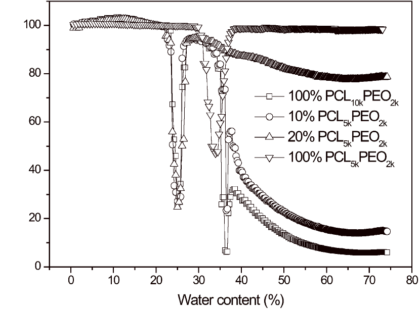
These results are in contrast to those obtained for mixtures of PCLPEO and PCLPEO in our previous publication Schuetz et al. (2011). There, vesicles were formed up to approximately sphere-former, whereas in the current system the transition from vesicles to micelles occurs between and PCLPEO. Increasing the length of the lamella-forming copolymer is therefore seen to favor the formation of more curved structures.
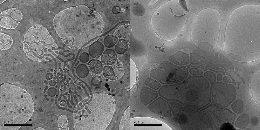
To gain more detailed insights into the system, we now consider cryo-TEM images taken at a range of sphere-former concentrations. In mixes with PCLPEO and PCLPEO, this technique reveals the presence of a large variety of structures, as can be seen in Figure 2. First, we note that a significant number of vesicles is still present, accounting for the high turbidity of this mixture seen in Figure 1. In addition, wormlike micelles and rings (end-to-end joined worms) can be seen in the left image. The wormlike micelles here often form branched network structures showing multiple three-way connections. The individual branches of the network also tend to be rather short, terminating in enlarged end-caps. These images closely resemble some of those shown by Jain and Bates Jain and Bates (2004) for PEO--PB block-copolymer mixtures as well as those of Chen et al. Chen et al. (1999) for PS--PAA. In different regions of the same TEM grid, very unusual vesicles could also be seen that on drying deformed to create a space-filling tessellated pattern (Figure 2, right image). The majority of these structures clearly show the dark outer ring of the vesicle wall confirming that they are indeed closed vesicles. However, on the top right there are some structures that do not have this pronounced darker rim and may therefore be unwrapped bilayer sheets. Such structures have been proposed as intermediate stages in the self-assembly of vesicles Lasic (1988); Antonietti and Forster (2003).
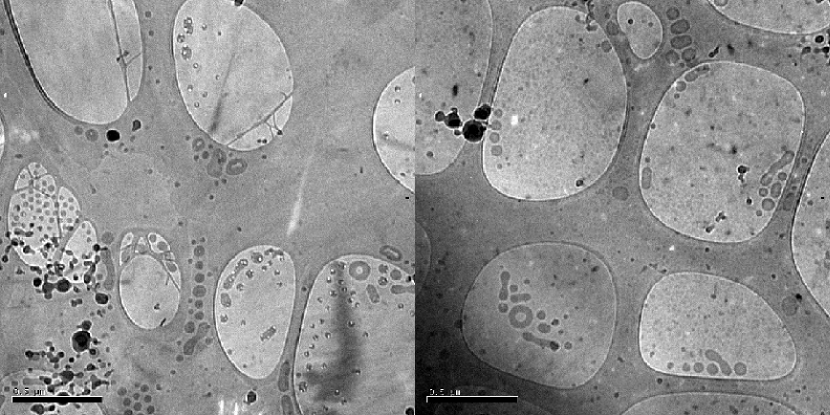
At a mixing ratio of PCLPEO and PCLPEO (Figure 3), the TEM shows a mix of spherical micelles, short worms and toroidal rings. The vesicles and sheet-like structures shown in Figure 2 no longer appear at this concentration. This is in line with the turbidity trace results: this solution is clear at high water concentrations, consistent with the presence of small micelles such as rings and short rods Adams et al. (2009).
We now present our self-consistent field theory calculations on our simple model of our experimental systems. For each set of calculations, we first consider the PCLPEO-PCLPEO mixture studied in our previous paper Schuetz et al. (2011), modeled, as described in the methods section, by a polymer blend including relatively short symmetric lamella-formers containing monomers and sphere-formers containing monomers. Next, we move on to our model of the PCLPEO-PCLPEO system of the current paper, in which the SCFT lamella-formers are still symmetric but now contain monomers, and look at how the self-assembly is altered by the change in polymer architecture.
To begin, we calculate the free-energy densities of ideal spheres, infinite cylinders and infinite bilayers using the method of variable subsystem size described above, and determine how these vary as the volume fraction of sphere-former is increased. To ensure that the system is relatively dilute and that aggregates are surrounded by a large volume of solvent, we fix the overall volume fraction of copolymer to . All free energy densities are plotted with respect to that of the homogeneous solution with the same composition ; that is, we plot the quantity . Since the free-energy densities of the three shapes of aggregate are quite close together, they are plotted normalized with respect to the magnitude of the free-energy density of the cylindrical micelle to show the shape transitions clearly. The cylinder free-energy density then appears as a horizontal line at , and is approached from above and below by the sphere and lamella free-energy densities as the sphere-former concentration increases.
Figure 4 shows the free-energy densities of spherical, cylindrical and planar aggregates plotted against , where is the volume fraction of sphere-former and is the total volume fraction of copolymer (sphere formers plus lamella-formers). Panel a shows the results for the system with the shorter lamella-former of monomers Schuetz et al. (2011). In this system, the lamella-former has the lowest free energy at lower sphere-former volume fractions. At around , the lamellar and cylindrical free energies cross, and the cylinder has the lowest free energy until , when the spherical micelle finally becomes most energetically favorable. This reproduces the series of transitions from vesicles to cylindrical micelles (worms and rings) to spherical micelles seen in our experiments Schuetz et al. (2011), although the values of at which the transitions occur are slightly shifted, as would be expected in view of the simplicity of our model. In these TEM images Schuetz et al. (2011), vesicles were seen at and sphere-forming copolymer and spherical micelles are seen at sphere-former, in line with our calculations. However, a mixture of worms, rings and vesicles is seen at , where our calculations predict spherical micelles. It is interesting to note that both our calculations and experiments demonstrate that a blend of sphere-forming and lamella-forming amphiphiles can form cylindrical micelles, even though this structure is favored by neither of these molecules individually Greenall and Gompper (2011).

In panel b of Figure 4, we plot the corresponding free-energy densities for the same system but with the degree of polymerization of the symmetric lamella former increased to monomers: a simple representation of the longer PCLPEO molecules of the current system. Again, we find the same sequence of morphologies as the sphere-former concentration is increased. However, as in the experiments, the vesicle-cylinder and cylinder-sphere transitions are shifted to lower sphere-former volume fractions with respect to the previous system Schuetz et al. (2011). Specifically, the former transition now occurs at approximately , whilst the latter is moved to around . Again, due to the simplicity of our theoretical approach, these transitions are not perfectly aligned with those in the experimental system. For example, the TEM images taken at a mixing ratio of (Figure 3) show a mixture of rings, short rods and spherical micelles, whereas our theory predicts that this mixture will form vesicles, or, equivalently, that the region of coexistence of morphologies would be expected at a mixing ratio closer to .
We believe that the shift in the shape transitions to lower sphere- former volume fractions for increasing chain length of the lamella-former is primarily due to the greater shape asymmetry of the lamella former when its length is increased whilst keeping the ratio of the hydrophilic and hydrophobic blocks constant. This can be seen from the following heuristic model of the shape asymmetry of diblock copolymers. We assume that the hydrophilic block A of the lamella-former is swollen by solvent and so has an end-to-end distance Jones (2002) of and effective volume . In contrast, the hydrophobic B-blocks are taken to be in a collapsed, brush-like state Safran (1994) and so have effective volume . Since we keep the ratio of the hydrophilic and hydrophobic blocks constant, we can also write and , where is the total length of the lamella-former. Defining the shape asymmetry of the lamella-former to be , we have . Therefore, as we increase at constant hydrophilic to hydrophobic ratio, will also increase and the lamella-former will become more asymmetric. In consequence, we expect it to have an greater tendency to form curved structures, with the swollen A-blocks on the outside of the curved surface. This shift towards more curved aggregates as the overall copolymer length is increased has indeed been seen in experiments on symmetric diblock copolymers in solution Kaya et al. (2002).
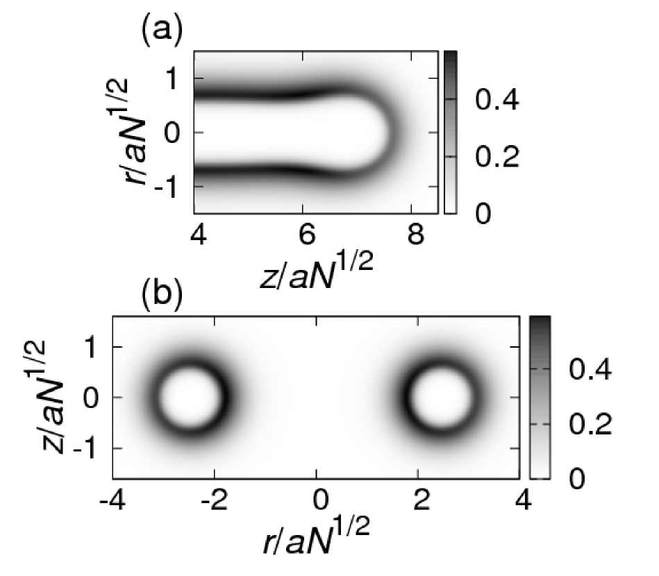
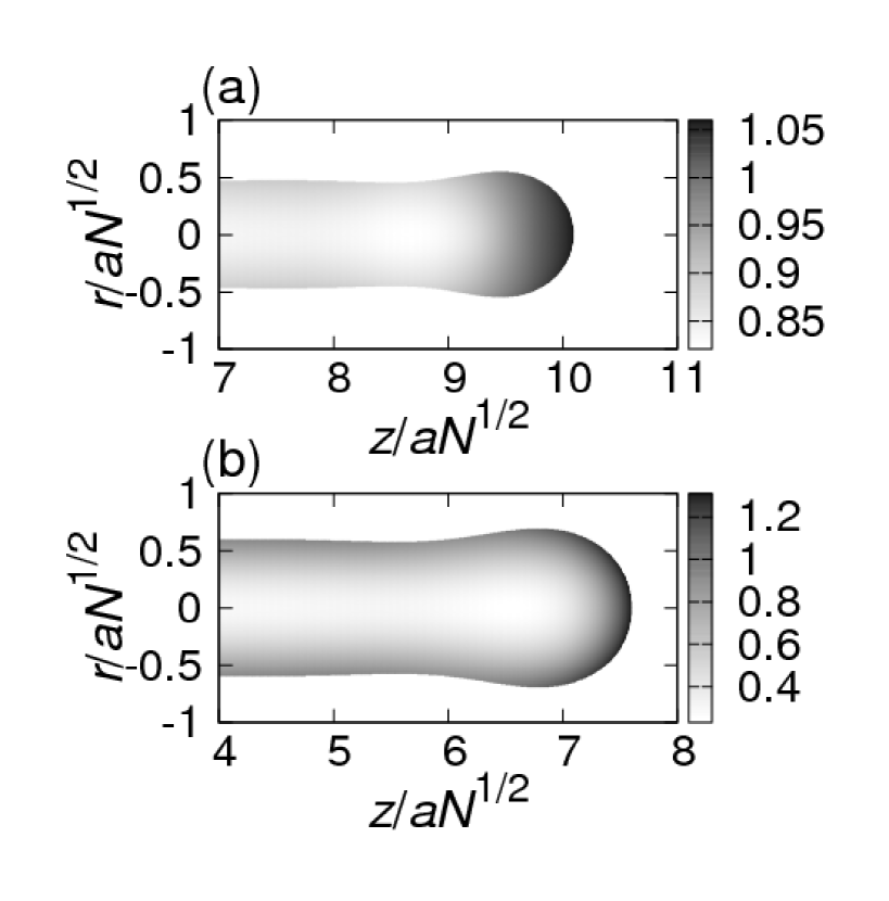
In the above calculations, we have studied only infinite cylinders. However, the cylindrical micelles seen in the experiments have the form of rings or short, bulbous-ended rods. We now use effectively 2d SCFT calculations in cylindrical polar coordinates Schuetz et al. (2011); Greenall and Gompper (2011) to study these structures in more detail. First, in Figure 5, we demonstrate that these structures can indeed be reproduced within our model’s parameter space. We focus on a system of lamella former with monomers mixed with sphere-former in a ratio of 2 to 1: a blending ratio where cylindrical micelles have the lowest free energy in our calculations (Figure 4). To show the form of the structures, the sum of the hydrophilic block densities of the two species is plotted. Panel a of Figure 5 shows the bulbous end of a rod Bernheim-Groswasser et al. (2000), preceded by a thinner neck region of negative curvature Jódar-Reyes and Leermakers (2006a, b). We find also that ring-like structures exist as local solutions to SCFT, and plot a cross-section through one of these aggregates in panel b.
We now turn our attention to the distribution of the two copolymer species within the rod structures. To this end, we introduce an enhancement factor Schuetz et al. (2011); Greenall and Gompper (2011) , which we define as
| (1) |
Here, is the local volume fraction of lamella-former hydrophobic blocks and is the corresponding quantity for the sphere-former hydrophobic blocks. The mean volume fractions of the two species are denoted by and . The enhancement factor tells us how much the concentration of sphere-former is enhanced with respect to that of the lamella-former at a given point in the system. We define such that it is normalized with respect to the mean volume fractions of the two core species, so that values greater than one represent enhancement of the sphere-former concentration and values less than one represent depletion. The enhancement factor is plotted only within the core of the micelle, defined as the region where the total density of the hydrophobic species is greater than that of the hydrophilic species . To locate this region accurately, we apply simple bilinear interpolation to our SCFT data to make the grid finer before plotting . In Figure 6 (a), we show the enhancement factor for the blend of sphere-former and shorter lamella-former ( monomers) Schuetz et al. (2011). In this system, the sphere-formers segregate to the end of the rod Schuetz et al. (2011), which is the most strongly curved part of the aggregate and, in fact, closely resembles a spherical micelle. Since the hydrophobic blocks of the two species are matched and so can both reach the center of the micelle, the ratio of the concentrations of the two species varies rather little in the main cylindrical body of the aggregate away from the endcaps. This is in contrast to the system shown in panel b of Figure 6, where a longer lamella-former of monomers is used. Here, the sphere-formers have shorter hydrophobic blocks than the lamella-formers, and no longer reach the central axis of the cylindrical micelle. This means that the sphere-former concentration is enhanced on the surface of the body of the cylinder as well as in the endcap, and is strongly depleted in the center of the rod. We note also that this mismatch between the hydrophobic blocks means that segregation is a stronger effect than in the previous system, with the range of values taken by significantly increased.
To study the distribution of the sphere- and lamella-formers within the micelles in more detail, we now plot cuts through the density profiles of the various species both along and perpendicular to the axis of the cylinder for the two systems shown in Figure 6. The plot for the system with shorter lamella-formers (Figure 7) confirms two points made above. First, it can be seen from the cut along the rod axis shown in the main panel of Figure 7 that the segregation of the sphere-formers to the endcap is relatively weak and that the peak in sphere-former concentration here is quite small. Furthermore, from the inset to Figure 7, which shows a cut perpendicular to the rod axis at the center of the micelle, we see that there is no segregation of the two polymer species in the main body of the aggregate. In corresponding plots for the system with longer lamella-formers (Figure 8), we see strong segregation of the sphere-formers to both the endcaps and surface of the cylinder. In addition, plotting the data in this way also shows us that, despite the new effect of the enhancement of sphere-former concentration on the surface of the cylinder, the strongest segregation is still to the most highly-curved region: the endcaps. To see this, note that the peak in sphere-former concentration at the end of the rod (main panel of Figure 8) is higher and more pronounced than that at the surface of the cylindrical section of the rod (inset to Figure 8). We note also that the sphere-formers have a dip in concentration just before the peak at the cylinder endcap. This feature corresponds to the negatively-curved neck of the micelle Jódar-Reyes and Leermakers (2006b) visible in Figure 6. Since the sphere-formers naturally prefer positive curvature, they migrate away from this region.


We believe that the stronger segregation of the sphere-formers in the mixture with the longer lamella-former is due to the entropic elasticity of the core blocks. Specifically, if the sphere-formers were homogeneously mixed with the longer lamella-formers, the hydrophobic cores would have to be strongly stretched, restricting the number of configurations they can access and so leading to a loss of entropy. In consequence, the sphere-formers move to the endcaps, leading to the formation of short cylinders or network structures with many bulbous endcaps such as those seen in Figure 2.
The larger number of Y-junctions Dan and Safran (2006) in the PCLPEO system compared to the previous PCLPEO system Schuetz et al. (2011) may be explained in a similar way Jain and Bates (2003): in the system containing the shorter lamella-former PCLPEO, the relatively short core blocks can adopt a smaller number of conformations and so are less able to pack into a more complex structure such as a Y-junction. The increased number of branched structures at higher molecular weights is in agreement with the work of several other groups Jain and Bates (2003); Won et al. (2002); Chen et al. (1999); Dan et al. (2006) on single-component systems.
III.2 Blends of two lamella-formers
Having studied the effect of blending two copolymers that individually form different structures, we now investigate a system where the two species are lamella-formers but of different lengths. The two copolymers considered are the shorter PCLPEO Schuetz et al. (2011) and the longer PCLPEO molecule introduced above. As in our previous publication Schuetz et al. (2011), we mixed the two polymers both before and after their individual self-assembly.
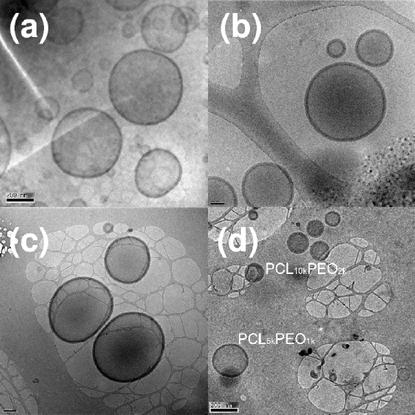

Figure 9 shows representative cryo-TEM images of the vesicles formed by PCLPEO (panel a) and PCLPEO (panel b) on their own. In the second row of the figure the vesicles are shown that result when the two block-copolymers are mixed before assembly (in pure THF solution) (panel c) and after assembly i.e. after dilution to THF (panel d). This latter case corresponds to mixing the solutions shown in panels a and b of Figure 9. As the chain lengths of the two copolymers differ by a factor of two, the resulting vesicles have different wall thickness ( and respectively). In the image d of the sample mixed post-assembly, the wall thicknesses of the individual vesicles remain unchanged and two different populations can be clearly distinguished, while in the sample mixed pre-assembly an intermediate wall thickness of ca. is found. This result indicates that, as before Schuetz et al. (2011), the exchange of material between the different structures is suppressed at THF concentrations below .
We now wish to see whether the result of the blending of two types of lamella-former leading to the formation of vesicles of intermediate wall thickness can be reproduced in our simple SCFT model. In panel a of Figure 10, we plot the density profiles of the hydrophobic B-blocks, hydrophilic A blocks and solvent of a bilayer formed of relatively short lamella-formers of monomers calculated, as above, from the method of variable subsystem size. We also show the total density profile of the copolymers (A blocks plus B blocks) and the bilayer thickness, defined as the distance from the origin at which the densities of copolymer and solvent are equal). Panel c shows the corresponding profiles for the longer lamella former with monomers. As expected from the respective lengths of the molecules, this latter polymer forms thicker bilayers than those shown in panel a. In the central panel b, we show the density profiles for a bilayer formed of an equal-parts mixture of the two species. In excellent agreement with the experiments, this mixed bilayer has a thickness approximately halfway between those of the pure bilayers. By comparing the density profiles in panels a and b we see that the addition of the longer species (in panel b) causes the core blocks of the shorter species to stretch outwards from their equilibrium state in a single-component structure (panel a).
III.3 Strongly hydrophobic copolymer mixed with micelle former
Finally, we investigate an extreme case of two strongly mismatched copolymers. The first of these, PCLPEO, is so hydrophobic that, in isolation, it fails to assemble into vesicles and precipitates instead. This polymer was mixed with the micelle-former considered above, PCLPEO. The two species were mixed before their individual self-assembly in a mass ratio of PCLPEO to PCLPEO. When self-assembly was triggered by the addition of water, the solution became turbid, indicating the presence of large aggregates.
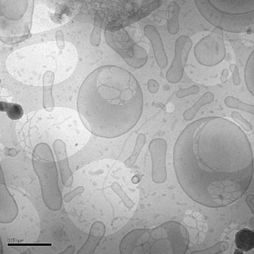
This was confirmed by cryo-TEM (Figure 11), which revealed the presence of a large fraction of vesicles with smaller populations of wormlike and spherical micelles. Remarkably, many of these vesicles are not spherical but have a novel elongated shape, which could be due to segregation of the two copolymer species within the individual aggregates. This is likely to be an especially strong effect in the current system, as the solubilities of the two copolymer species are very different and the PCLPEO chains start to aggregate at much lower water fractions than the more hydrophilic PCLPEO. This produces a phenomenon analogous to the intermediate mixing discussed in our earlier work Schuetz et al. (2011): the strongly hydrophobic chains are partially self-assembled at the time they encounter the sphere-formers. In consequence, the two species mix less efficiently, and regions of different curvature can coexist within the same aggregate Schuetz et al. (2011). The negative curvature regions around the centers of the elongated vesicles are likely to contain higher concentrations of the strongly hydrophobic copolymer, whereas the curved ends of these structures will probably contain more of the micelle-former. Due to the small difference in curvature between the different regions of the aggregate, testing this hypothesis is unfortunately beyond the scope of our current SCFT methods.
IV Conclusion
In this paper, we have used a combination of experiment and theory to show that varying the chain geometry of binary copolymer mixtures in solution can give us precise control over the form of the self-assembled aggregates and lead to the formation of new structures. We investigated three distinct situations. First, we studied a mixture of sphere-forming and lamella-forming copolymers, and found in both electron microscopy experiments and coarse-grained mean-field theory that increasing the chain length of the lamella-former whilst keeping the ratio of its hydrophilic and hydrophobic components constant leads to the formation of highly-curved structures at lower sphere-former volume fractions. We presented an explanation of this behavior in terms of the volume asymmetry of the two sections of the diblock. Using more detailed SCFT calculations, we found the rings and bulbous-ended rods seen in the experiments, and observed a strong segregation of the sphere-forming copolymers to the curved ends of the cylindrical micelles. We explained this effect by suggesting that sphere-forming copolymers would pay a large free energy penalty if they were dispersed evenly through the aggregates, as their relatively short core blocks would need to be strongly stretched to fit in with those of the lamella-former. In consequence, the sphere-formers tend to de-mix from the lamella-formers, leading to the formation of cylinders with highly-curved endcaps. This segregation between species may be accentuated by other effects such as enthalpy of crystallization. We also studied a mixture of two lamella-forming copolymers of different molecular weights. In both experiment and theory, we found that the bilayers formed in this system have a wall thickness that is in between those observed in systems containing only one of the two types of lamella-former. Finally, a mixture of sphere-forming copolymer and a strongly hydrophobic copolymer that precipitates in isolation was shown to form a range of structures, including novel elongated vesicles.
The results presented above demonstrate the power of a combined experimental and theoretical approach to the investigation and design of self-assembling block copolymers. Self-consistent field theory can map out the broad phase diagram of block copolymer mixtures and suggest experimental parameter spaces to search for new morphologies. Furthermore, it yields insights into the aggregates observed in the experiments, reproducing details of the structures and the distribution of the different polymer species within these. The wealth of morphologies observed in our work highlights the fine balance of forces governing the self-assembly behavior of block-copolymer systems. Further investigation of the different factors could open up a new zoo of self-assembled aggregates with the distribution and magnitude of local curvature differences as additional design parameters, and several avenues for extension of this work suggest themselves. In particular, the precise role of the sphere-former architecture could be investigated, with the aim of producing structures of a specified curvature. The various components could also be mixed at different stages in their self-assembly Schuetz et al. (2011), to access further new aggregates and to gain insight into the intermediate steps in micelle and vesicle formation. On the theoretical side, our SCFT calculations could be extended to investigate the favorability of the more complex structures, particularly the Y-junctions, in different copolymer mixtures.
V Acknowledgements
This work was performed in Project 264 of the Micro and Nanotechnology Scheme part-funded by the UK Technology Strategy Board (formerly DTI). Unilever is thanked for permission to publish this work. MJG is currently funded by the EU under a FP7 Marie Curie fellowship.
References
- Jain and Bates (2003) S. Jain and F. S. Bates, Science 300, 460 (2003).
- Jain and Bates (2004) S. Jain and F. S. Bates, Macromolecules 37, 1511 (2004).
- Smart et al. (2008) T. P. Smart, H. Lomas, M. Massignani, M. V. Flores-Merino, L. R. Perez, and G. Battaglia, Nano Today 3, 38 (2008).
- Edwards (1965) S. F. Edwards, Proc. Phys. Soc. 85, 613 (1965).
- Schmid (1998) F. Schmid, J. Phys.: Condens. Matter 10, 8105 (1998).
- Matsen (2006) M. W. Matsen, Soft Matter (Wiley-VCH, Weinheim, 2006), chap. 2.
- Maniadis et al. (2007) P. Maniadis, T. Lookman, E. M. Kober, and K. O. Rasmussen, Physical Review Letters 99, 048302 (2007).
- Drolet and Fredrickson (1999) F. Drolet and G. H. Fredrickson, Phys. Rev. Lett. 83, 4317 (1999).
- Kim et al. (2005) Y. Kim, P. Dalhaimer, D. A. Christian, and D. E. Discher, Nanotechnology 16, S484 (2005).
- Discher et al. (1999) B. M. Discher, Y. Y. Won, D. S. Ege, J. C. M. Lee, F. S. Bates, D. E. Discher, and D. A. Hammer, Science 284, 1143 (1999).
- Discher and Eisenberg (2002) D. E. Discher and A. Eisenberg, Science 297, 967 (2002).
- Kinning et al. (1988) D. J. Kinning, K. I. Winey, and E. L. Thomas, Macromolecules 21, 3502 (1988).
- Kaya et al. (2002) H. Kaya, L. Willner, J. Allgaier, and D. Richter, Applied Physics A-Materials Science and Processing 74, S499 (2002).
- Adams et al. (2009) D. J. Adams, C. Kitchen, S. Adams, S. Furzeland, D. Atkins, P. Schuetz, C. M. Fernyhough, N. Tzokova, A. J. Ryan, and M. F. Butler, Soft Matter 5, 3086 (2009).
- Sorre et al. (2009) B. Sorre, A. Callan-Jones, J. B. Manneville, P. Nassoy, J. F. Joanny, J. Prost, B. Goud, and P. Bassereau, Proceedings of the National Academy of Sciences of the United States of America 106, 5622 (2009).
- Zidovska et al. (2009) A. Zidovska, K. K. Ewert, J. Quispe, B. Carragher, C. S. Potter, and C. R. Safinya, Langmuir 25, 2979 (2009).
- Vinson et al. (1989) P. K. Vinson, Y. Talmon, and A. Walter, Biophys. J. 56, 669 (1989).
- Oberdisse et al. (1998) J. Oberdisse, O. Regev, and G. Porte, J. Phys. Chem. B 102, 1102 (1998).
- Schuetz et al. (2011) P. Schuetz, M. J. Greenall, J. Bent, S. Furzeland, D. Atkins, M. F. Butler, T. C. B. McLeish, and D. M. A. Buzza, Soft Matter 7, 749 (2011).
- Li et al. (2009) F. Li, A. T. M. Marcelis, E. J. R. Sudholter, M. A. C. Stuart, and F. A. M. Leermakers, Soft Matter 5, 4173 (2009).
- Greenall and Gompper (2011) M. J. Greenall and G. Gompper, Langmuir 27, 3416 (2011).
- Fattal et al. (1995) D. R. Fattal, D. Andelman, and A. Ben-Shaul, Langmuir 11, 1154 (1995).
- Andelman et al. (1994) D. Andelman, M. M. Kozlov, and W. Helfrich, Europhysics Letters 25, 231 (1994).
- Müller (2006) M. Müller, Soft Matter (Wiley-VCH, Weinheim, 2006), chap. 3.
- Matsen (2004) M. W. Matsen, J. Chem. Phys. 121, 1938 (2004).
- Press et al. (1992) W. H. Press, B. P. Flannery, S. A. Teukolsky, and W. T. Vetterling, Numerical Recipes in C (Cambridge University Press, Cambridge, 1992), 2nd ed.
- Greenall et al. (2009a) M. J. Greenall, D. M. A. Buzza, and T. C. B. McLeish, Macromolecules 42, 5873 (2009a).
- Greenall et al. (2009b) M. J. Greenall, D. M. A. Buzza, and T. C. B. McLeish, J. Chem. Phys. 131, 034904 (2009b).
- Cristobal et al. (2008) G. Cristobal, J. F. Berret, C. Chevallier, R. Talingting-Pabalan, M. Joanicot, and I. Grillo, Macromolecules 41, 1872 (2008).
- Schuhmacher et al. (2001) E. Schuhmacher, V. Soldi, and A. T. N. Pires, Journal of Membrane Science 184, 187 (2001).
- Chen et al. (1999) L. Chen, H. W. Shen, and A. Eisenberg, Journal of Physical Chemistry B 103, 9488 (1999).
- Lasic (1988) D. D. Lasic, Biochemical Journal 256, 1 (1988).
- Antonietti and Forster (2003) M. Antonietti and S. Forster, Advanced Materials 15, 1323 (2003).
- Jones (2002) R. A. L. Jones, Soft Condensed Matter (Oxford University Press, Oxford, 2002).
- Safran (1994) S. A. Safran, Statistical Thermodynamics of Surfaces, Interfaces, and Membranes (Westview Press, Boulder, 1994).
- Bernheim-Groswasser et al. (2000) A. Bernheim-Groswasser, R. Zana, and Y. Talmon, J. Phys. Chem. B 104, 4005 (2000).
- Jódar-Reyes and Leermakers (2006a) A. B. Jódar-Reyes and F. A. M. Leermakers, J. Phys. Chem. B 110, 6300 (2006a).
- Jódar-Reyes and Leermakers (2006b) A. B. Jódar-Reyes and F. A. M. Leermakers, J. Phys. Chem. B 110, 18415 (2006b).
- Dan and Safran (2006) N. Dan and S. A. Safran, Advances in Colloid and Interface Science 123, 323 (2006).
- Won et al. (2002) Y. Y. Won, A. K. Brannan, H. T. Davis, and F. S. Bates, Journal of Physical Chemistry B 106, 3354 (2002).
- Dan et al. (2006) N. Dan, K. Shimoni, V. Pata, and D. Danino, Langmuir 22, 9860 (2006).