Electron-conformational transformations in nanoscopic RyR channels govern both the heart’s contraction and beating
Abstract
We show that a simple biophysically based electron-conformational model of RyR channel is able to explain and describe on equal footing the oscillatory regime of the heart’s cell release unit both in sinoatrial node (pacemaker) cells under normal physiological conditions and in ventricular myocytes under Ca2+ SR overload.
Calcium (Ca2+) dynamics is of a principal importance for functioning of different heart’s cells from atrial and ventricular cardiomyocites to sinoatrial node cells (SANC) though the former are responsible for the heart’s contraction while the latter for primary heart’s pacemaking, respectively Bers . Cardiac contraction in cardiomyocites is activated by an increase in intracellular calcium concentration (Ca), most of which comes from a specific calcium cistern of sarcoplasmic reticulum (SR). Ca2+ is released via the ryanodine receptors (RyR) in response to Ca2+ entering the cell via the L-type channels (see Fig. 1). The cardiac type RyR is the common major Ca2+ release channel type in SANC and ventricular myocytes. It has been experimentally documented in chemically skinned and voltage-clamped SANC, in which effects of voltage-activated sarcolemmal ion currents are excluded, that the isolated SR is capable to spontaneously and rhythmically release Ca2+ via RyRs Fabiato ; Vinogradova-04 . These spontaneous, rhythmic, local subsarcolemmal Ca2+ releases (Ca2+ clock), which occur in SANCs, interact somehow with the classic sarcolemmal voltage oscillator (membrane clock membrane-clock ). At present there is a general consensus about the importance of Ca2+ oscillator for SANC rate Lipsius , however, an important discussion still remains whether it is a dominant or critical factor for cardiac pacemaker cell functioning. Furthermore, the very existence of the intracellular Ca2+ clock is not captured by the most part of existing essentially membrane-delimited cardiac pacemaker cell numerical models.
Recently, Maltsev and Lakatta Maltsev-09 have developed a new numerical SANC model (ML-model) featuring interactions of SR-based Ca2+ and membrane clocks to explore novel mechanistic insights into cardiac impulse initiation. They started with a well-known simplified model of the cell structure consisting of four compartments: sub-sarcolemmal space (subspace), cytosol, network SR (nSR), and junctional, or luminal SR (jSR) (Fig. 1). As in most existing models the authors used an effective medium theory, where Ca2+ concentrations in the subspace and in jSR (CaSS and CajSR) are main governing parameters that obey standard reaction-diffusion equations, while RyR gating is usually considered in a simplified manner through a dependence of the release on the Ca2+ concentrations. The ML-model adopted the formulation of cardiac RyR function developed by Shannon et al. Shannon-04 ) and the Kurata et al. model Kurata-02 of primary rabbit SANC. Finally the model was formulated in terms of a system of 29 first-order differential equations. The isolated SR can indeed operate as a self-sustained Ca2+ oscillator, described by a simple ”release-pumping-delay” mechanism: a small spontaneous Ca2+ release from jSR to the subspace occurs as the primary or initiating event. When CaSS increases to a sufficient level, it amplifies the Ca2+ release via the mechanism of the Ca2+-induced Ca2+ release (CICR) Bers ; this relatively strong, secondary Ca2+ release simultaneously depletes (i.e., resets) jSR. The released Ca2+ is pumped into the nSR. The delay between releases is determined by the Ca2+ pumping rate and Ca2+ diffusion from the subspace to cytosol and also from nSR to jSR. As CajSR slowly increases, RyRs are restituted, and the next release is ultimately initiated, etc.
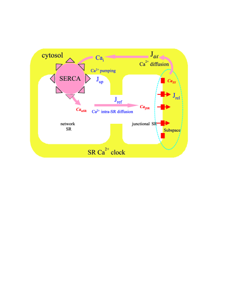
The ML-model Maltsev-09 of coupled oscillators seems to reproduce basically all recently discovered new behavioral details of cardiac pacemaker cell function, however, this phenomenological integrative model ignores many important physiological features of the cardiac cells, in particular, the fine spatiotemporal structure of the Ca2+ release. The model of integrated Ca2+ dynamics does not describe stochastic, locally propagating Ca2+ releases within the subsarcolemmal space. Indeed, RyRs in SANC, as in other cardiomyocites, seem to be arranged in clusters under sarcolemma (Fig. 1) and thus probably form subsarcolemmal calcium release units (CRUs). In this instance, RyRs release Ca2+ into a relatively small volume of subspace where individual jSRs of CRUs approach sarcolemma Bers . Thus a realistic modeling of the Ca2+ oscillator and SANC function should include a stochastic mechanism of a local Ca2+ release generation by CRUs. Furthermore, main assumption of the ML-model Maltsev-09 , that is the Ca2+ released from RyR channels activates the RyR channels as like as the trans-sarcolemmal Ca2+ from the L-type channel in a close apposition, seems to be questionable.
Recently we have applied well-known electron-conformational (EC) model (see, e.g.,Ref.Rubin ) for a single RyR channel ECM1 ; ECM2 ; ECM3 that was shown to capture important features of the individual and cooperative behaviour of RyRs in ventricular myocytes. The EC model of RyR functioning under Ca2+ stimuli is based on a biophysical adaptation of the well-known theory of photo-induced structural phase transitions, which has been successfully applied to different solids Koshino . Hereafter, in the Letter we will show that EC model of RyR channel is able to explain and describe on equal footing a puzzling spontaneous oscillatory regime of the release unit both in SANC under normal physiological conditions and in ventricular myocytes under Ca2+ SR overload.
The ion-activated RyR channel is a giant ( nm) macromolecular protein complex comprising 4 subunits of 565 000 Daltons each Bers . As other ion channels it has a great many of internal electron and conformational degrees of freedom and exhibits remarkable complexities that need to be considered when developing realistic models of ion permeation. Nevertheless, until recently most modelling efforts for RyR channels were focused on a simple ”hole in the wall” type model with a set of different (open, closed) states. Our knowledge of molecular mechanisms of RyR channel functioning is limited; hence we are forced to start with the most general ”physicists’” approach, which is typical for protein biophysics. Such an approach to the modelling of biomolecular system implies its simplifying to bare essentials with guidance from experimental data.
Modelling the RyR we start with a simple and a little bit naive picture of the massive nanoscopic channel like an elastic rubber tube with a varying cross-section governed by a conformational coordinate and a light ”electronic” plug switched due to Ca2+-RyR binding/unbinding in the subspace remark . This electronic plug interacts with the conformational coordinate and acts as a trigger to stimulate its change and related channel cross-section/conductivity.
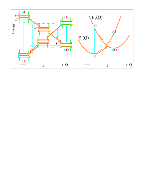
In other words, we reduce a large variety of RyR degrees of freedom to only two: a fast and a slow one, conventionally termed as electronic and conformational one, respectively. Both degrees of freedom are implied to be coupled to realize an EC transformation that is the electronic control of the slow conformational motion. Bearing in mind the main function of RyR channels, we assume only two actual electronic RyR states: ”open” and ”closed”, and a single conformational degree of freedom, , described by a classical continuous variable. Figure 2 illustrates the model with a set of representative states of the system.
Change in the electronic and conformational states regulate the main RyR channel function, i.e. determines whether the channel is ”open” and permeable for Ca2+ ions or ”closed” and impermeable to ions. Hereafter we assume that the conformational variable Q specifies the RyR channel ”cross-section” or, more precisely, a permeability for Ca2+, while the dichotomic electronic variable determines its opening and closure. This allows us to describe the Ca2+ flux through the RyR as follows:
| (1) |
if the channel is electronically open, and = 0, if it is closed. Here, the permeability coefficient reflects the ease with which Ca2+ passes through an open RyR. Its functional dependence on the conformational coordinate should be one of the essential model assumptions. is assumed to be an increasing function of the conformational coordinate, varying from zero or small leakage value to some saturated value , when varies from large negative to large positive magnitudes, passing through some subconductive state at . As a simplest limiting case, we may consider the step-like dependence .
Hereafter, we shall assume a simple harmonic approximation for the conformational energy and use a Hookes harmonic law for elastic potential energy: , where is the effective ”elastic” constant and relates to a base state with ”unstrained tube” and a bare cross-section. It is worth noting that namely the EC model introduces the energy to be an important factor of the RyR functioning.
As a starting point of the EC model algebra we introduce a simple effective Hamiltonian for a single RyR channel as follows ECM1 ; ECM2 ; ECM3
| (2) |
where and are well-known Pauli matrices, and the first term describes the bare energy splitting of ”up” and ”down” (electronically ”open” and ”closed”) states with an energy gap , while the second term describes their mixing. It is worth noting that given we arrive at a classical approach with a dichotomic electronic variable. The third and fourth terms in (2) describe the linear and quadratic contributions to the conformational energy. Here, the linear term formally corresponds to the energy of an external conformational stress, described by an effective stress parameter . The last term describes the EC interaction with the coupling parameter . Hereafter we make use of the dimensionless conformational variable ; therefore all of the model parameters (, ) are assigned energy units. Two eigenvalues of our Hamiltonian
| (3) |
define two branches of the adiabatic, or conformational potential (CP), attributed to electronically closed () and electronically open () states of the RyR, respectively (see Fig. 2).
The classical dynamics of the conformational coordinate we assume to obey a conventional Langevin equation of motion
| (4) |
where first term describes a total systematic conformational force with being an effective RyR mass (below let to be unity), is an effective dimensionless friction damping constant (relaxation rate), and is the thermal fluctuation force (Gaussian-Markovian noise). The last two terms reflect the coupling to an external environment of channels.
The thermo-activated transitions are caused by the thermal fluctuating force and free from the Franck-Condon principle. As a result of thermal transitions the whole system will finally relax to the thermal equilibrium. The temperature plays an important role in overcoming the energy barriers. In fact we deal with a hybrid ”over” (thermal activation) and ”through” (quantum tunneling) barrier transfer transitions (reactions). Quantum tunneling is often addressed to specify the low-temperature limit of the barrier transfer transition probability that points to the possible way to uncover non-classical behaviour for the RyR channel. We assumed the resonant quantum tunneling takes place between two branches of conformational potential starting within a tunneling zone centered at the CP minimum with the probability obeyed the effective Gamov law
| (5) |
where is the width, is the height of the energy barrier, or the energy separation between the tunneling points and the point of the two branch intersection (see Fig. 2), and effective tunneling attempt frequency.
At present there is no sufficient understanding of the mechanisms that regulate local Ca2+ signaling in heart’s cell despite persistent efforts to discriminate between the cytosolic and luminal Ca2+ activation hypotheses. Calcium enters the subspace by two pathways: across the sarcolemma via L-type channels and from the SR via RyR channels. Activation of RyRs by CajSR has been attributed to either Ca2+ feedthrough to high-affinity cytoplasmic Ca2+ activation sites or to Ca2+ regulatory sites on the luminal side of the RyR. However, most of experimental observations on cardiac RyRs more difficult to reconcile with the Ca2+ feedthrough effect Gyorke . A luminal Ca2+ sensor appears to continuously regulate the functional activity of the SR Ca2+ stores by linking SR Ca2+ content to the activity of the RyRs Gyorke . However, in contrast with purely electronic effect of CaSS, the effect of relatively slowly varying CajSR on the RyR channels is likely to be purely ”mechanical” one, through the respective conformational strain applied to RyR channels. The effect can be naturally incorporated into EC model, if we assume the strain parameter in the RyR Hamiltonian to be a function of CajSR or the jSR-to-subspace Ca2+ gradient. We assume to rise with the luminal Ca2+ concentration in accordance with the Hill curve ECM2 :
| (6) |
where is the half maximal value, is a Hill coefficient. Rise of in the interval (-1,+1) leads to a crucial modification of CP from that of stabilizing closed RyR state to that of stabilizing open RyR state. It should be noted that in terms of the EC model one might introduce a critical SR load that specifies a critical effective strain when the minimum of the CPc branch for the electronically closed RyR state crosses CPo branch thus destroying the RyR bistability conditions and making RyRs stay close to their activation threshold where their cCoO activation can presumably be easily provoked.
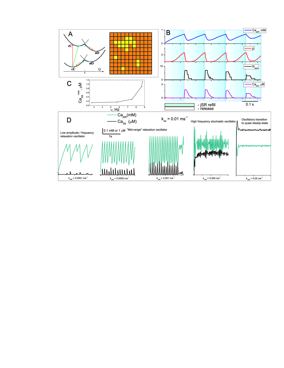
Cooperative dynamics of the RyR clusters in CRU has been studied in a series of model simulations for 1111 square RyR lattice in our prior paper ECM3 . We simulated both ”in vitro” dynamics of RyR lattice when the calcium surrounding was assumed to be simply as a source of effective external fields, such as effective strain, and ”in vivo” situation when the RyR lattice dynamics is incorporated into the whole cell calcium dynamics (EC-CRU model). In such a case the EC dynamics of RyR lattice is assumed to specify the number of open channels which in its turn specifies the release flux from the SR to subspace through RyR channels: , where is a single RyR channel release velocity coefficient. The system demonstrated four different modes of behaviour, depending on the SR load ECM3 : inactivation mode I at a rather small SR load (heavily underloaded SR); single channel activated mode II (Ca synapse, or quark mode) at underloaded SR; domino-like firing-termination mode III with high degree of cooperativity (cluster bomb mode) at optimally loaded SR. The cluster bomb mode is characterized by a step-by-step opening of the neighboring RyRs with formation of a cluster composed of up to 40% open RyRs. The process results in an effective high-gain Ca2+ release from the SR. However, the decrease in SR load leads to a lowering of the effective strain, accompanied by the shift of the system to preferable channel’s closure. This negative feedback effect results, first, in a slowing down of the nucleation process and, second, in an evolution of the inverse domino-like effect with full collapse of the cluster of open RyRs and termination of Ca2+ release. Puzzlingly, the CRU simulation ECM3 showed that SR overload can result in the excitation of the RyR lattice auto-oscillations when the domino-like opening of a cluster of RyR channels resulted in effective Ca2+ release that, in turn, caused a lowering of the effective strain and simultaneous closure of RyRs before the SR load started rising. However, until CajSR approached the initial value, close to a critical concentration all channels re-opened simultaneously, and Ca2+ release repeated spontaneously. This behaviour is repetitive, i.e. the system turned out to behave in an auto-oscillatory mode IV with a spontaneous SR Ca2+ release. It should be emphasized that in contrast with the ML model, the auto-oscillation mode IV occurs as a result of the CajSR-dependent shape of the CP and a purely conformational transformation without any L-type channel activity and Ca2+-induced electronic transitions. To the best of our knowledge it was a first quantitative model for the CRU oscillatory mode.
Obviously the abnormal Ca2+ automaticity in SR overloaded ventricular myocytes suggests the normal pacemaker SANC activity may have some similar features and actually proceed with elevated Ca2+ level. Indeed, Vinogradova et al. Vinogradova-04 found a minimum diastolic Ca2+ level in rabbit SANC of 200 nM, that is twice the diastolic Ca2+ level in resting ventricular myocytes.
Hereafter in the paper we address a more realistic EC-CRU model relevant for the SANCs which incorporates two main features of the Ca2+ machinery, that is a local and stochastic character of the Ca2+ release. As in our prior paper ECM3 we address the CRU model with 1111 square lattice of RyRs, however, for simplicity we neglect any RyR-RyR coupling. At variance with ECM3 each RyR channel was described by a diabatic CP with non-FC tunneling transition between two CP branches. The calcium fluxes in a simplified cell model (Fig. 1) were assumed to obey a standard system of four differential equations Sobie :
| (7) |
| (8) |
| (9) |
| (10) |
where , , are diffusion fluxes between the nSR and the jSR, between the jSR and subspace, between subspace and cytosol, respectively, is uptake flux; are volume ratio constants between and cell compartments (). It should be noted that all the parameters were chosen rather as typical for integrative cell models than for a single CRU model. In other words, all the cell RyRs we consider to form a system of identical CRU’s functioning in concert. We set initial conditions as follows: Ca, Ca, Ca, Ca, for other Ca2+ dynamics parameters:. As in the ML model the CaSS and CajSR time courses strongly depend on the refill and release rate constants, that are expected both to change from their original values of ventricular myocytes and likely mediate the regulation of SR Ca2+ clock ticking speed Maltsev-09 . Main results of numerical simulations are presented in Fig. 3.
As in ML model, three types of the steady-state were found: 1) steady rhythmic oscillations; 2) no oscillations or damped oscillations; and 3) chaotic oscillations. The highest rates are reached when the oscillator approaches to dynamic equilibrium (steady release, Fig. 3d, right part). The lowest rates are reached when the oscillator approaches to static equilibrium. Static equilibrium occurs when either the release rate is too small or Ca2+ pumping rate is too fast, i.e., jSR becomes highly loaded. Fig. 3 distinctly shows that CRU can operate as a self-sustained Ca2+ oscillator, however, the triggering of the Ca2+ release is determined by the bringing CajSR near the critical concentration, or activation threshold rather than by a small initial spontaneous Ca2+ release from jSR as in the ML-model Maltsev-09 . Maximal CajSR appears to slightly rise with , while its oscillation amplitude strongly depends on , however, in an absolutely different way than in ML-model. Indeed, for a rather wide range of the CajSR oscillation amplitude rises with the refill rate, however, without full depletion of the jSR as it occurs in ML-model. Furthermore, in contrast with the ML-model the EC-CRU model reveals the Bowditch-like behavior: ”the faster rate, the stronger release”. Indeed, within oscillatory regime both the frequency and amplitude of CaSS do increase with . A detailed analysis shows this can be explained as a result of some effect due to a slow conformational dynamics near the CP minima; optimal condition for the inter-branch tunneling and Ca2+ release are realized at higher and consequently, at higher CajSR. Indeed, a high-speed increase of the CajSR results in a rapid rise of effective stress with a shift of the CP branches with its minima up and right (see Fig. 3). Given rather slow conformational dynamics the CP shift leads to a tunneling retardation effect as the conformational coordinate needs a time to reach the new tunneling zone in the vicinity of new CP minimum. In other words, the EC system is not in time for adjusting to the rapid shift of the CP branches. However, the higher CajSR the slower the CP shift, that allows conformational coordinate to run tunneling zone down, and provide the optimal condition for the inter-branch tunneling and Ca2+ release. Despite the tunneling retardation, the bigger the shorter time to reach the critical CajSR level.
Just the opposite behavior, ”the higher the rate, the lower the amplitude,” represents the major limitation of the isolated Ca2+ oscillator in frames of ML-model: it is unable to generate high-amplitude oscillations at higher rates. However, the authors Maltsev-09 have shown that the interactions of the SR and sarcolemma clocks overcomes the limitation of the isolated SR Ca2+ clock, i.e., the oscillation amplitude of the full system Ca2+ clock raises as the oscillation rate increases, i.e., the Bowditch phenomenon restores. At the same time the ”Bowditch-like” behavior (the faster the stronger) for the both CaSS and CajSR appears to be a distinct feature of the EC-CRU model. In other words, it seems the SR Ca2+ oscillator can serve as a dominant source of persistent heart’s beating. Thus the interrelation between two types of cell oscillators needs in a futher examination.
In summary, at variance with the integrative model by Maltsev and Lakatta simple biophysically based EC model of RyR channel describes stochastic local Ca2+ releases within the subsarcolemmal space as a result of conformational transformations followed by a tunneling between two CP branches. The model is able to explain and describe the spontaneous oscillatory regime of the CRU both in pacemaker cells under normal physiological conditions and in ventricular myocytes under Ca2+ SR overload including its subtle features such as the amplitude and frequency fluctuations. At variance with the integrative ML-model, the oscillation amplitude of the intracellular Ca2+ clock raises as the oscillation rate increases, thus providing the Bowditch law functioning of the pacemaker cell without any membrane clock assistance. Despite the EC model is intentionally simplistic, it offers novel insight into potential mechanisms governing by the Ca2+ fluxes and may thus provide a starting point for further exploration of physical principles guiding cardiac cell functioning in vitro and in vivo.
We acknowledge the support by Ural Branch of RAS under grant No.09-M-14-2001.
References
- (1) D.M. Bers, Excitation-Contraction Coupling and Cardiac Contractile Force. Second Edition, Kluwer Academic Publishers, New York, 2002, 420 pp.
- (2) A. Fabiato, Basic Res. Cardiol., 80, suppl.2, 83 (1985).
- (3) T.M. Vinogradova, Y.Y. Zhou, V. Maltsev, A. Lyashkov, M. Stern, E.G. Lakatta, Circ Res 94, 802, (2004).
- (4) D. Noble., J. Physiol. 353, 1 (1984).
- (5) R.W. Tsien, R.S. Kass, R. Weingart, J. Exp. Biol. 81, 205, (1979); S.L. Lipsius, D.M. Bers, J. Mol. Cell Cardiol. 35, 891 (2003).
- (6) V.A. Maltsev and E.G. Lakatta, Am. J. Physiol. Heart Circ. Physiol. 296, 594, (2009)
- (7) T.R. Shannon, F. Wang, J. Puglisi, C. Weber, D.M. Bers, Biophys. J. 87, 3351 (2004).
- (8) Y. Kurata, I. Hisatome, S. Imanishi, T. Shibamotop, Am. J. Physiol. Heart. Circ. Physiol. 283 H2074 (2002).
- (9) A.B. Rubin, Biofizika. Teoreticheskaya biofizika, V.I, URSS, 2004, 464 p.(in Russian).
- (10) A.S. Moskvin, M.P. Philipiev, O.E. Solovyova, P. Kohl, V.S. Markhasin, Doklady Biochemistry and Biophysics, 400, 32 (2005).
- (11) A.S. Moskvin, M.P. Philipiev, O.E. Solovyova, V.S. Markhasin, Journal of Physics: Conference Series, 21, 195 (2005).
- (12) A.S. Moskvin, M.P. Philipiev, O.E. Solovyova, P. Kohl, V.S. Markhasin, Progress in Biophysics and Molecular Biology, 90, 88 (2006).
- (13) K. Koshino., T. Ogawa, Journal of Luminescence, 87-89, 642 (2000).
- (14) At variance with the ion-activated RyR channel the potential-activated channels can be described by a single conformational coordinate governed by the electric field.
- (15) I. Gyorke and S. Gyorke, Biophys. J. 75 (6), 2801 (1998).
- (16) E.A. Sobie, K.W. Dilly, J. dos Santos Cruz, W.J. Lederer, M.S. Jafri, Biophys. J. 83, 59 (2002).