Resonant Photoelectron Diffraction with circularly polarized light
Abstract
Resonant angle scanned x-ray photoelectron diffraction (RXPD) allows the determination of the atomic and magnetic structure of surfaces and interfaces. For the case of magnetized nickel the resonant excitation with circularly polarized light yields electrons with a dichroic signature from which the dipolar part may be retrieved. The corresponding and Auger electrons carry different angular momenta since their source waves rotate the dichroic dipole in the electron emission patterns by distinct angles.
pacs:
79.60.-i, 61.05.js, 75.25.-j, 79.60.BmThe quest for atomic scale structure determination at surfaces and interfaces lead to the development of a large number of powerful methods Van Hove (1999). Among those, x-ray photoelectron spectroscopy (XPS) with angular resolution (XPD) allows structure determination paired with chemical and magnetic sensitivity Fadley (2010). The signal is best, when the x-ray absorption coefficient is at maximum. These maxima occur in resonant excitation and have so far been exploited for the probing of defect states in TiO2 Krüger et al. (2008), for looking inside an endofullerene Treier et al. (2009), or for the investigation of the mixed valence structure of magnetite Magnan et al. (2010).
In this letter, angle scanned resonant photoelectron diffraction (RXPD) is applied to nickel, the ”fruit fly” of resonant photoemission Guillot et al. (1977); Feldkamp and Davis (1979); Tjeng et al. (1993); van der Laan (1994); Weinelt et al. (1997). Circularly polarized light is used for the precise measurement of the dipole induced by the x-ray magnetic circular dichroism which is largest at resonance. After this proof of principle we show that the angular momentum of the outgoing electrons can be measured with RXPD. This extends the results of Daimon et al., who demonstrated that forward scattering XPD patterns rotate due to the angular momentum of circularly polarized photons Daimon et al. (1993); Daimon (2001); Matsui et al. (2010). Here, the electron source wave Greber et al. (1992a, b) rotates the magnetisation direction in the final state of the emitted electrons. It is also found that Auger electrons may carry angular momenta opposite to that of the exciting photon and larger than .
Circular magnetic dichroism is the difference between the absorption coefficient of right and left circularly polarized light. It is proportional to the scalar product of the magnetization and the angular momentum of the incoming photon Stöhr and Siegmann (2006).
| (1) |
For right circularly polarized light () is parallel to the propagation direction of the photon, and for left circularly polarized light ( ) is antiparallel. is a dipole, i.e. proportional to , where is the angle between and . From this follows that the absolute orientation of is determined from three or more non-coplanar light incidences.
Photoemission yields , under the assumption that the photoemission current is proportional to the x-ray absorption coefficient. This must not hold for partial measurements like in angular resolved photoemission where is measured Westphal et al. (1989). The different photoelectron source waves of electrons excited with differently polarized light lead to different photoemission final states that comprise information on the magnetism and the surrounding of the emitter Chassé et al. (2005). Here we show that contributions of the atomic and the magnetic structure can be disentangled with a spin integrated experiment.
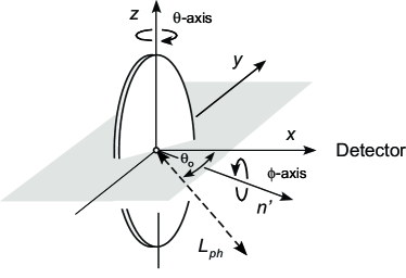
The experiments have been performed at the SIM beamline at the Swiss Light Source (SLS) Flechsig et al. (2010) in an endstation dedicated for x-ray photoelectron spectroscopy (XPS) and angle-resolved x-ray photoelectron diffraction (XPD) with a base pressure below mbar. The x-rays impinge perpendicular to the polar rotation axis with an angle between the x-rays and the electron energy analyzer of 55∘ (see Figure 1). The degree of polarization is better than 98%. All measurements are done at room temperature. The Ni(111) yoke crystal Okuda et al. (2009) was cleaned by repeated cycles of argon sputtering and annealing. It is magnetized by passing a current of 2 A for 30 s through the yoke coil. The resulting magnetization was inferred from an x-ray magnetic circular dichroism (XMCD) spectrum at the Ni L2,L3-edges in the total-electron-yield mode. Comparison with corresponding spectra of Chen et al. Chen et al. (1991) indicate a magnetization of 40%, which is not 100 % due to a multidomain structure.
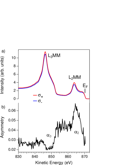
Resonant photoemission on 3d transition metals is most intense at the L3 absorption edge Chen et al. (1991); Krüger et al. (2008). Here we investigate the L2 resonance since it provides L2MM and L3MM emission which allows for direct comparison and consistency checks. Figure 2 shows resonant x-ray photoelectron spectra from magnetized Ni(111) of right and left circularly polarized light. The photon energy is set on the Ni L2-resonance (2p3d) at eV. The Fermi level at 870.5 eV electron energy, the (863.8 eV) 6 eV satellite (see Hüfner (2003) and references therein), and the (846.2 eV) Auger deexcitation peak are most prominent. The spectra have been normalized with the photon flux. Figure 2b) demonstrates circular dichroism in these resonantly excited electron emission spectra. The asymmetry ))/( between right and left circularly polarized light exhibits a maximum at and a minimum at . The asymmetry can be reversed by switching the magnetization or by the rotation of the sample by 180∘ Morscher et al. (2011). Off resonance, at eV the dichroic asymmetry in the Auger line is 1.4 0.8 % (data not shown). The extrema at 848.4 eV and at 863.6 eV do not exactly coincide with the and the intensity maxima, which indicates multiplet structure Magnuson et al. (1998). In the following we use the labels for electrons at the energies of and , excited with and polarized radiation, respectively.
If we perform on these resonances angle scanned x-ray photoelectron diffraction (RXPD), the experiment yields information on the atomic and the magnetic structure. Briefly, the sample frame is rotated in the lab frame . The photoelectron intensity is mapped in polar coordinates , where the polar angle and the azimuthal angle define the sample orientation with respect to the electron detection direction (see Figure 1) Osterwalder et al. (1991). This leads for to a dipolar function in the XPD map that depends on , the electron detection direction in the lab frame and in the sample frame, where the amplitude is a measure for the magnitude of the dichroism Morscher et al. (2011).
Figure 3a) shows RXPD data for . The XPD map is dominated by the information on the atomic structure which corresponds to that of a face centered cubic crystal which is cut along the (111) plane Wider et al. (2001). Below the obvious atomic structure dichroic information must be hidden. In order to visualize the dichroism, we form the asymmetry between the and the XPD scans (see Figure 3b). These data contain information on the dipolar (magnetic) nature of dichroism and higher order multipoles, which are related to differences in the diffraction patterns due to different source waves Daimon et al. (1993); Greber (2001). The dipolar part has the symmetry , as it is expected for in plane magnetisation. Figure 3c) shows the fit of a dipolar function , which determines . We find , and , where is set to the direction. This result is consistent with spin polarized photoemission Okuda et al. (2009), though the magnetization is not aligned along the second easy axis as it was the case for an other Ni(111) picture frame crystal Donath (1994). The rotation of the sample and the incidence of the light impose on two nodal lines (): A circle at , and a diameter perpendicular to . In Figure 3d) the residuum of the asymmetry and is shown. It has the C3 symmetry of the substrate and indicates further differences in the diffraction patterns due to different source waves created by and photons, respectively. Such effects have been pioneered by Daimon et al., where they showed that the angular momenta of the photons are transferred to the photoelectrons, which in turn lead to an emitter scatterer distance dependent rotation of the forward scattering peak Daimon et al. (1993); Daimon (2001).
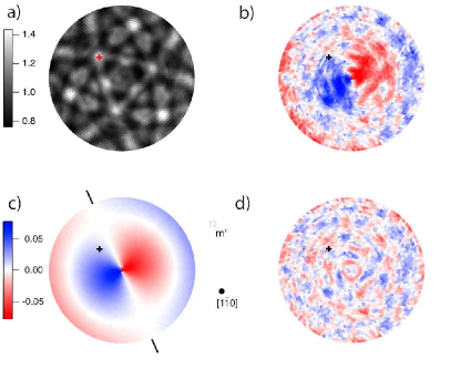
For the data in Figure 3 b) we expect no photon induced rotation because in the asymmetry between and any such effect should be canceled. This changes, when a dipolar function is fitted to an individual XPD scan with either or radiation. If we fit a dipolar function to the data in Figure 3a), or the ones recorded with polarization, we find magnetisation directions which are within consistent with the magnetization direction as found from Figure 3 b). Although this has the practical advantage that the magnetization is determined without switching the light polarization, it is not very accurate since the -function is much weaker than the forward scattering induced XPD patterns.
If we want to extract more quantitative information on the rotation of the -functions upon use of photons with plus or minus angular momentum we have to perform a normalisation that removes the forward scattering intensity modulations but preserves, in contrast to the asymmetry used in Figure 3 the angular momentum of or . We do so in using the -averaged data and form and vice versa.
As and electrons are expected to have very similar XPD patterns Greber et al. (1992b) - the wavelength-difference between the two selected electron energies of and is 1% - most XPD information on the atomic structure should be cancelled, though the are expected to show a polarization dependent rotation of the observed magnetisation direction. For the 6 eV satellite, i.e. MM transition, we find a rotation of around the value of Figure 3b). It is related to the Daimon effect, i.e. forward scattering peak rotation Daimon et al. (1993). Essentially, the angular momentum of an outgoing photoelectron induces a rotation of all features in the XPD patterns with respect to the crystal lattice. For single scattering the maximum angle of rotation is given by , where is the distance between emitter and scatterer, the momentum of the outgoing electron and the angle between the light incidence and the electron detection (see Figure 1) Daimon (2001); Chassé and Rennert (1997). For nickel, , an electron kinetic energy of 850 eV and =55∘, gets . Of course, the angular shift is not isotropic, it depends on the angle between the electron angular momentum and the nearest neighbour directions. However, for an material as it is nickel, the 12 nearest neighbours of an emitter in the bulk sit on a sphere with radius , on the vertices of a cuboctahedron and must lead to a fairly isotropic rotation of the XPD patterns around the axis of the incoming photons. The ’s have the same sense of rotation as the corresponding photon angular momentum and are compatible with the transfer of 2 of angular momentum to the emitted electrons.
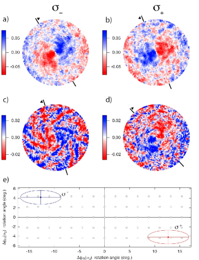
Figure 4 shows the and the XPD scans for and radiation. Dipolar functions () as shown in Figure 3c) appear, where the sign changes upon change of the polarization. The -functions for and indicate ’s of and , respectively. We want to note that the use of more than 3000 different photon incidence angles allows a very accurate determination of the ’s and permits for single quantum assignments 2 (-2). In the patterns with a lower asymmetry (see Figure 2) the error increases by a factor of 3 and makes it compatible with angular momenta of or (see Figure 4 e)). This surprising result implies that a Auger electron channel produces electrons with large, opposite angular momentum compared to that of the photons. These results emphasise that the information on the angular momentum of an electron source wave may not only be accessed by a forward scattering peak rotation Daimon et al. (1993), but for magnetic systems also by the precise measurement of the source wave dependent circular magnetic dichroism.
In summary it has been shown that resonant x-ray photoelectron diffraction (RXPD) is suited to extract the atomic and magnetic structure of surfaces and interfaces. Furthermore it is demonstrated that the method directly accesses the angular momenta of the emitted electrons.
Fruitful discussions with J. Osterwalder and the support of the Swiss National Science Foundation are gratefully acknowledged. The experiments have been performed at the Swiss Light Source.
References
- Van Hove (1999) M. A. Van Hove, Surface and Interface Analysis 28, 36 (1999).
- Fadley (2010) C. S. Fadley, Journal of Electron Spectroscopy and Related Phenomena 178, 2 (2010).
- Krüger et al. (2008) P. Krüger, S. Bourgeois, B. Domenichini, H. Magnan, D. Chandesris, P. Le Fèvre, A. Flank, J. Jupille, L. Floreano, A. Cossaro, et al., Physical Review Letters 100, 055501 (2008).
- Treier et al. (2009) M. Treier, P. Ruffieux, R. Fasel, F. Nolting, S. Yang, L. Dunsch, and T. Greber, Physical Review B 80, 081403 (2009).
- Magnan et al. (2010) H. Magnan, P. Le Fevre, D. Chandesris, P. Kruger, S. Bourgeois, B. Domenichini, A. Verdini, L. Floreano, and A. Morgante, Physical Review B 81, 085121 (2010).
- Guillot et al. (1977) C. Guillot, Y. Ballu, J. Paignè, J. Lecante, K. Jain, P. Thiry, R. Pinchaux, Y. Pètroff, and L. Falicov, Physical Review Letters 39, 1632 (1977).
- Feldkamp and Davis (1979) L. A. Feldkamp and L. C. Davis, Physical Review Letters 43, 151 (1979).
- Tjeng et al. (1993) L. Tjeng, C. Chen, P. Rudolf, and G. Meigs, Physical Review B 48, 13378 (1993).
- van der Laan (1994) G. van der Laan, International Journal of Modern Physics B 8, 641 (1994).
- Weinelt et al. (1997) M. Weinelt, A. Nilsson, M. Magnuson, T. Wiell, A. Wassdahl, O. Karis, A. Föhlisch, N. Mårtensson, J. Stöhr, and M. Samant, Physical Review Letters 78, 967 (1997).
- Daimon et al. (1993) H. Daimon, T. Nakatani, S. Imada, S. Suga, Y. Kagoshima, and T. Miyahara, Japanese Journal of Applied Physics Part 2 - Letters 32, L1480 (1993).
- Daimon (2001) H. Daimon, Physical Review Letters 86, 2034 (2001).
- Matsui et al. (2010) F. Matsui, T. Matsushita, and H. Daimon, Journal of Electron Spectroscopy and Related Phenomena 178, 221-240 (2010).
- Greber et al. (1992a) T. Greber, J. Osterwalder, S. Hüfner, and L. Schlapbach, Physical Review B 45, 4540 (1992a).
- Greber et al. (1992b) T. Greber, J. Osterwalder, D. Naumovic, A. Stuck, S. Hüfner, and L. Schlapbach, Physical Review Letters 69, 1947 (1992b).
- Stöhr and Siegmann (2006) J. Stöhr and H. Siegmann, Magnetism - From Fundamentals to Nanoscale Dynamics (Springer, 2006).
- Westphal et al. (1989) C. Westphal, J. Bansmann, M. Getzlaff, and G. Schönhense, Physical Review Letters 63, 151 (1989).
- Chassé et al. (2005) A. Chassé, W. Kuch, M. Kotsugi, X. Gao, F. Offi, S. Imada, S. Suga, H. Daimon, and J. Kirschner, Physical Review B 71, 014444 (2005).
- Flechsig et al. (2010) U. Flechsig, F. Nolting, A. Fraile Rodriguez, J. Krempasky, C. Quitmann, T. Schmidt, S. Spielmann, and D. Zimoch, AIP Conf. Proc. 1234, 319 (2010).
- Okuda et al. (2009) T. Okuda, J. Lobo-Checa, W. Auwärter, M. Morscher, M. Hoesch, V. N. Petrov, M. Hengsberger, A. Tamai, A. Dolocan, C. Cirelli, et al., Physical Review B 80, 180404 (2009).
- Chen et al. (1991) C. Chen, N. Smith, and F. Sette, Physical Review B 43, 6785 (1991).
- Hüfner (2003) S. Hüfner, Photoelectron Spectroscopy: Principles and Applications (Springer, Berlin, 2003).
- Morscher et al. (2011) M. Morscher, F. Nolting, T. Brugger, and T. Greber, Manuscript in Preparation (2011).
- Magnuson et al. (1998) M. Magnuson, N. Wassdahl, A. Nilsson, A. Föhlisch, J. Nordgren, and N. Mårtensson, Physical Review B 58, 3677 (1998).
- Osterwalder et al. (1991) J. Osterwalder, T. Greber, A. Stuck, and L. Schlapbach, Physical Review B 44, 13764 (1991).
- Wider et al. (2001) J. Wider, F. Baumberger, M. Sambi, R. Gotter, A. Verdini, F. Bruno, D. Cvetko, A. Morgante, T. Greber, and J. Osterwalder, Physical Review Letters 86, 2237 (2001).
- Greber (2001) T. Greber, Journal of Physics - Condensed Matter 13, 10561 (2001).
- Donath (1994) M. Donath, Surface Science Reports 20, 251 (1994).
- Chassé and Rennert (1997) A. Chassé and P. Rennert, Physical Review B 55, 4120 (1997).