Active split-ring metamaterial slabs for magnetic resonance imaging
Abstract
In this work, it is analyzed the ability of split-ring metamaterial slabs with zero/high permeability to reject/confine the radiofrequency magnetic field in magnetic resonance imaging systems. Using an homogenization procedure, split-ring slabs have been designed and fabricated to work in a 1.5T system. Active elements consisting of pairs of crossed diodes are inserted in the split-rings. With these elements, the permeability of the slabs can be automatically switched between a unity value when interacting with the strong excitation field of the transmitting body coil, and zero or high values when interacting with the weak field produced by protons in tissue. Experiments are shown for different configurations where these slabs can help to locally increase the signal-to-noise-ratio.
pacs:
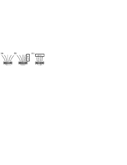
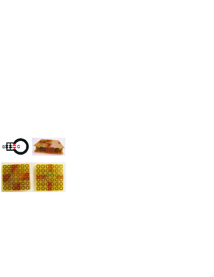
42.30.-d,41.20.Jb,78.70.Gq,78.20.CiApplication of metamaterials in magnetic resonance imaging (MRI) has been previously explored in several works making use of devices based on swiss-rolls Wiltshire -Mathieu , wires Radu and capacitively-loaded split rings Freire-APL-2008 -Freire-APL-2011 . Most of these works have explored the sub-wavelength imaging ability of metamaterials with negative permeability (). In previous works of the authors, metamaterials slabs with has been fabricated and tested in MRI systems to show the ability of these slabs to increase the sensitivity of surface coils Freire-APL-2008 ; Freire-JMR-2010 and to improve the field localization of these coils, a fact that may find applications in parallel MRI Freire-APL-2011 . Although metamaterials can be engineered to tailor whatever value of permeability at the desired frequency, little attention has been paid to permeability values different from negative ones. In the present work, it is explored the application in MRI of capacitively-loaded split-ring metamaterials which show zero permeability () or high permeability () at the operating frequency. and slabs can reject (see Fig. 1.b) and confine (see Fig. 1.c), respectively, the radiofrequency (RF) magnetic field. These properties can help to locally increase the signal-to-noise-ratio (SNR) of surface coils in certain configurations which have been experimentally investigated in this work. Typical MRI acquisition consists of the excitation of tissue with a strong and uniform RF field generated by a transmitting body coil, and then the detection of the weak field generated by hydrogen nuclei in tissue by means of surface coils. The split-ring device previously reported by the authors consisted of a slab Freire-APL-2008 -Freire-APL-2011 , which does not distort the uniform excitation field. However, and slabs can actually distort this field. Therefore, it is necessary to implement these slabs as active slabs which can be automatically switched to show under the strong field of excitation and and under the weak field coming from tissue. This can be accomplished in the practical implementation of the slabs by inserting active elements in the split-rings that allow to switch between different responses under strong or weak fields. In particular, a pair of crossed diodes inserted in each split-ring can help to switch off them under the strong excitation field. Following the homogenization procedure previously reported by some of the authors Baena-PRA-2008 , two split-ring slabs of unit cells with a periodicity of 15 mm were designed to exhibit and at the frequency of 63.6 MHz. This frequency corresponds to the Larmor frequency of 1.5T Siemens Avanto MRI system sited in the Department of Experimental Physics 5 (Biophysics) of the University of Wrzburg (Germany), where the experiments reported in this work were done. The fabricated split-rings have 12 mm in diameter and 1.87 mm of strip width for the slab, 11.8 mm in diameter and 1.7 mm of strip width for the slab. Each split-ring in the array contains a 470 pF non-magnetic capacitor (American Technical Ceramics Corp., NY, USA) for resonance at a specific frequency below 63.6 MHz, and a pair of crossed diodes (Microsemi Corp., CA, USA) in parallel with the capacitor (see Fig. 2) in order to switch off the slab in transmission. Under the strong excitation field, the high electromotive force induced in the rings makes the diodes to drive and then the capacitors are short-circuited, so that the split-rings behave like simple closed metallic rings. Following the homogenization procedure Baena-PRA-2008 , the calculated permeability for this system of simple metallic loops is , a value which is closed to the value of the permeability of air. Once the sample is excited, the tissue reradiates a weak field which is unable to drive the diodes, so that the rings behave like resonant circuits. The frequency of resonance has been chosen so that from the homogenization model Baena-PRA-2008 the system has and at the working frequency. Since the capacitance of each split-ring is fixed, the frequency of resonance was fitted by adjusting the dimensions of the rings. A small correction to the value predicted by the homogenization model was necessary due to the parasitic reactance of the diodes.
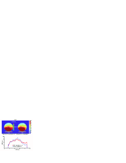
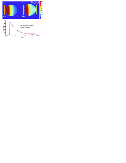
For the experiments, a 90 mm in diameter receive-only single loop coil was used and a cylindrical bottle 16 cm in diameter, filled with a H2O solution doped with 5 g/l NaCl and 1.25 g/l NiSO4, was used as a load for the experiments. The loop was tuned to 63.63 MHz and matched to in the presence of the slabs and the phantom. It was actively decoupled by a tuned trap circuit including a PIN diode in transmission. The active decoupling for the loop was -25dB with and without metamaterial slabs. All the experiments were performed in a 1.5 T whole body scanner. In the experiment, the metamaterial slab is perpendicular to the loop and it is positioned at one side of the phantom (see Fig. 3), so that the magnetic flux is rejected by the slab and then confined inside the phantom. This will increase the signal coming from this region of the phantom. In the experiment, the metamaterial slab is placed parallel to the loop in the opposite side of the phantom (see Fig. 4) in order to guide the flux lines through the phantom.
SNR maps were calculated from a series of identical phantom measurements Ohliger-MRM-2004 for both the and the slabs and compared with the situation where the slabs were removed. In Fig. 3 top, the calculated SNR maps are shown for both the presence and the absence of the slab and profiles are compared (see Fig. 3 bottom). In the side of the phantom where the slab is placed, the signal increases (approx. 15%). The calculated SNR maps for the slab are shown in Fig. 4 top, and the corresponding profiles in Fig. 4 bottom. Again, the signal presents an increment of approximately 15% with the presence of the slab.
This work demonstrates how split-ring metamaterial slabs designed with specific permeability values can increase the SNR in different configurations. The SNR gain in the demonstration was moderate, but it could be improved with a smart design of the configuration by suitably choosing both the coild and phantom size. Moreover, although the SNR gain could not be comparable to that provided by another loop coil positioned in the same as the slab, the metamaterial slabs could be useful in limited channel systems or as complement of an array. Some artifacts appear in the phantom s surface due to the discrete nature of the split-rings, but it can be easily removed by taking the salb 1 cm far from the surface of the phantom.
This work was supported by the Spanish Ministerio de Ciencia e Innovacion under project Consolider CSD2008-00066. The authors want to thank the company NORAS MRI Products for the advice.
References
- (1) M.C.K. Wiltshire, J.B. Pendry, I.R. Young, D.J. Larkman, D.J. Gilderdale, J.V. Hajnal, Microstructured magnetic materials for RF flux guides in magnetic resonance imaging, Science 291 (2001) 849-851.
- (2) V.C. Behr, A. Haase, P.M. Jakob, RF flux guides for excitation and reception in 31P spectroscopic and imaging experiments at 2 Tesla, Concepts in Magnetic Resonance Part B (Magnetic Resonance Engineering) 23B(1) (2004) 44-49.
- (3) M.C.K. Wiltshire, Radio frequency (RF) metamaterials, Phys. Stat. Sol. (b) 244 (2007) 1227-1236.
- (4) M. Allard, M.C.K. Wiltshire, J.V. Hajnal, R.M. Henkelman, Improved signal detection with metamaterial magnetic yoke, Proc. Intl. Soc. Mag. Reson. Med. 13 (2005) 871.
- (5) A. Mathieu, R.M. Henkelman, Using metamaterial yokes in NMR measurements, J. Magn. Reson. 182 (2006) 200-207.
- (6) X. Radu, D. Garray and C. Craeye, Metamaterials, 3, 90 (2009).
- (7) M.J. Freire, R. Marques, L. Jelinek, Appl. Phys. Lett., 93, 231108 (2008).
- (8) M. J. Freire, L. Jelinek, R. Marques and M. Lapine, J. Magn. Res., 203, 81 (2010).
- (9) J.M. Algarin, M.J. Freire, M.A. Lopez, M. Lapine, P.M. Jakob, V.C. Behr, R. Marques, Appl. Phys. Lett., 98, 014105 (2011).
- (10) J. D. Baena, L. Jelinek, R. Marques and M. G. Silveirinha, Phys. Rev. A, 78, 013842 (2008).
- (11) M.A. Ohliger et al., Magn. Reson. Med., 52, 628 (2004).
CAPTION TO FIGURES
Figure 1. a) Magnetic field lines for a single coil. b) Magnetic field lines with a slab perpendicular to the coil. c) Magnetic field lines with a slab placed parallel to the coil.
Figure 2. Sketch of the constituent element and photographs of the slabs. The slabs are unit cells and the periodicity is 15 mm. The has been fabricated with rings of 12 mm in diameter and 1.87 mm of strip width, and the has been fabricated with 11.8 mm in diameter and 1.7 mm of strip width. For both cases, 470 pF capacitors were used to tune the loops. A pair of crossed diodes in parallel with the capacitors provides the active response which is different for strong or weak fields.
Figure 3. SNR map for the experiment with the slab (up) in a axial plane. Profile of the SNR over the white line in the map (down).
Figure 4. SNR map of the experiment with the slab(up) in a axial plane. Profile of the SNR over the transversal axis (down).