Dynamics of Bacteriophage Genome Ejection In Vitro and In Vivo
Abstract
Bacteriophages, phages for short, are viruses of bacteria. The majority of phages contain a double-stranded DNA genome packaged in a capsid at a density of mg/ml. This high density requires substantial compression of the normal B form helix, leading to the conjecture that DNA in mature phage virions is under significant pressure, and that pressure is used to eject the DNA during infection. A large number of theoretical, computer simulation and in vitro experimental studies surrounding this conjecture has revealed many — though often isolated and/or contradictory — aspects of packaged DNA. This prompts us to present a unified view of the statistical physics and thermodynamics of DNA packaged in phage capsids. We argue that the DNA in a mature phage is in a (meta)stable state, wherein electrostatic self-repulsion is balanced by curvature stress due to confinement in the capsid. We show that in addition to the osmotic pressure associated with the packaged DNA and its counterions, there are four different pressures within the capsid: pressure on the DNA, hydrostatic pressure, the pressure experienced by the capsid, and the pressure associated with the chemical potential of DNA ejection. Significantly, we analyze the mechanism of force transmission in the packaged DNA, and demonstrate that the pressure on DNA is not important for ejection. We derive equations showing a strong hydrostatic pressure difference across the capsid shell. We propose that when a phage is triggered to eject by interaction with its receptor in vitro, the (thermodynamic) incentive of water molecules to enter the phage capsid flushes the DNA out of the capsid. In vivo, the difference between the osmotic pressures in the bacterial cell cytoplasm and the culture medium similarly results in a water flow that drags the DNA out of the capsid and into the bacterial cell.
1 Introduction
Bacteriophages, or phages, are viruses of bacteria. Phages consist of a protein capsid that encapsidates their genome, and a tail — a hollow tube connected to the capsid via a portal complex. During infection, the phage tail attaches to a host bacterium, punctures the cytoplasmic membrane and its genome translocates through the portal and the tail into the bacterial cytoplasm. Infection initiates the phage life-cycle: within the bacterial cytoplasm the genome is transcribed and replicated, phage proteins are synthesized, and new genome copies are packaged into newly assembled capsids. The cycle ends with lysis of the host cell and the release of multiple progeny. Understanding the mechanism(s) of phage genome ejection is important, not only in the insights it provides on DNA structure but also to provide a model for how eukaryotic viruses may release their nucleic acid in to the cytoplasm or nucleus of an infected cell.
The genome of a mature phage virion is usually a B-form, double-stranded DNA (dsDNA). A common phage may have 35-50 kb DNA (DNA and genome will be used interchangeably) packed into a capsid of nm diameter, i.e., the phage DNA is packaged tightly within the capsid in a condensed state, at a linear compression factor , or at a density mg/ml [1, 2, 3, 4, 5, 6, 7, 8, 9, 10, 11, 12, 13, 14, 20, 21]. More than five decades ago, this compression led to the conjecture (henceforth referred to as the “pressure-conjecture”) that DNA in a mature phage capsid is under significant pressure, and is kept in place by means of a “plug”: protein(s) in the tail tube. When the plug is opened by the action of the appropriate receptor, the pressure of the DNA causes its release into the cytoplasm of an infected cell in a biologically passive manner [22, 23, 24, 25]. The difficulty of obtaining experimental kinetic data for the ejection of any phage genome into an infected cell resulted in the conjecture becoming “fact”. The few published examples that were inconsistent with the theory elicited little response and most textbooks simply refer to the “DNA injection” step without further elaboration. However, at the beginning of this millennium, the pressure conjecture resurfaced in the biophysics community. A single molecule study reported that the packaging motor of phage is capable of packaging DNA into a phage capsid against a force of magnitude pN [26]. The force, which was assumed to be totally conserved, and thus available for the subsequent ejection step, and which therefore can be termed as the “ejection force”, was simply defined as the pressure on the DNA within the capsid, multiplied by the cross-sectional area of the unit cell corresponding to the hexagonal lattice arrangement of the packaged DNA. It was estimated that the pressure on the packaged DNA, as well as the pressure on the inside of the capsid is in the order of 60 atm for a mature phage virion. This work was quickly followed by a large number of computer simulations [27, 28, 29, 30], theoretical analyses [31, 32, 33, 34, 35], and in vitro experimental studies [36, 37, 38, 39, 40, 41, 44, 45, 46, 47, 48] surrounding the pressure-conjecture, a significant fraction of these studies stating the 60 atm pressure on the packaged DNA for a mature virion as a matter of fact.
Many recent theoretical, computer simulation and in vitro experimental studies have been principally directed to quantify how the pressure on the DNA can be understood from a thermodynamic perspective. However, to the best of our knowledge, the issue of the mechanical transmission of the pressure along the packaged DNA helix has received no attention. Putting aside that question for the moment, a general consensus has been reached on a thermodynamic description of the pressure on the DNA. DNA is a charged polymer with persistence length nm; DNA confined inside a capsid of diameter nm has a large free energy cost , relative to the state of the DNA outside the capsid. The pressure on the packaged DNA can be thermodynamically derived from this free energy. However, fundamental disagreements remain between different theories on the thermodynamic origin of the pressure acting on the packaged DNA. In light of these disagreements, together with the question of how mechanical transmission of force along a flexible polymer, naturally raises the question: “Does the pressure-conjecture necessitate a biologically passive ejection force”?
In order to appreciate the outstanding issues more easily, we provide a brief overview of the structure of packaged phage DNA, and existing approaches to describe phage DNA packaging and ejection.
1.1 Structure of the packaged DNA in mature phage virions
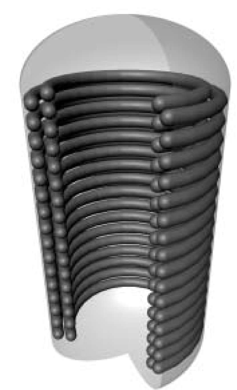
Experiments on the structure of the packaged DNA within phage capsids have a long history: preliminary evidence of hexagonal packaging of the DNA for T2 and T7 phages was first obtained in 1961 [1], and X-ray diffraction was also used to establish that DNA forms concentric layers within the capsid [2, 3, 4]. Several theoretical models have been proposed for the topology of the packaged DNA: wound into a spool [49, 50, 51], a liquid crystal with hairpins [52, 53] or defects [54], to name but a few, subjecting the topic to much debate. The first unambiguous high-quality images of DNA organization in mature phage capsids were obtained using cryo-electron microscopy in 1997. Averaged over many mature T7 tail-deletion mutant virions, viewed along the portal axis, the packaged DNA showed patterns of circular striations, spaced nm apart (Fig. 1) [4]. A computer-modeled projection (side views) of spooled DNA within the capsid, again averaged over many virions, strongly suggested that T7 DNA is wrapped axially in concentric shells of a toroid. In each shell, DNA is coiled with an axial rise of nm per turn, and the diameter of each turn is imposed by the previous shell. Following this pioneering study, a succession of cryo-electron microscopy reconstructions have provided support for a toroidal structure: [5], both isometric and prolate T4 [6, 7], P22, , [8, 9, 10, 11, 12, 13], K1E and K1-5 [14], P-SPP7 [15] and N4 [20] all reveal consecutive layers of toroidal DNA spaced by nm. Together, these observations suggest that, when averaged over many virions, the well-defined hexagonal lattice for the packaged DNA may be a generic feature of all phages (Fig. 2). Separately, it should be noted that, where it was resolved in these reconstructions, the leading end (first end to enter the infected cell) is seen to extend into the narrow portal channel. Furthermore, in earlier studies, the leading end of the genome could be cross-linked to the tail of several mature phage virions [16, 17, 18, 19]. Portal/tail insertion of the leading DNA end during virion morphogenesis is also likely a general feature as it ensures that the subsequent DNA ejection step is efficient.
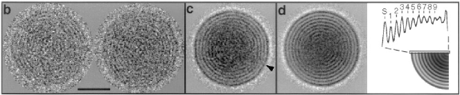
It is of paramount importance to emphasize that the hexagonal toroidal spool structure of the packaged DNA is an average property. This point is illustrated beautifully in a recent paper on phage T5 [55]. Further, to be packed within a capsid the DNA helix has to cross itself and therefore cannot lie on a perfect lattice. The toroidal structure has also been suggested to hold only for the part of the DNA close to the inner wall of the capsid, and not for the entire DNA (e.g., Refs. [21, 29]).
1.2 Existing approaches for the thermodynamics of packaging and ejection forces
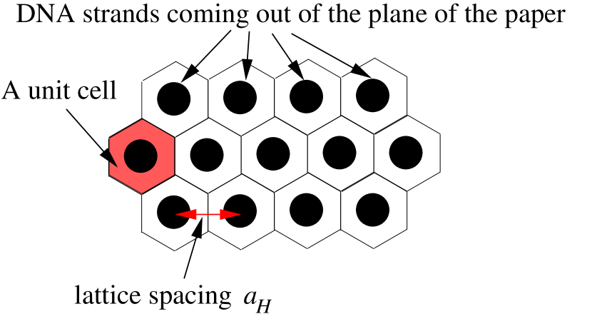
There have been two main — and mutually conflicting — approaches that address the thermodynamic origin of packaging and ejection forces: continuum mechanics and coarse-grained molecular mechanics models. In the continuum mechanics model [31, 32, 33, 34, 35], packaged DNA is seen as a uniformly charged rod, with a certain persistent length , organized in a toroidal spool on a hexagonal lattice (Fig. 2). The continuum mechanics model is simple enough to allow analytical calculations: packaged DNA is assumed to repulsively self-interact (with electrostatic Coulomb interaction), allowing calculation of the stored electrostatic energy . Similarly, the bending energy is obtained from the toroidal spool arrangement of DNA on a hexagonal lattice. Both the electrostatic and bending energies are assumed to be zero for free DNA outside the capsid. Thereafter, with the assumption that the difference between the entropy of free and packaged DNA is negligible, the difference between the Helmholtz free energy of packaged and free DNA is obtained as . Note that in a given buffered solution and a capsid of volume , made of a perfectly rigid material, is only a function of the length of the DNA within the capsid, i.e., . In particular, increases as a function of , and it is , the pressure associated with the chemical potential of DNA ejection (the chemical potential of DNA ejection is simply defined as the negative rate of change of the free energy with respect to the length of DNA within the capsid) that manifests as the ejection pressure, where is the cross sectional area of the DNA. Note that has the units of force; consequently, in the continuum mechanics model, is viewed as the ejection force, and as the packaging force (note that is also the chemical potential of DNA ejection, as defined above).
In the in vitro experiments [36, 37, 38, 39], phage virions are immersed in a solution containing PEG and/or DNA condensing agents, and DNA is ejected when triggered by the LamB receptor protein. The length of the DNA that remains within the capsid at the end of the ejection process depends on the concentration of the PEG and/or DNA condensing agent. (The basic principles underlying these in vitro experiments were first presented in a theoretical paper more than four decades ago [25]). The continuum mechanics model has been widely advertised to explain the in vitro ejection data, not just for but for all phages — both in vitro and in vivo. A somewhat deeper look, however, reveals that the agreement between theory and the in vitro experiments using T5 (one of only three phages studied in osmotic suppression experiments), remains poor [40, 41]. The model also neglects how the ejection of proteins from infecting virions (which is a feature of most phages) into the cell is accomplished, and it cannot therefore describe the complete infection process. Further, there is also disagreement between the continuum mechanics theory and the experimental kinetics of T7 DNA translocation into the bacterial cytoplasm in vivo [56, 57]. All but the leading 0.5 kb of the 70 kb genome of the unrelated phage N4 is also known to be internalized by transcription in the cell [20, 42], and marker rescue experiments with phage SP82 suggest that the phage genome enters a cell at a constant rate [43]. None of the in vivo experiments that show kinetics of genome internalization are consistent with theories derived from in vitro ejection studies.
The continuum mechanics model suffers from fundamentally unrealistic assumptions that diminish its value. In particular, free energy is an equilibrium concept; obtaining an ejection pressure by , in order to explain the experimental data on the irreversible ejection process, has the underlying assumption that DNA remains at equilibrium in a toroidal spool configuration on a hexagonal lattice at all stages of ejection. Not only is this assumption unsubstantiated by polymer physics theory, it is also inconsistent with actual experimental data on T5 [55]; the latter is illustrated in Fig. 3 below. In order to circumvent some of these problems, the continuum mechanics model invokes two free parameters to fit experimental data.

In coarse-grained molecular mechanics models [27, 28, 29, 30], DNA is modeled as a polymer, with a persistence length , and polymer dynamics is allowed to take its own course in simulations of phage genome packaging and ejection. The model does not allow analytical calculations — it is effected only by computer simulations, and in work published to date, the DNA is modeled without surrounding water and counterions. However, there is no assumption that DNA remains at equilibrium in a toroidal spool configuration on a hexagonal lattice during packaging or ejection. The main limitation of coarse-grained molecular mechanics model is that because processes are only materialized by computer simulation, they are difficult to correlate with experimental data.
1.3 This paper
In summary, our understanding of the thermodynamics of phage DNA packaging and ejection, provided by the existing literature, is far from satisfactory. In this paper we present a comprehensive treatise of the thermodynamics of packaged DNA and the mechanism of genome ejection, independent of any specific model. In Sec. 2 we show that there are four distinct pressures within the capsid: pressure on DNA, hydrostatic pressure, pressure experienced by the capsid, and pressure associated with the chemical potential of DNA ejection. These are all different thermodynamic quantities. We also show that for amature phage the hydrostatic pressure within the capsid is much higher than outside, and that the pressure on the DNA is a lot smaller than commonly envisaged. In Sec. 3 we take up the issue of force transmission along the DNA helix, and show that the pressure on DNA is not important for genome ejection. Instead, in Sec. 4, we suggest that when a phage is triggered in vitro to eject by interaction with its receptor, the (thermodynamic) incentive for water to enter the capsid flushes the DNA out. We argue that in vivo, the difference between the osmotic pressure within the cell cytoplasm and the outside culture medium initiates, and importantly maintains, a flow of water from the culture medium into the capsid, and then from the capsid down the phage tail-tube into the bacterial cell cytoplasm, dragging the DNA out of the capsid and into the cell. We show that this theory is consistent with both studies of in vitro ejection and observations of infection in vivo.
2 Thermodynamics of phages in vitro
2.1 Entropy of DNA ejection in vitro
We consider an experiment with an ensemble of realizations, each containing a phage capsid initially placed in a certain buffer solution: for each realization a DNA of length is packaged within a capsid, whose volume we denote by . The DNA is allowed to exit the phage in vitro, and at the end of the experiment the entire genome ends up in the buffer. The experimental system is kept at a temperature at all times. The only interaction of the system with the environment is to exchange (thermal) energy. For the system in its entirety, we define the initial (DNA within the capsid) free-energy and the entropy ; the final (DNA in the buffer) free-energy and entropy . The quantities , and are then the free energy and entropy of ejection, respectively.
The above isothermal experiment, when actually performed in a laboratory, is exothermic [58]. Earlier experiments using differential scanning calorimetry led to the same conclusion [59, 60, 61]; i.e., the system releases an amount of heat, of magnitude to the environment. This tells us that ; however, the amount of heat released to the environment cannot give us the value because the ejection process is spontaneous and irreversible. All we can say, using the second law of thermodynamics, is that .
The result that the entropy of ejection is negative, from the point of view of DNA confinement in the capsid, is counter-intuitive. One would expect the configurational entropy of the DNA to be strongly reduced due to confinement when compared to the same DNA in solution; in other words, from the configuration entropy of the DNA in isolation one would expect . This is, in fact, correct; however, the DNA cannot be considered in isolation. The configurational entropy of the DNA is only a very small part of the entropy of the entire system; most of the change in entropy associated with ejection of DNA from its confined state within the capsid into the environment involves the ordering of water molecules around the free DNA [62]. DNA, being a charged molecule, orders the dipole orientation of nearby water molecules. There are many fewer water molecules surrounding the DNA inside the capsid when compared to the same DNA in solution. Consequently, although the DNA molecule does gain entropy following ejection from the capsid, so many more water molecules lose entropy that the entropy of the entire system decreases as DNA is ejected from the capsid.
This issue can be used to evaluate the current approaches for analyzing the physics of phage ejection. DNA configurational entropy is not considered a significant parameter in the continuum mechanics model, while molecular mechanics models [27, 28, 29, 30] show that the configurational entropy constitutes a sizable fraction of the free energy of the packaged DNA.
2.2 Thermodynamics for the capsid content of a mature phage in a buffer solution
Most phage capsid shells are fully permeable to water and small ions or molecules, but are impermeable to large molecules in the buffer or to the encapsidated DNA. Some phage capsids, like those of T4 and its relatives, are much less permeable, even to small molecules or ions, including cesium chloride and ammonium acetate. Such capsids can easily be broken by osmotic shock, a phenomenon that resulted in the classic electron micrograph showing phage T2 DNA outside a ruptured virion [63].
We assume that the entire system, with DNA packaged within the capsid, can be thermodynamically separated into two subsystems separated by the capsid shell. The subsystem within the capsid consists of DNA (perhaps in addition to protein molecules, which we will not specifically refer to further), and an aqueous environment containing small solutes (including ions) that can permeate the capsid shell, while the subsystem outside usually consists of water and solute molecules that can or cannot permeate the capsid shell. Apart from exchanges of solutes that can permeate the capsid shell, these two subsystems do not interact. This assumption allows us to express , where and are, respectively, the free energy of the contents of the capsid and that of the environment outside the capsid. Clearly, is a function of the equilibrium capsid volume , DNA length , number of water molecules and the number of solute molecules of all species within the capsid. Similarly, is a function of number of water molecules and the number of solute molecules of all species in the solution. Both subsystems are “open” in the thermodynamic sense, since they exchange water and small solute molecules. We assume (realistically) that the volume of the buffer in which the phage is immersed is , so for analyzing the thermodynamics of the phage and its contents, the external solution can be considered to be an infinite reservoir for water and small permeable solute molecules. The number of solute molecules within the phage capsid is then fixed by the chemical potential of the small solute molecules in the buffer, collectively denoted by (for the case of counterions, via the Donnan equilibrium). This simply means that . The free energy can be further dissociated into the interaction free energy of the ions, DNA and water molecules within the capsid, and the DNA configurational free energy (this includes bending, or curvature, energy and the configurational entropy of the DNA) of the DNA is then given by
| (1) |
We also assume that water is incompressible and that small solutes do not occupy any physical volume. With these assumptions, in equilibrium, the number of water molecules within the capsid is then determined by minus the physical volume of the DNA, and we can now explicitly focus on a number of thermodynamic issues.
2.2.1 Pressure associated with the chemical potential for DNA ejection
If we assume that the capsid is made of a perfectly rigid material, then from the above definitions , we can define the pressure associated with the chemical potential for DNA ejection. Consider two situations using the same buffer conditions (i.e., at fixed chemical potentials of permeable ions), one where DNA of length and the other a length of , is packaged within the capsid. We assume that Donnan equilibrium conditions are satisfied for both lengths of DNA within the phage capsid. One can then define the pressure associated with the chemical potential of DNA ejection by comparing the free energy of the capsid content under these two situations, viz. and , as:
| (2) |
where is the cross-sectional area of the DNA. The pressure is the pressure that the plug at the end of the tail tube feels from within the phage.
2.3 Pressure on the capsid from within, hydrostatic pressure imbalance across the capsid and pressure on the DNA
In Sec. 2.2, we considered the thermodynamics of the capsid content as a subsystem, under a fixed chemical potential of permeable small solute molecules in the buffer, while assuming that the number of water molecules within the capsid is simply determined by the capsid volume. This is however an incomplete description of the thermodynamics of the entire system (namely the phage immersed in the buffer solution), as we did not consider the thermodynamic penalties associated with exchange of water molecules across the capsid shell. If the capsid is made of a perfectly rigid material without the possibility of expansion or contraction, such a description would be correct, but in reality, the capsid is comprised of protein molecules, and therefore has a finite, albeit high, rigidity [65]. Keeping the assumption that water is incompressible, rather than a fixed capsid volume determining the number of water molecules internalized, the capsid volume should actually be determined by the thermodynamics of water molecule exchange across the capsid shell, which is equivalent to a semi-permeable membrane.
We now argue that for a given length of the DNA within the capsid, the equilibrium volume is determined by the buffer composition. We presuppose that an expansion/contraction of the capsid allows for an exchange of a small number of water molecules, relative to the total number of water molecules present in the external buffer. We can then safely assume that the chemical potential of permeable small solutes in the buffer remains unaltered by the exchange of water molecules across the capsid shell.
First, the pressure on the capsid from within, , can be calculated from the -dependence of the free energy as follows. Imagine a virtual, uniform expansion of the capsid leading to a volume increase from to (the extra volume will be filled by water from the buffer and permeable solute molecules as dictated by — for the case of ions, via the Donnan equilibrium). For a given length of DNA, under fixed , this leads to a new value of the free energy of the inside material of the capsid, , but also, since the buffer solution loses a volume of water, the free energy of the buffer solution increases by an amount , where is the osmotic pressure of the buffer solution. The pressure on the capsid from within, is therefore given by
| (3) |
Secondly, water’s thermodynamic incentive to enter the capsid is given by the osmotic gradient across the capsid shell, namely , where is the osmotic pressure within the capsid shell, and is defined by [see Eq. (1)]
| (4) |
Simultaneously, when entering the capsid, the water molecules exit a zone of hydrostatic pressure , the hydrostatic pressure in the solution outside the capsid, and enter the capsid which may have, in principle, a different pressure, . The equilibrium capsid volume is then determined by the condition that is counter-balanced by the pressure-volume work done by water molecules in entering the capsid
| (5) |
It is interesting to note that Eq. (5) is well-known to hold for bacterial and plant cells: the osmotic pressure gradient is counter-balanced by a hydrostatic pressure differential (turgor) [68]. Turgor allows cells to enlarge and thus facilitates growth.
Further, an appreciation for Eq. (5) is afforded by a straightforward gedanken experiment that connects to elementary physical chemistry. Consider a vertical U-tube with a membrane at the lowest point separating the two arms. The membrane is permeable to water, but impermeable to, say, sugar molecules. We fill up the two arms of the U-tube to equal height: the left arm with pure water, and the right arm with sugar solution. As time progresses, water from the left arm will permeate into the right arm, reducing the height of the water column on the left, while increasing the height of the sugar solution column on the right. At equilibrium, the height of water in the left column will be lower than the sugar solution, meaning that there is a hydrostatic pressure difference across the membrane. Moreover, at equilibrium the concentration of sugar in the right column is non-zero, while it remains zero in the left column by construction. There is therefore an osmotic gradient across the membrane, which is precisely counter-balanced by the hydrostatic pressure . Equation (5) describes the same equilibrium, where the difference between the osmotic pressures inside and outside the capsid is counter-balanced by the hydrostatic pressure difference across its protein shell.
Equations (3) and (5) allow us to appreciate not only how the pressure on the capsid of finite rigidity is mechanically materialized, but also how the pressure that the capsid mechanically transmits to the DNA (and by action-reaction, the pressure the DNA transmits to the capsid). First, the force balance on a small capsid surface tells us that the capsid pressure equals the hydrostatic pressure gradient across the capsid plus the pressure that is mechanically transmitted to the DNA inside; i.e.,
| (6) |
Incorporating Eqs. (1) and (4-6), this reads
| (7) |
Note in Eq. (7) that the osmotic pressure within the capsid is derived from the interaction energy of the ions, DNA and water molecules within the capsid, while the same interaction energy appears in , but with opposite sign [Eq. (4)]. Consequently, the net contribution of this interaction energy to the r.h.s. of Eq. (7) is zero. This conclusion implies that the pressure on the DNA mechanically transmitted by the capsid comes entirely from the stiffness of the DNA. We verify this conclusion in Sec. 3 for a single turn of the DNA. Therein we also discuss why Eq. (7) is not in conflict with the experiment by Smith et al. [26].
It is straightforward to show that Eqs. (3-7) are consistent with the Gibbs free energy minimization of the entire system (the mature phage plus the buffer solution). Moreover, we note that due to the cancellation of the interaction energy term in Eq. (7) as explained above, the compressive forces on the DNA mechanically transmitted by the capsid are small compared to : the bulk of is mechanically transmitted to the water inside. This is in fact confirmed from the structural and interaction properties of both the packaged DNA and the structure of the protein molecules comprising the capsid shell. Raman spectral studies of mature P22 and T7 phages have not found any evidence of any structural alteration of protein or DNA that would be expected if they were subjected to high pressure [66, 67]. Only the configuration of the phosphodiester groups in packaged DNA are perturbed from that found in free DNA solutions, and the capsid protein structures of mature virions are indistinguishable from empty, DNA-free, particles.
Finally, we note from Eqs. (2-7) that the osmotic pressure within the capsid, the pressure associated with the chemical potential of DNA ejection, and the pressure imparted by the capsid on the DNA are entirely different thermodynamic quantities. Importantly, it must be appreciated that these pressures are all equilibrium quantities, and they cannot be used to describe a non-equilibrium situation.
3 Can and play any role in DNA ejection dynamics?
In this section, we investigate whether and play a role in DNA ejection. We first discuss the case of within the context of the continuum mechanics model; in order to do so we start by analyzing the stability of the DNA within a phage capsid.
3.1 Stability of the DNA within a phage capsid
As described in Secs. 1.2, continuum mechanics model starts with the assumption that the DNA, within a capsid made of a perfectly rigid material, takes a toroidal spool configuration on a hexagonal lattice at all stages of ejection. Given that the outer radius of the spool is fixed by the capsid dimensions, all structural aspects of the spool are then determined by the spacing of the hexagonal lattice. Phages with internal cores constitute a special case: the radius of the innermost ring of the spool is simply given by the radius of the core. For a given ionic condition inside the capsid — which in turn is fixed by the chemical potential of the small solutes in the external buffer (for the case of counterions, via the Donnan equilibrium) — and DNA of length within a capsid of volume , the free energy of the material inside the capsid is a function of alone, where is the hexagonal lattice spacing; i.e., . Note that the configurational entropy of the DNA is not a meaningful quantity in the continuum mechanics model, hence in this section only contains curvature (bending) energy of the DNA; i.e., .
Within the continuum mechanics model, one can argue that is fixed, via a trade-off between and [see Eq. (1)]. The interaction free energy can be lowered by increasing . However, since the “volume constraint” (meaning that the total available volume within the capsid is ) must be obeyed, increasing will reduce the minimum radius of the DNA spool, because within the inner part of the spool the DNA will be much more tightly bent, which will increase its bending energy. In this manner, a reduction in interaction free energy increases curvature energy and vice versa. In other words the trade-off between these two quantities dictates that:
| (8) |
which minimizes the free energy . We denote the value of , obtained from Eq. (8) by , and the corresponding minimum of the free energy of the inside content of the capsid by . If it is further assumed that most of the interaction free energy variation due to variations in , caused by a virtual uniform expansion of the capsid [as in Eq. (4)], involves the DNA helix, then the osmotic pressure within the capsid can also be defined as
| (9) |
where the volume that the DNA toroid occupies within the hexagonal lattice is given by . This analysis for the specific case of the phage T7 — a phage with an internal proteinaceous core — can be found in Ref. [64].
3.2 Can transmit down the tail tube?
The only way can play a role in DNA ejection is if it can transmit from within the capsid, where the bulk of the DNA is located, down the tail tube. To evaluate this idea, we begin with the appreciation that favors a uniform expansion of the capsid. However, since DNA is not a continuum material, does not trivially transmit along the helical axis into the tail tube. Nevertheless, when the DNA organization within the capsid is averaged over many phage particles, the picture that emerges is that pressure on the toroidal spool of DNA does translate into a compressive force acting along the contour of the DNA. Following Sec. 3.1, we start by analyzing this force within the continuum mechanics model, and we then ask whether this force can transmit down the tail tube.
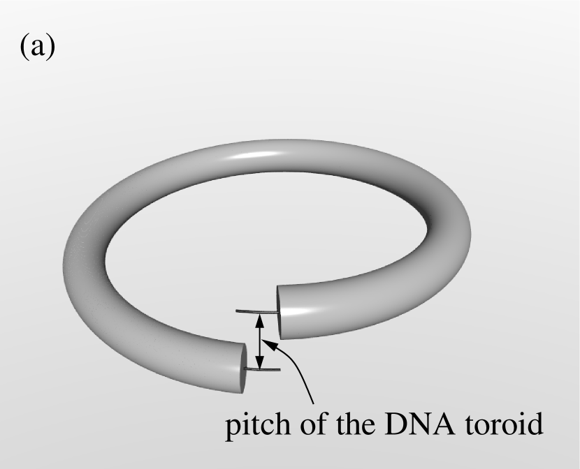
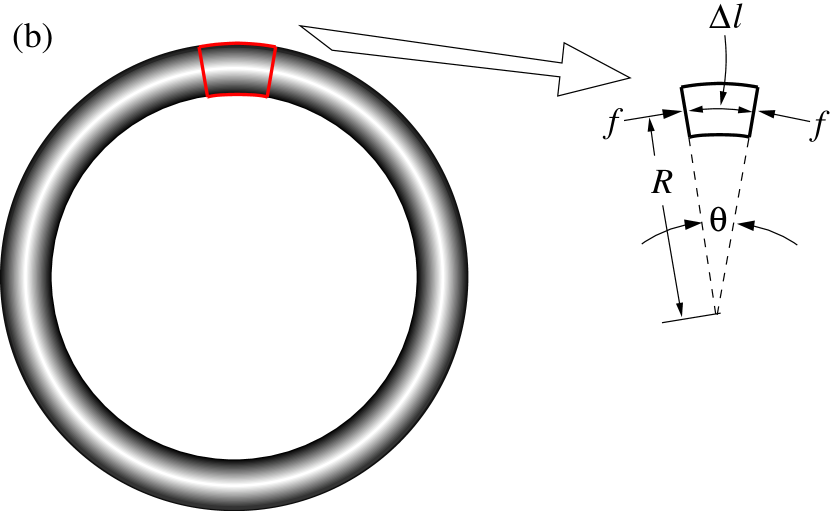
We consider one full turn of DNA around the axis of the idealized toroidal spool [Fig. 4(a)]. For simplicity, we ignore the pitch of this turn, i.e., it is idealized as a DNA ring of radius , and we focus on a differential length segment . The DNA ring and the compressive force along the DNA contour are shown in Fig. 4(b). The fact that the entire DNA spool is under a pressure within the phage capsid implies that the isolated ring itself is under a pressure , but whose relation to we have not specified.
Within the scheme of continuum mechanics, the force balance equation for this differential length segment along the transverse direction (along a line joining the differential length element and the center of the ring) is given by
| (10) |
where is the cross-sectional diameter of the DNA. Further, the geometrical consideration from Fig. 4(b) tells us that
| (11) |
i.e.,
| (12) |
We now note that, just like in Eq. (8), the DNA ring stays in its configuration by the balance of two competing thermodynamic forces, one derived from its curvature energy , the other from the energy of self-interaction with nearby DNA strands. The thermodynamic force related to curvature energy favors an increase of the radius of the ring, while the thermodynamic force related to the self-interaction free energy favors a decrease. The magnitude of this force is given by
| (13) |
implying that
| (14) |
At room temperature ( K) Joules. If we take nm, the widely used value, then
| (15) |
Thus, depending on the radius of the DNA ring, the magnitude of the compressive force along the contour of the DNA can be a few pN. Equation (15) tells us that the pressure on this DNA ring is only in terms of the stiffness of the DNA, as we argued in Sec. 2.3.
How much of this force can transmit down the tail tube? We should remember that force is a vector quantity. Although the above exercise shows that there is a compressive force along the contour of the DNA ring that make up the toroidal spool, it is necessary to follow the actual turns of the DNA leading into the tail tube in order to identify how much of the force is actually being transmitted. However, the definition of persistence length in polymer physics means that while a DNA molecule of length is semi-flexible, a DNA molecule with length is simply a flexible polymer. It is therefore impossible to transmit compressive forces along the contour of a DNA that is nm. This consideration alone ensures that any compressive force along the contour of, e.g., an m long genome will not transmit into the tail tube. Therefore, compressive forces on the packaged phage genomic DNA cannot be important for DNA ejection.
Our estimate of having a magnitude of a few pN is, at first blush, in apparent contradiction with the experiment by Smith et al. [26]. A deeper look however immediately reveals that Eq. (15) and the forces exerted by the packaging motor are actually compatible. Thermodynamically, we have identified four different pressures within the capsid: pressure on the DNA , hydrostatic pressure , the pressure experienced by the capsid , and the pressure associated with the chemical potential of DNA ejection. These are all different quantities. Smith et al. [26] measure ; they assumed that the work done against by the packaging motor is used to pressurize the DNA and is conserved in the capsid. However, most of the work done against by the packaging motor is expended in increasing the osmotic pressure within the capsid. This increase is due to the expulsion of water molecules out of the capsid — a reverse osmosis process — in order to allow the phage genome being packaged to condense, while only a small part of the work is used to increase The energy expended during water expulsion is not conserved by the packaged genome, which explains our lower estimate of inside the capsid. In this context it is important to remember that osmotic pressure is only a measure for the chemical potential of water: the fact that the osmotic pressure inside the capsid is high does not imply that the DNA is under enhanced pressure.
3.3 Is important for DNA ejection dynamics?
Equation (2) shows that for a mature phage in a given buffer, the tail plug feels a thermodynamic pressure from inside the capsid. Given that is a thermodynamically derived quantity (i.e., derivative of with respect to ), it is clear that if genome ejection were a quasi-equilibrium process, it would indeed govern the ejection dynamics. However, the underlying assumption of quasi-equilibrium is that at all stages of ejection the DNA remaining within the phage head stays in a toroidal loop, with an ever-increasing spacing between the toroidal strands as ejection proceeds. This is the major thesis of the continuum mechanics model, which uses to explain the in vitro experimental data using phage .
Whether this thesis and its underlying assumption are correct or not lies in the question of time scales. If the time-scales associated with the equilibration of DNA (in the form of a toroidal spool on a hexagonal lattice) and the small solute molecules within the phage capsid (maintained at a fixed chemical potential by the external buffer), are much smaller than the ejection time, equilibrium pressures and forces can be used to describe genome ejection. However, recent experimental evidence with T5 clearly demonstrates that as the DNA leaves the capsid, it goes through a series of phase transitions [55]. The cryo-electron microscopic images of T5 virions during DNA ejection strongly suggest that the process is non-equilibrium, which invalidates the use of to describe its dynamics. Comparable experiments have not yet been conducted with other phages, but there is no justifiable reason to assume that the genome of other phages, in particular those lacking a defined internal core structure, whose DNA is packaged to the same density, would behave otherwise. We are not aware of experimental studies that probe the equilibration time-scales of confined DNA, such as within a phage capsid. However, the high velocity of phage DNA ejection measured in vitro: up to 75 kb/sec (T5) with long pauses between distinct steps [44], and up to 60 kb/sec () in 10 mM Na+ buffer or kb/sec in 10 mM Mg2+ [45], makes it highly unlikely that these are quasi-equilibrium processes. These experimental data suggest that cannot be used to describe DNA ejection dynamics even in vitro.
4 An alternative mechanism of DNA ejection from phage virions
Sections 2 and 3 illustrate the fundamental problems with the idea that the thermodynamically derived ejection pressure (or the corresponding ejection force ) causes DNA ejection from phages. There is therefore a clear need for a mechanism that can explain the physics of DNA ejection, one that is consistent with both in vitro and in vivo experimental data. We suggest such a mechanism below.
4.1 DNA ejection in vitro
Of the two main approaches for DNA ejection: continuum mechanics and coarse-grained molecular mechanics models, only the former addresses, using , the DNA ejection mechanism in a way that can be compared to in vitro experimental data. In these creative experiments, phages are immersed in a solution containing PEG and/or DNA condensing agents, and DNA is ejected from the virion when triggered by the receptor protein. The length of DNA that remains within the capsid at the end of the ejection process depends on the concentration of the PEG and/or DNA condensing agent.
With the caveats we have discussed concerning the validity of using to describe phage DNA ejection, and remembering that also involves fitted parameters, the continuum mechanics theory does provide good agreement with in vitro genome ejection data for and SPP1 (e.g., Refs. [36, 37, 38, 47, 48]). However, the data obtained with just two phage systems, along with the physics of the continuum mechanics model, have often been extrapolated to all phages and in addition to explain all phage DNA ejection in vivo. These generalizations ignore data from the third phage system that has been studied in vitro, which provides rather different conclusions. At the presumed pressures internal to the largely full T5 capsid, in vitro experimental data can be fitted to the continuum mechanics model, but at low to moderate pressures, when approximately half or less of the genome remain in the capsid, multiple populations are found to co-exist [40]: some phages have completely ejected their genomes, whereas others have ejected a varying amount of DNA that is not dependent on the external osmotic pressure. Furthermore, at atm external osmotic pressure, most T5 virions completely eject their DNA. In contrast, the same external pressure prevents ejection of of the genome [37]. Theories dependent on are clearly unable to explain how all phages eject their DNA, even in vitro.
4.1.1 Hydrodynamic model of phage DNA ejection
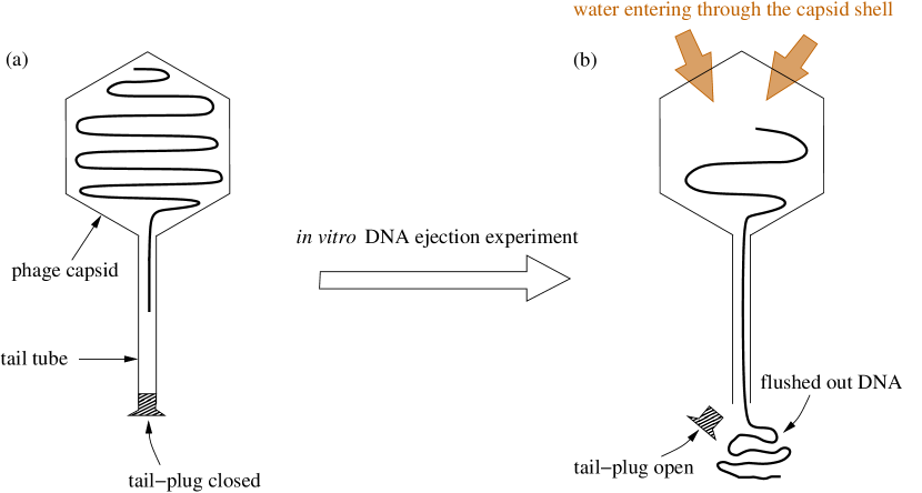
As discussed in Sec. 3.3, in an in vitro experiment when a mature phage is equilibrated in a certain buffer solution, the difference between and the osmotic pressure of the buffer solution determines the thermodynamic incentive for water molecules to enter the capsid. Equation (5) shows that if the phage tail is plugged, this incentive is counter-balanced by a hydrostatic pressure gradient across the capsid shell (and the capsid, being of high but not infinite rigidity, will be slightly expanded from its relaxed state under the enhanced hydrostatic pressure from inside). We propose that it is this thermodynamic incentive of water molecules to enter the capsid that flushes the DNA out when the tail plug is opened by the action of the appropriate receptor. We refer to this idea as the hydrodynamic model of DNA ejection.
When the tail plug is opened by the appropriate receptor, e.g., LamB protein in the case of , FhuA for T5, or YueB780 for SPP1, it is likely that the excess hydrostatic pressure within the capsid is partially relieved by a transient water flow down the tail tube and, and as a result, the capsid will reduce in size. Although water flow down the tail-tube will exert a hydrodynamic force that is likely to drag along the DNA, one end of which is already inserted into the tail-tube, this process will not last long enough to drag the entire genome out of the virion. While a very small contraction of the capsid is unlikely to be detected even by state-of-the-art experiments, we can provide a “guesstimate” for how much DNA could be dragged out by this transient water release. With a typical capsid diameter nm (assumed spherical), let the capsid diameter contract by nm; the water released is nm3. With a typical tail tube inner diameter nm, its cross-sectional area is nm2 (ignoring the cross-section of the DNA within the tail tube) and the length of the water column passing into the tail-tube nm nm. Assuming the DNA flows freely with this water column down the tail, in this example the DNA, which occupies 25% of the cross-sectional area of the tube, can be moved 60 nm, corresponding to base pair of a phage genome.
Once the water pressure difference across the capsid shell is relieved by this transient water flow, so long as the difference between the osmotic pressure of the remaining DNA within the capsid and stays positive, water will have a thermodynamic incentive to move into the capsid using whichever path it can find. However, since the capsid has a finite volume, any water movement into the capsid can only occur at the expense of an equal volume of water, now containing DNA (as a solute) being ejected through the tail tube. In other words, following the initial transient water release down the tail-tube, continued water movement from the buffer into the capsid up an osmotic gradient will cause the DNA to be ejected. This process will stop when the osmotic pressure of the leftover amount of DNA in the capsid equals . The physics behind this in vitro DNA ejection experiment (omitting the transient water flow down the tail tube due to the excess hydrostatic pressure) is schematically shown in Fig. 5.
The hydrodynamic mechanism for DNA ejection commensurate with in vitro experimental data. All that is required to drive DNA ejection is an incentive for the water molecules to enter the capsid, which is decided dynamically in a non-equilibrium manner. There is no a priori assumption that phage genome ejection is a quasi-equilibrium process, and thus the model is not dependent on thermodynamic analyses.
The difference between the osmotic pressure of the residual DNA in the capsid and is controlled by two aspects: (i) the presence of any small DNA condensing agents that reduces the osmotic pressure of the capsid content without altering , and (ii) the presence in the external buffer of large solute molecules like PEG that increase , but do not affect the inside osmotic pressure as they cannot penetrate into the capsid. Both achieve the same thermodynamic incentive differential for water between inside and outside the capsid, and are qualitatively in agreement with in vitro DNA ejection data [36, 37, 38, 39, 40, 41, 44, 45, 46, 47, 48]. When the osmotic pressure of any DNA remaining in the capsid (such as at the end of the ejection process), there will be no further flow of water into the capsid, and thus no more DNA ejection.
The hydrodynamic model requires that the time-scale of water flow into the capsid be sufficiently fast to match the time-scales for DNA ejection in vitro. To this end, we refer to an experiment where a mature wild-type capsid was locally deformed (compressed) by up to 25% of its original volume with an AFM tip [65]. Compression of the capsid without it rupturing has to drive out internal water and small solutes. When the AFM tip was removed, the virion capsid returned to its normal size and shape within ms. This suggests that the large surface area to volume ratio of the capsid allows copious amounts of water to diffuse — a slow process relative to hydrodynamic processes — across the capsid shell in a very short time. Extrapolating these data to yield a complete exchange of the contents of the capsid with the external buffer gives 20 ms; assuming that DNA moves freely, this value results in a rate of phage genome ejection in vitro at least 50 times the maximum measured to date.
By itself, the hydrodynamic model cannot explain either the stepwise ejection of DNA from T5 particles or the coexistence of virions containing different amounts of DNA at low external pressures. A partial ad hoc solution for T5 (one that is also necessary for the model of DNA ejection) is that the tail tube becomes temporarily blocked by protein conformational changes, thereby impeding water flow — and thus stopping DNA ejection. Note that water flow from the external buffer into the capsid interior will be simultaneously impeded. Release of the blockage by a further conformational change in the tail proteins then allows resumption of water flow and thus of DNA ejection. Interestingly, the apparent activation energy for releasing the block in the step-wise ejection process of T5 DNA ejection has been experimentally shown to be independent of the amount of DNA remaining in the capsid [37, 70].
In summary, the hydrodynamic model is fully compatible with the experimental data for and for SPP1 DNA ejection in vitro. Like thermodynamics-based models (e.g., continuum mechanics or molecular dynamics), the hydrodynamic model requires an ad hoc assumption to explain the step-wise process of T5 DNA ejection. However, the hydrodynamic model is not dependent on the critical assumption — used in -based models — that the encapsidated DNA remains at equilibrium at all times during the ejection process. Furthermore, as we argue below, only the hydrodynamics model explains how complete genome ejection can be achieved in the face of an opposing force. This is the situation in natural infections of bacterial cells.
4.2 DNA ejection in vivo
Before discussing specific mechanisms of phage DNA ejection in vivo, it is instructive to consider some salient points about phage infections.
4.2.1 Phage infection of bacteria
Largely because of influential textbooks and, until recently, of only little experimental information, it is not widely appreciated that all phages eject proteins into the cell [69, 71]. At a minimum, the protein(s) comprising the tail plug must be removed, and for long-tailed phages (like T4, T5, SPP1 and ) the tapemeasure protein, which determines the precise length of the tail, must be ejected in order to allow the phage genome to pass through the tail tube. Short tailed phages may eject internal proteins into the cell to extend — at least functionally — their tail so that it can span the infected cell envelope [57].
Some phages eject many different protein molecules from their capsid, some necessarily before, others perhaps after, genome ejection. To give just two examples, T7 virions ejects molecules representing five different protein species into the cell prior to DNA penetration of the cell cytoplasm [72], and T4 virions eject IP (internal protein) molecules into the cell [73]. Clearly, any model purporting to explain even dsDNA phage genome ejection in vivo must also accommodate ejection of virion proteins.
Phage virions can also eject more than one DNA molecule [74, 75]. However, as the diameter of the channel through the portal complex and tail tube (inner diameter Å) cannot accommodate more than a single DNA helix, the leading end of the second (and perhaps subsequent molecules) must find the exit channel without a built-in guiding vectorial force. Genome ejection by single-stranded DNA (ssDNA) or RNA viruses, whose nucleic acid is packaged at much lower densities than dsDNA phages, should also be explained by models describing phage DNA ejection. Packaged genomes of these phages are not thought to be under pressure, and their mode of genome ejection has therefore not been considered by continuum mechanics or molecular mechanics models.
The E. coli cytoplasm has a positive osmotic pressure of several atm above the environment (under various growth conditions the pressure has been estimated to vary between 2 and 15 atm, with 3.5-5 atm being commonly accepted values [76, 77]). This osmotic pressure gradient is counter-balanced by a hydrostatic pressure differential (turgor) [68] that enables the cell to enlarge during growth [78]. Gram-positive cells, such as Bacillus subtilis, the host for phage SPP1, have much higher turgor: atm [79]. If turgor is assumed to provide an opposing force to phage genome ejection into the cytoplasm, then a -based mechanism cannot possibly explain complete genome ejection into cells. This problem was recognized in the first osmotic suppression of genome ejection experiments in vitro [38], but has often been ignored in subsequent general statements about how phages infect cells in vivo. Interestingly, the osmotic pressure of the cytoplasm declines as bacteria enter stationary phase, which should allow better genome penetration by a -based ejection mechanism. However, stationary phase bacteria are not readily infected by most phages [94], which infect exponentially growing cells, with their higher turgor pressure, much more efficiently.
A bacterial cytoplasmic membrane is composed of a phospholipid bilayer that is impermeable to most molecules other than water and glycerol. An electrical potential is maintained across this membrane. A strong osmotic gradient also exists between the external medium and the cell cytoplasm. The higher internal pressure (turgor) is necessary for bacteria to enlarge and undergo cell division. In addition, at all times, a K+ concentration gradient [K+], with [K+] high inside, is maintained in cells (a reverse gradient of Na+ concentration also usually exists). These conditions are perturbed when a bacterium is infected by a phage as a direct connection between the cytoplasm and the external medium is opened. This connection passes through the phage capsid and tail, and if it is open, and, as we have argued, water will flow from the external medium into the cell to neutralize the overall osmotic gradient. Simultaneously, K+ will flow from the cytoplasm into the external medium. Furthermore, the membrane potential can no longer be maintained.
Advocates of -based genome ejection in vivo have made various ad hoc suggestions to explain how an entire phage genome can enter the cell cytoplasm, including the involvement of cytoplasmic, non-sequence-specific, DNA-binding proteins or condensation of the phage DNA in the crowded cell cytoplasm. There is however, no direct experimental support for any of these ideas. Condensing the entering phage genome in the bacterial cytoplasm would effectively prevent its transcription, but efficient — and immediate — gene expression is precisely the primary strategy of phage infections. Furthermore, with the specific exception of second-step transfer of T5 DNA, it is hard to imagine how entering phage DNA could effectively compete with the 100-fold higher DNA concentration of the bacterial chromosome for those proteins in order to complete genome internalization in a kinetically reasonable time frame. T5 second-step transfer requires the prior synthesis of two T5-encoded DNA-binding proteins, and early T5-encoded nucleases completely degrade the bacterial chromosome [80], thereby removing competitor DNA. Nevertheless, sequence-specific DNA-binding proteins, which have no or fewer binding sites on bulk chromosomal DNA, and can therefore effectively bind to incoming phage DNA in vivo, have been shown to catalyze genome internalization of phage T7 and its relatives [69], and also of N4 [20].
4.2.2 The hydrodynamics model is consistent with the physiology of phage infection
DNA ejection in vivo and in vitro are actually quite different. The major distinction is that when the tail is unplugged in vitro, the environment at the distal end of the tail tube is the same buffer that surrounds the capsid. In contrast, during a natural infection the distal end of the tail tube is in the cytoplasm of the infected cell, while the environment of the capsid is the growth medium. Consequently, the osmotic pressure of the medium that surrounds the phage capsid is different from that at the opening of the tail tip.
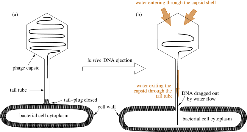
We denote the osmotic pressure of the culture medium by , and that of the cytoplasm of the infected cell by ; note that as the bacteria need to maintain positive turgor in order to grow [77]. Although in -based model of phage genome ejection turgor is assumed to provide a resisting force against , it is the bacterial Achilles heel that actually promotes complete genome ejection by a hydrodynamics mechanism. When the tail plug is removed by interaction with its receptor, water will flow up the osmotic gradient — one that exists between the growth medium and the cell cytoplasm — from the growth medium, through the capsid shell and tail tube into the cell cytoplasm. This directional water flow can drag the DNA into the infected cell. This mechanism is schematically shown in Fig. 6. It should be noted that bacterial cells respond immediately to changes in and they will always maintain some turgor even during phage infection.
When bacteria enter stationary phase, their turgor pressure drops considerably. This reduces the osmotic gradient between the external medium and the cytoplasm, in turn this reduces the strength of water flow from the culture medium into the cytoplasm. This may explain why bacteria in stationary phase are not easily infected by most phages. Notably, T7 is an exception, infecting stationary (and starved) cells at normal efficiency [94], even though subsequent phage development may be compromised by a lack of biosynthetic capacity in the cell.
Following infection by all but one of the phages we are aware of that have been tested, there is a transient drop in membrane potential and a transient release of intra-cytoplasmic ions into the external medium [81, 82]. Both occur during the period that DNA is thought to be ejected from the virion into the cell cytoplasm. In order to evaluate the role of the membrane potential during T4 infection, an extensive series of electrochemical studies that monitored ion fluxes were conducted (Reviewed in Refs. [83, 84]). Experiments were often combined with infective center assays after Hershey-Chase blending of phage-cell complexes, allowing estimates of - bp/sec for the rate of T4 genome internalization in vivo [81, 83, 85, 84]. Furthermore, T5 releases its DNA in two distinct steps [80]. During each step, cytoplasmic K+ leaks into the external medium, while during the pause between the steps no leakage occurs [86]. Thus, ion leakage is associated with phage DNA ejection, and if ions are leaking out, water should be flowing from the external medium through the phage virion into the cell cytoplasm to reduce the osmotic gradient. This is exactly what is expected if hydrodynamic forces are dragging the phage genome into the cell.
There are conflicting published data on membrane depolarization and ion leakage for phage T7, but it is now clear that T7 infection does not result in either phenomenon [87, 88], indicating that no open channel that could allow water flow exists between the environment and the infected cell cytoplasm. These are significant observations, because, unlike other phages, the internalization of T7 and related phage genomes is entirely catalyzed by molecular motors. These motors are enzymes that utilize cellular energy to transport the entire genome into the cell, each functioning at a constant rate regardless of how much DNA remains in the phage head [56, 89, 90, 91, 92, 93]. Thus, in the absence of hydrodynamic forces, energy-requiring enzymes are necessary to effect phage genome internalization by the cell.
The hydrodynamic model of DNA ejection does not require secondary ad hoc mechanisms to complete genome transfer into the cell. Because the water flow is determined by the values of and , the ejection process is (relatively) independent of DNA condensing agents. A bacterial cell responds immediately to changes in and will maintain some turgor even during phage infection. Thus, hydrodynamic flow from the external environment through the phage particle into the cell cytoplasm will continue until genome internalization is complete and the channel is closed. (It is not known how this occurs in any phage system, but all models of phage DNA ejection must invoke this step in order to prevent a permanent loss of the cellular membrane potential. If the potential is totally collapsed, the cell would die and no phage progeny would ever result.) Furthermore, although low concentrations of DNA condensing agents have a major effect on , they have little effect on either or (e.g., intracellular levels of polyamines, which are critical for ribosome and chromosome stability are tightly regulated). This means that phage DNA ejection in vivo should be relatively independent of the presence of DNA condensing agents in the growth medium. Indeed, ongoing experiments in the laboratory of one author (IJM) suggest that the latent periods and burst sizes of various phages (including parallel experiments with and the deletion mutant b221, the latter having a substantially reduced value, relative to wild-type , of ) are not affected by the presence of 1 mM spermine in the bacterial growth medium.
The hydrodynamic model also provides a mechanism for the ejection of single-stranded genomes and proteins into the cell during infection. This is important as all phages eject protein molecules into the infected cell, and a significant fraction of phages do not contain dsDNA packaged at the high density of mg/ml. Any molecule in the path of water flowing along the osmotic gradient between the external medium and the cell cytoplasm can be driven down the tail tube and into the cell. As this water flow is necessarily vectorial, nucleic acids or proteins to be ejected do not need to be positioned in the tail tube in the mature phage particle. Neither do they need to possess internal energy to drive their ejection from the capsid.
In summary, with the caveat that, to explain the multi-step process of T5 and perhaps other phages ( is one known example [95]), an additional ad hoc mechanism of protein conformational changes that temporarily block water flow from the external fluid into the cell cytoplasm, in turn temporarily stopping DNA ejection, the hydrodynamic model of phage DNA ejection is consistent with — and importantly can explain — all the observations we are aware of that have been made using any phage-host combination in vivo.
However, theories need to be tested experimentally. Determining the kinetics of phage genome internalization into infected cells would go a long way to supporting or refuting competing models. The internal capsid pressure models predict that the late stages of genome ejection should slow towards a zero rate as the residual pressure equals that in the cell cytoplasm, and that a secondary process using, for example, polyamines or DNA-binding proteins to interact with the entering the DNA completes the process. A secondary process is likely to occur with different kinetic parameters and may be experimentally detectable. Conversely, the hydrodynamic model would predict a largely constant rate of genome internalization, as the osmotic pressure gradient between the cell cytoplasm and the environment is unlikely to change substantially while viral DNA enters the cell. Unfortunately, it has proved very hard to measure directly the in vivo kinetics of genome entry outside the T7 family of phages, although some measurements have been made with phage N4 [96], and experiments with both and T5 are being initiated in the Molineux laboratory. However, it may be possible, using a combination of polyamines and external osmolytes, to increase the cytoplasmic osmotic pressure above that inside the mature phage head. If phage infection still occurs normally under those conditions, it would then provide irrefutable evidence that internal capsid pressure is not important for infection, although it would not provide any direct evidence for the hydrodynamic model.
Acknowledgements
Work in the laboratory of I. J. M. has been supported by grant GM32095 from the National Institutes of Health (USA). The authors are grateful to Anton Petrov and Steve Harvey for their extensive and constructive critiques of the paper. We also thank Theo Odijk for discussions, and Marco Bosch for help with Fig. 4.
References
- [1] North A C T and Rich A 1961 X-ray diffraction studies of bacterial viruses Nature 191 1242-1245
- [2] Earnshaw W C and Harrison S C 1977 DNA arrangement in isometric phage heads Nature 268 598-602
- [3] Earnshaw W C and Casjens S R 1980 DNA packaging by the double-stranded DNA bacteriophages Cell 21 319-331
- [4] Cerritelli M E, Cheng B, Rosenberg A H, McPherson C E, Booy F P and Steven A C 1997 Encapsidated conformation of bacteriophage T7 DNA Cell 91 271-280
- [5] Hud N V and Downing K H 2001 Cryoelectron microscopy of phage DNA condensates in vitreous ice: the fine structure of DNA toroids Proc Natl. Acad. Sci. USA 98 14925-14930
- [6] Olson N H, Gingery M, Eiserling F A and Baker T S 2001 The structure of isometric capsids of bacteriophage T4 Virology 279 385-391
- [7] Fokine A, Chipman P R, Leiman P G, Mesyanzhinov V V, Rao V B and Rossmann M G 2004 Molecular architecture of the prolate head of bacteriophage T4 Proc. Natl. Acad. Sci. USA 101 6003-6008
- [8] Agirrezabala X, Martin-Benito J, Caston J R, Miranda R, Valpuesta J M and Carrascosa J L 2005 Maturation of phage T7 involves structural modification of both shell and inner core components EMBO J. 24 3820-3829
- [9] Chang J, Weigele P, King J, Chiu W and Jiang W 2006 Cryo-EM asymmetric reconstruction of bacteriophage P22 reveals organization of its DNA packaging and infecting machinery Structure 14 1073-1082
- [10] Jiang W, Chang J, Jakana J, Weigele P, King J and Chiu W 2006 Structure of bacteriophage reveals genome organization and DNA packaging/injection apparatus Nature 439 612-616
- [11] Lander G C, Tang L, Casjens S R, Gilcrease E B, Prevelige P, Poliakov A, Potter C S, Carragher B and Johnson J E 2006 The structure of an infectious P22 virion shows the signal for headful DNA packaging Science 312 1791-1795
- [12] Xiang Y, Morais M C, Battisti A J, Grimes S, Jardine P J, Anderson D L and Rossmann M G 2006 Structural changes of bacteriophage upon DNA packaging and release EMBO J. 25 5229-5239
- [13] Tang J, Olson N, Jardine P J, Grimes S, Anderson D L and Baker T S 2008 DNA poised for release in bacteriophage Structure 16 935-943
- [14] Leiman P G, Battisti A J, Bowman V D, Stummeyer K, Mühlenhoff M, Gerardy-Schahn R, Scholl D and Molineux I J 2007 The structures of bacteriophages K1E and K1-5 explain processive degradation of polysaccharide capsules and evolution of new host specificities J. Mol. Biol. 371 836-849
- [15] Liu X, Zhang Q, Murata K, Baker ML, Sullivan MB, Fu C, Dougherty MT, Schmid MF, Osburne, MS, Chisholm SW and Chiu W 2010 Structural changes in a marine podovirus associated with release of its genome into Prochlorococcus Nat. Struct. Mol. Biol. 17 830-837
- [16] Chattoraj D K and Inman R B 1974 Location of DNA ends in P2, 186, P4, and lambda bacteriophage heads J. Mol. Biol. 87 11-22
- [17] Saigo K 1975 Tail-DNA connection and chromosome structure in bacteriophage T5 Virology 68 154-165
- [18] Saigo K, and Uchida H 1974 Connection of the right-hand terminus of DNA to the proximal end of the tail in bacteriophage Virology 61 524-536
- [19] Thomas J O 1974 Chemical linkage of the tail to the right-hand end of bacteriophage lambda DNA J. Mol. Biol. 87 1-9
- [20] Choi K H, McPartland J, Kaganman I, Bowman V D, Rothman-Denes L B and Rossmann M G 2008 Insight into DNA and protein transport in double-stranded DNA viruses: the structure of bacteriophage N4 J. Mol. Biol. 378 726-736
- [21] Comolli L R, Spakowitz A J, Siegerist C E, Jardine P J, Grimes S, Anderson D L, Bustamante C and Downing K H 2008 Three-dimensional architecture of the bacteriophage packaged genome and elucidation of its packaging process Virology 371 267-277
- [22] Hershey A D and Chase M 1952 Independent functions of viral protein and nucleic acid in growth of bacteriophage J. Gen. Physiol. 36 39-56
- [23] Stent G S 1963 Molecular Biology of Bacterial Viruses (San Francisco, CA: W.H. Freeman and Co.)
- [24] Hayes W 1964 The Genetics of Bacteria and Their Viruses (New York, NY: Wiley & Sons Inc.)
- [25] Zaríbnický V 1969 Mechanism of T-even DNA ejection J. Theoret. Biol. 22, 33-42
- [26] Smith D E, Tans S J, Smith S B, Grimes S, Anderson D L and Bustamante C 2001 The bacteriophage portal motor can package DNA against a large internal force Nature 413 748-752
- [27] Locker C R, Fuller S D and Harvey S C 2007 DNA organization and thermodynamics during viral packing Biophys. J. 93 2861-2869
- [28] Petrov A S, Boz M B and Harvey S C 2007 The conformation of double-stranded DNA inside bacteriophages depends on capsid size and shape J. Struct. Biol. 160 241-248
- [29] Petrov A S and Harvey S C 2008 Packaging double-helical DNA into viral capsids: structures, forces, and energetics Biophys. J. 95 497-502
- [30] Petrov A S, Locker C R and Harvey S C 2009 Characterization of DNA conformation inside bacterial viruses Phys. Rev. E 80 021914
- [31] Tzlil S, Kindt J T, Gelbart W M and Ben-Shaul A 2003 Forces and pressures in DNA packaging and release from viral capsids Biophys. J. 84 1616-1627.
- [32] Purohit P K, Inamdar M M, Grayson P D, Squires T M, Kondev J and Phillips R 2005 Forces during bacteriphage DNA packaging and ejection Biophys. J. 88 851-866
- [33] Kindt J, Tzlil S, Ben-Shaul A and Gelbart W M 2001 DNA packaging and ejection forces in bacteriophage Proc. Natl. Acad. Sci. USA 98 13671-13674
- [34] Inamdar M M, Gelbart W M and Phillips R 2006 Dynamics of DNA ejection from bacteriophages Biophys. J. 91 411-420
- [35] Purohit P K, Kondev J and Phillips R 2005 Mechanisms of DNA packaging in viruses Proc. Natl. Acad. Sci. USA 100 3173-3178
- [36] Evilevitch A, Gober J W, Phillips M, Knobler C M and Gelbart W M 2005 Measurements of DNA lengths remaining in a viral capsid after osmotically suppressed partial ejection Biophys. J. 88 751-756
- [37] Grayson P, Evilevitch A, Inamdar M M, Purohit P K, Gelbart W M, Knobler C M and Phillips R 2006 The effect of genome length on ejection forces in bacteriophage . Virology 348 430-436
- [38] Evilevitch A, Lavelle L, Knobler C M, Raspaud E and Gelbart W M 2003 Osmotic pressure inhibition of DNA ejection from phage Proc. Natl. Acad. Sci. USA 100 9292-9295
- [39] Jeembaeva M, Castelnovo M, Larsson F and Evilevitch A 2008 Osmotic pressure: resisting or promoting DNA ejection from phage? J. Mol. Biol. 381 310-323
- [40] Leforestier A, Brasiles S, de Frutos M, Raspaud E, Letellier L, Tavares P and Livolant F 2008 Bacteriophage T5 DNA ejection under pressure J. Mol. Biol. 384 730-739
- [41] de Frutos M, Letellier L and Raspaud E 2005 DNA ejection from bacteriophage T5: analysis of the kinetics and energetics Biophys. J. 88 1364-1370
- [42] Kazmierczak K and Rothman-Denes L 2006 Bacteriophage N4. In: Calendar R (ed) The Bacteriophages (Oxford: Oxford University Press) 302-314
- [43] McAllister W T 1970 Bacteriophage infection: which end of the SP82G genome goes in first? J. Virol. 5 194-198
- [44] Mangenot S, Hochrein M, Rädler J and Letellier L 2005 Real-time imaging of DNA ejection from single phage particles Curr. Biol. 15 430-435
- [45] Grayson P, Han L, Winter T and Phillips R 2007 Real-time observations of single bacteriophage DNA ejections in vitro Proc. Natl. Acad. Sci. USA 104 14652-14657
- [46] Rickgauer J P, Fuller D N, Grimes S, Jardine P J, Anderson D L and Smith D E 2008 Portal motor velocity and internal force resisting viral DNA packaging in bacteriophage Biophys. J. 94 159-167
- [47] São-José C, de Frutos M, Raspaud E, Santos M A and Tavares P 2007 Pressure built by DNA packing inside virions: enough to drive DNA ejection in vitro, largely insufficient for delivery into the bacterial cytoplasm J. Mol. Biol. 374 346-355
- [48] Evilevitch A, Fang L T, Yoffe A M, Castelnovo M, Rau D C, Parsegian V A, Gelbart W M, Knobler C M 2008 Effects of salt concentrations and bending energy on the extent of ejection of phage genomes Biophys J. 94 1110-1120
- [49] Riemer S C and Bloomfield V A 1978 Packaging of DNA in bacteriophage heads: some considerations on energetics Biopolymers 17 785-794
- [50] Harrison S C 1983 Packaging of DNA into bacteriophage heads: a model J. Mol. Biol. 171 577-580
- [51] Gabashvili I S and Grosberg A Yu 1992 Dynamics of double stranded DNA reptation from bacteriophage J. Biomol. Struct. Dyn. 9 911-920
- [52] Black L W 1989 DNA packaging in dsDNA bacteriophages Annu. Rev. Microbiol. 43 267-292
- [53] Serwer P 1986 Arrangement of double-stranded DNA packaged in bacteriophage capsids. An alternative model J. Mol. Biol. 190 509-551
- [54] Lepault J, Dubochet J, Baschong W and Kellenberger E 1987 Organization of double-stranded DNA in bacteriophages: a study by cryo-electron microscopy of vitrified samples EMBO J. 6 1507-1512
- [55] Leforestier A and Livolant F 2009 The bacteriophage genome undergoes a succession of intracapsid phase transitions upon DNA ejection J. Mol. Biol. 396 384-395
- [56] Kemp P, Gupta M and Molineux I J 2004 Bacteriophage T7 DNA ejection into cells is initiated by an enzyme-like mechanism Mol. Microbiol. 53 1251-1265
- [57] Molineux I J 2001 No syringes please, ejection of T7 DNA from the virion is enzyme-driven Mol. Microbiol. 40 1-8
- [58] Jeembaeva M, Jönsson B, Castelnovo M and Evilevitch A 2010 DNA heats up: energetics of genome ejection from phage revealed by isothermal titration calorimetry J. Mol. Biol. 395 1079-1087
- [59] Mdzinarashvili T J, Mrevlishvili G M, Khvedelidz M M, Ivanova A T, Janelidz N, Kiziria E L, Tushishvili D G, Tediashvili M I and Kemp R B 2006 Pycnometric, viscometric and calorimetric studies of the process to release the double-stranded DNA from the Un bacteriophage Biophys. Chem. 124 43-51
- [60] Serwer P 2003 Models of bacteriophage packaging motors J. Struct. Biol. 141 179-188
- [61] Serwer P 2004 T3/T7 DNA packaging. In Viral genome packaging machines: genetics, structure and mechanism Catalano C Ed (New York, NY: Kluwer Academic/Plenum Publishers)
- [62] Leikin S, Rau D C and Parsegian V A 1991 Measured entropy and enthalpy of hydration as a function of distance between DNA double helices Phys. Rev. A 44 5272-5278
- [63] Kleinschmidt A K, Lang D, Jacherts D and Zahn R K 1989 Preparation and length measurements of the total deoxyribonucleic acid content of T2 bacteriophages Biochim. Biophys. Acta 1000 41-48. This is a reprint of the original article in German (Kleinschmidt A K, Lang D, Jacherts D and Zahn R K 1962 Darstellung und längenmessungen des gesamten desoxyribonucleinsäure-inhaltes von T2-bakteriophagen Biochim. Biophys. Acta 61 857-864
- [64] Odijk T 1998 Hexagonally packed DNA within bacteriophage T7 stabilized by curvature stress Biophys. J. 75 1223-1227
- [65] Ivanovska I, Wuite G, Jönsson B and Evilevitch A 2007 Internal DNA pressure modifies stability of WT phage Proc. Natl. Acad. Sci. USA 104 9603-9608
- [66] Aubrey K L, Casjens S R and Thomas Jr. G J 1992 Secondary Structure and Interactions of the Packaged dsDNA Genome of Bacteriophage P22 Investigated by Raman Difference Spectroscopy Biochemistry 31 11835-11842
- [67] Overman S A, Aubrey K L, Reilly K E, Osman O, Hayes S J, Serwer P and Thomas Jr. G J 1998 Conformation and Interactions of the Packaged Double-Stranded DNA Genome of Bacteriophage T7 Biospectroscopy 4 S47-S56
- [68] Koch A L 1998 The biophysics of gram-negative periplasmic space Crit. Rev. Microbiol. 24 23-59
- [69] Molineux I J 2005 Fifty-three years since Harshey and Chase; much ado about pressure but which pressure is it? Virology 344 221-229
- [70] Raspaud E, Forth T, São-José C, Tavares P and de Frutos M 2007 A kinetic analysis of DNA ejection from tailed phages revealing the prerequisite activation energy. Biophys. J. 93 3999-4005.
- [71] Grayson P and Molineux I J 2007 Is phage DNA ‘injected’ into cells-biologists and physicists can agree Curr. Op. Microbiol. 10 401-409
- [72] Kemp P, Garcia L R and Molineux I J 2005 Changes in bacteriophage T7 virion structure at the initiation of infection Virology 340 307-317
- [73] Onorato L, Stirmer B, Showe M K 1978 Isolation and characterization of bacteriophage T4 mutant preheads J. Virol. 27 409-426
- [74] Leffers G and Rao V B 1996 A discontinuous headful packaging model for packaging less than headful length DNA molecules by bacteriophage T4 J. Mol. Biol. 258 839-850
- [75] Coren J, Pierce J and Sternberg N 1995 Headful packaging revisited: the packaging of more than one DNA molecule into a bacteriophage P1 head J. Mol. Biol. 249 176-184
- [76] Stock J B, Rauch B and Roseman S 1977 Periplasmic space in Salmonella typhimurium and Escherichia coli J. Biol. Chem. 252 7850-7861
- [77] Koch A L The surface stress theory of microbial morphogenesis 1983 Adv. Microbiol. Physiol. 24 301-336
- [78] Koch A L 1984 Shrinkage of growing Escherichia coli cells by osmotic challenge J Bacteriol. 159 919-924
- [79] Whatmore A M and Reed R H 1990 Determination of turgor pressure in Bacillus subtilis: a possible role for K+ in turgor regulation J. Gen. Microbiol. 136 2521-2526
- [80] McCorquodale D J and Warner H R 1988 Bacteriophage T5 and related phages. In The bacteriophages Calendar R Ed (New York, NY, Plenum Press)
- [81] Kalasauskaitė E V, Kadiăitė D L, Daugelavic̆ius R J, Grinius L and Jasaitis A A 1983 Studies on energy supply for genetic processes Eur. J. Biochem. 130 123-130
- [82] Dreiseikelmann B 1994 Translocation of DNA across bacterial membranes Microbiol. Rev. 58 293-316
- [83] Grinius L 1987 Energy transduction and gene transfer in chemotrophic bacteria: macromolecules on the move (Zurich, Switzerland: Harwood Academic Publishers)
- [84] Goldberg E B Grinius L and Letellier L 1994 In Molecular biology of bacteriophage T4 Karam J D Ed. 347-356 (Washington DC, USA: American Society for Microbiology)
- [85] Grinius L and Daugelavic̆ius R 1988 Depolarization of Escherichia coli cytoplasmic membrane by bacteriophages T4 and lambda: evidence for induction of ion-permeable channels Bioelectrochem Bioenerg. 19 235-245
- [86] Boulanger P and Letellier L 1992 Ion channels are likely to be involved in the two steps of phage T5 DNA penetration into Escherichia coli cells J. Biol. Chem. 267 3168-3172
- [87] Kuhn A and Kellenberger E 1985 Productive phage infection in Escherichia coli with reduced internal levels of the major cations J. Bacteriol. 163 906-912
- [88] Ramanculov E and Molineux I J unpublished data
- [89] García L R and Molineux I J 1995 Rate of translocation of bacteriophage T7 DNA across the membranes of Escherichia coli J. Bacteriol. 177 4066-4076
- [90] García L R and Molineux I J 1996 Transcription-independent DNA translocation of bacteriophage T7 DNA into Escherichia coli J. Bacteriol. 178 6921-6929
- [91] García L R and Molineux I J 1999 Translocation and cleavage of bacteriophage T7 DNA by the type I restriction enzyme EcoKI in vivo Proc. Natl. Acad. Sci USA 96 12430-12435
- [92] Struthers-Schlinke J S, Robins W P, Kemp P and Molineux I J 2000 The internal head protein gp16 of bacteriophage T7 controls DNA ejection from the virion J. Mol. Biol. 301 35-45.
- [93] Chang C-Y, Kemp P and Molineux I J 2010 Gp15 and gp16 cooperate in translocating bacteriophage T7 DNA into the infected cell Virology 398 176-186.
- [94] Schrader H S, Schrader J O, Walker J J, Wolf T A, Nickerson K W, Kokjohn T A 1997 Bacteriophage infection and multiplication occur in Pseudomonas aeruginosa starved for 5 years Can. J. Microbiol. 43 1157-1163
- [95] González-Huici V, Salas M, Hermoso J M 2004 The push-pull mechanism of bacteriophage DNA injection. Mol Microbiol 52 529-540
- [96] Demidenko A 2010 personal communication