Chemical Raman Enhancement of Organic Adsorbates on Metal Surfaces
Abstract
Using a combination of first-principles theory and experiments, we provide a quantitative explanation for chemical contributions to surface-enhanced Raman spectroscopy for a well-studied organic molecule, benzene thiol, chemisorbed on planar Au(111) surfaces. With density functional theory calculations of the static Raman tensor, we demonstrate and quantify a strong mode-dependent modification of benzene thiol Raman spectra by Au substrates. Raman active modes with the largest enhancements result from stronger contributions from Au to their electron-vibron coupling, as quantified through a deformation potential, a well-defined property of each vibrational mode. A straightforward and general analysis is introduced that allows extraction of chemical enhancement from experiments for specific vibrational modes; measured values are in excellent agreement with our calculations.
pacs:
78.30.-j, 31.15.A, 33.20.Fb, 68.43.PqThe ability to detect and characterize chemical species at the single-molecule level requires probes at the limits of present experimental resolution, and is a fundamental challenge to nanoscience. Since its discovery over three decades ago, surface enhanced Raman spectroscopy (SERS) has shown significant promise for sensing individual molecules adsorbed near metal nanostructures or substrates with nanoscale roughnessFleischman et al. (1974); Jeanmaire and Duyne (1977); Albrecht and Creighton (1977). In SERS, the conversion of incident light into surface plasmons near asperities on metal surfaces, combined with chemical and resonant effects, has been reported to yield Raman cross sections increased by factors of up to 1014, enabling single-molecule detection Moskovits (2005); Kneipp et al. (1997); Michaels et al. (2000); Nie and Emory (1997); Haran (2010); Ward et al. (2008). While the enhancement associated with surface plasmons can reach 108 Willets and Duyne (2007), remaining increases and associated changes in mode frequencies have been reported to arise from chemical adsorption Moskovits (1985); Campion and Kambhampati (1998); Moskovits (2005); Jensen et al. (2008); Morton and Jensen (2009); Heller et al. (1982); Persson (1981), as well as resonant intra-molecular and metal-molecule charge transfer Adrian (1982); Arenas et al. (1996); Lombardi and Birke (2008). Despite a wealth of prior theoretical studies, these chemical enhancement (CE) mechanisms have remained poorly understood and difficult to quantify Campion and Kambhampati (1998); Moskovits (2005); Jensen et al. (2008); Morton and Jensen (2009). While for some vibrational modes CE is estimated Moskovits (1985); Campion and Kambhampati (1998); Moskovits (2005); Maitani et al. (2009) to be about 10-100, there is currently no clear picture for why certain modes are enhanced more than others. Previous theoretical studies have proposed mechanisms for this mode dependence Heller et al. (1982); Persson (1981); Morton and Jensen (2009); Adrian (1982); Arenas et al. (1996); Lombardi and Birke (2008), but none of these models have been validated by experiments or more rigorous first-principles calculations, leaving the origin of dominant chemical contributions to SERS an open question Campion and Kambhampati (1998); Moskovits (2005); Jensen et al. (2008); Morton and Jensen (2009).
In this Letter, we use a combination of first-principles calculations and experiment to demonstrate the origin of chemical contributions to SERS for benzene thiol (BT) molecules chemisorbed to Au surfaces. From our density functional theory (DFT) calculations of static contributions to the Raman tensor, we elucidate the vibrational mode dependence Campion and Kambhampati (1998); Moskovits (2005); Jensen et al. (2008); Morton and Jensen (2009) of the chemical enhancement, explicitly relating modes with the largest CE to those with the greatest mode-induced shift of the molecular frontier orbital energy, as quantified through a deformation potential. Relative CE of BT vibrational modes measured at different probe frequencies agree quantitatively with our static calculations for all binding sites considered. While the magnitude of CE for BT on Au is sensitive to binding motif, its relative value is not.
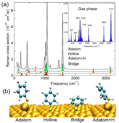
DFT calculations are performed using the Vienna Ab-initio Simulations Package (VASP) and within a generalized gradient approximationPerdew et al. (1996); Kresse and Furthmüller (1995). We model the BT adsorbate-Au substrate system with an ordered monolayer of one BT molecule per nm2 bonded to a flat periodic Au(111) slab. Our supercell consists of 5 atomic layers of Au stacked along [111] with 16 atoms per layer, with 30 Å of vacuum. The forces of the three upper layers and molecule are well converged to less than 1 meV/Å. The in-plane lattice parameters are kept fixed to their computed Au fcc bulk value of 4.17 Å. A 400 eV plane-wave cutoff and 2x2x1 Monkhorst-Pack k-point mesh is used for calculations involving Au slabs. Four binding geometries are considered, as shown in Fig. 1b: fcc hollow (EB=0.219 eV), adatom (EB=0.446 eV), hydrogenated adatom (EB=0.795 eV), and bridge (EB=0.192 eV), where EB is the calculated binding energy relative to a free Au surface and gas-phase in the dilute limit. The gas-phase BT molecule is simulated in the same large supercell as the slab geometry, but using the point only.
Static Raman tensors are constructed mode-by-mode using a finite-differences approach, in two steps. First, the dynamical matrix of the system is generated by displacing each atom along each Cartesian direction by 0.03 Å. Vibrational frequencies and corresponding phonon eigenvectors are obtained by diagonalization of a truncated dynamical matrix treating only BT atoms, and Au atoms directly bonded with sulfur. (The remainder of Au atoms is treated in an infinite mass approximation.) Second, we compute the static polarizability within a second-order finite-difference expression using a saw-tooth potential with a gradient of 1 mV/Å, and compute its derivative as a function of the amplitude for each vibrational eigenmode. (See Supplementary Information for additional details.) Throughout the paper, all modes are labeled with gas-phase frequencies for simplicity.
In Fig. 1a, we report Raman cross sections calculated from the dominant (non-evanescent) component of the Raman tensor, , where is the normal to the surface. Prominent Raman peaks in the computed spectra agree well with experimentGuieu et al. (2009). Upon adsorption, BT vibrational frequencies are altered on average by 10-20 cm-1, in agreement with experiments (Supplementary Information). From Fig. 1a, the presence of the Au substrate enhances Raman cross sections for some modes more than for others. The intensities of modes with larger enhancements exhibit a stronger dependence on binding site. For example, while the 1100 cm-1 mode varies by more than two orders of magnitude across the different sites considered, the 3151 cm-1 mode remains relatively unaltered and comparable to the gas-phase intensity.
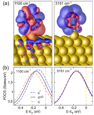
In Fig. 2a, we compare the computed charge density change induced by each mode, , where is the vibrational eigenvector. We find that the 1100 cm-1 mode induces significant charge redistribution within the Au surface, while the 3151 cm-1 mode does not. This behavior is entirely consistent with mode-induced changes observed in the DFT electronic structure (Fig.2b): The peak Kohn-Sham HOMO energy EHOMO shifts noticeably relative to the Fermi level EF with the 1100 cm-1 mode, whereas the 3151 cm-1 mode leaves the HOMO peak unchanged. Examining all modes, we find that modes that show larger enhancements induce a larger polarization response in the substrate (and larger shift in EF-EHOMO).
To rationalize our DFT results, we compare a model expression for the static polarizability and Raman tensor with our more rigorous first-principles calculations. We consider a single (dominant) term in an approximate single-particle form for the ground state electronic polarizability, one that includes just a lone virtual transition between the HOMO, , and a metallic state at the Au Fermi level, . Within this two-state approximation, an adsorbate-metal interfacial contribution to the Raman tensor RBT-Au can be expressed asJensen et al. (2008); Morton and Jensen (2009)
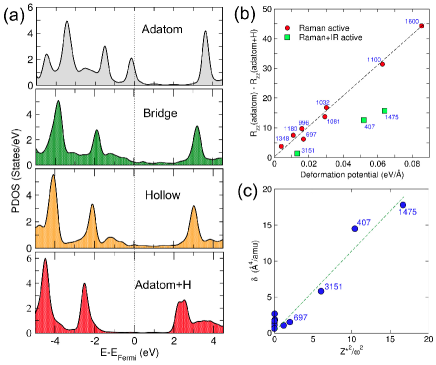
| (1) |
where is a dipole matrix element in the Cartesian direction , and is the difference between their eigenvalues. For modes that are not also IR-active, terms involving derivatives of induced dipole moments can be neglected, and Eq. (1) reduces to a term proportional to . As noted previouslyMorton and Jensen (2009), the factor in the denominator is consistent with a sensitivity of the overall spectral enhancement to the alignment of the frontier molecular electronic orbital with the metal Fermi energy. Indeed, this factor can be used to rationalize the binding site dependence of enhancements shown in Fig. 1a, and corresponding partial densities of states shown on Fig.3a. Calculated enhancements on the adatom site, for which the HOMO is close to EF, are significantly stronger than on other sites, where this level is broadened and further away from EF. However, this factor is the same for all modes, and cannot explain the mode-dependence of CE computed in Fig. 1a.
The strong modification of the BT Raman spectra by the Au substrate can be explained via computation of a deformation potential, , for each mode, i.e. the change in molecular electronic level alignment relative to the metallic Fermi level induced by a particular vibration mode. In Fig. 3b, we plot the difference against . Because the Kohn-Sham HOMO level of the adatom+H geometry is much further from the Fermi energy than for the adatom binding site (see Fig. 3a), is large, and the adatom+H geometry has a negligible interfacial contribution. Thus, to an excellent approximation, taking the difference removes intramolecular contributions to the Raman tensor unrelated to Eq. (1). Indeed, in Fig. 3b, we find a remarkable correlation between the interface contribution to the deformation potential and enhancement for most of the vibrational modes. The modes that deviate from the linear trend at 407 cm-1 (Au-S stretch), 1475 cm-1 (phenyl ring stretch), and 3151 cm-1 (C-H stretch) have significant IR activity. In the static limit used here, polarization induced by these IR active modes screens the electric field experienced by the molecule, leading to a reduction in their Raman cross sectionsCardona and Güntherodt (1982) by a factor proportional to , with the mode dynamical charge given by and where is the mode-induced interfacial dipole moment. In Fig. 3c, we show the deviation of the IR-active “outlier” modes from the linear trend observed for non-IR active modes versus . The correlation between and confirms that modes with larger contributions to the screening lead to greater deviations from the two-state model (Fig. 3b). We note however, that although important in our static calculations, this screening effect will have impact only below infrared frequencies, and thus will be inconsequential for typical probe frequencies, where the local fields will vary too rapidly for the IR-active vibrations to respond.
With this information, we can now connect the strong modification of Raman spectra by substrates to mode-specific changes of the electronic structure of the metal-adsorbate interface: modes with the largest interfacial contribution to the change in polarizability, as quantified through a deformation potential, result in the most substantial chemical enhancements. For BT, these modes are those that break the conjugation of the HOMO (Supplementary Information). Fig. 2a shows vividly how the 1100 cm-1 mode breaks the resonant character of the carbon ring, while the 3151 cm-1 mode leaves the -symmetry of the electrons on the phenyl ring intact. This suggests that for future adsorbates, the nature of modes with the largest CE might be intuitively rationalized a priori.
To validate our theory of chemical enhancement, we compare with experimental Raman and SERS measurements of BT on rough Au substrates (Fig. 4a,b). SERS substrates, consisting of roughened SiGe surfaces (Fig.4a) coated with 30 nm of Au, were incubated in 3mM BT solution (Sigma Aldrich W361607) in methanol overnight, then gently rinsed with methanol and dried by nitrogen gas. Raman (neat solution) and SERS spectra were collected at two wavelengths, 632.8 nm and 785 nm, using an inverted microscope set-up coupled to a spectrometer (Acton SpectraPro 2300i) equipped with a liquid-nitrogen-cooled charge-coupled device camera. To eliminate uncertainties associated with the number of molecular analytes in comparing Raman and SERS intensities, we normalize ratios of Raman and SERS spectra peak heights to the 996 cm-1 mode, which has only a modest enhancement from our calculations. We note that absolute CEs can be obtained if we normalize to a mode with zero deformation potential, , see mode 1348 cm-1 in Fig.3b, for example. As the 1348 cm-1 mode is not easily observed experimentally, we use 996 cm-1 in this case. Assuming the electromagnetic enhancement is the same for all modes, this relative enhancement will reflect CE. This assumption is acceptable for modes within a few hundred wavenumbers of the 996 cm-1 mode, based on the width on relatively low Q-factor for localized plasmon resonance in Au or Ag.
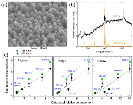
In Fig.4c, we compare directly Raman cross sections from Fig.1a (computed from ) to the averaged solution phase data from Fig.1a (inset). Binding geometries for BT on flat Au surfaces are the subject of debate in the literature; and for rough surfaces at room temperature, a variety of adsorption sites will be available, and binding geometries would be subject to thermal fluctuationsZhang et al. (2007); Ward et al. (2008). By comparing three very different, energy-minimized binding sites to the experiment, we sample different possibilities for time-averaged experimental binding morphologies. Surprisingly, all three geometries show good correlation, with the adatom site showing perhaps the most linear trend. (We note that SERS measurements do not observe the S-H stretching mode at 2600 cm-1 indicating the loss of hydrogen at the S-Au in the room-temperature experiments.) Importantly, the relative theoretical enhancements, also normalized to the 996 cm-1 mode, are in excellent quantitative agreement with the present experiments, as well as others in literature taken from different substrates Biggs et al. (2009); Aggarwal et al. (2009) (Supplementary Information).
In summary, through our calculations and comparison with experiment, we have demonstrated that the strong modification of Raman spectra by the substrate is a chemical effect, largely independent of laser probe frequency, and associated with the change in electronic structure of the molecule by the metal substrate. The mode dependence of chemical enhancement can be connected directly to interfacial contributions to the deformation potential, a well-defined intrinsic property of each vibrational mode and substrate. A new analysis of experimental SERS data is introduced that allows for direct comparison of theoretical calculations with experimental data. Comparing enhancements relative to a particular mode, we find excellent agreement between theory and experiment, indicating standard DFT approaches captures accurately and quantitatively dominant contributions to CE, even in the static limit. Additional support for our conclusions could be obtained by inelastic electron tunneling measurements, which provide direct access to interfacial contributions to the electron-vibron coupling. The quantitative connection between the deformation potential and chemical enhancement provides new opportunities for detection and control of adsorbate-metal interactions through SERS.
We thank L. Kronik, D. Prendergast, I. Tamblyn, and other colleagues at Molecular Foundry and UC Berkeley for helpful discussions. This work was supported by the AFOSR/DARPA Project BAA07-61 “SERS S&T Fundamentals” under contract FA9550-08-1-0257, and the Molecular Foundry through the Office of Science, Office of Basic Energy Sciences, of the U.S. Department of Energy under Contract No. DE-AC02-05CH11231. Computational resources were provided by DOE (LBNL Lawrencium, NERSC Franklin) and DOD (HPCMP ARL MJM).
References
- Fleischman et al. (1974) M. Fleischman, P. J. Hendra, and A. McQuillan, Chem. Phys. Lett. 26, 163 (1974).
- Jeanmaire and Duyne (1977) D. L. Jeanmaire and R. P. V. Duyne, J. Electroanal. Chem. 84, 1 (1977).
- Albrecht and Creighton (1977) M. G. Albrecht and J. A. Creighton, J. Am. Chem. Soc. 99, 5215 (1977).
- Moskovits (2005) M. Moskovits, J. Raman Spectroscopy 36, 485 (2005).
- Kneipp et al. (1997) K. Kneipp, Y. Wang, H. Kneipp, L. T. Perelman, I. Itzkan, R. R. Dasari, and M. S. Feld, Phys. Rev. Lett. 78, 1667 (1997).
- Michaels et al. (2000) A. M. Michaels, J. Jiang, and L. Brus, J. Chem. Phys. B 104, 11965 (2000).
- Nie and Emory (1997) S. Nie and S. R. Emory, Science 275, 1102 (1997).
- Haran (2010) G. Haran, Acc. Chem. Res. 43, 1135 (2010).
- Ward et al. (2008) D. R. Ward, N. J. Halas, J. W. Ciszek, J. M. Tour, Y. Wu, P. Nordlander, and D. Natelson, Nano Lett. 8, 919 (2008).
- Willets and Duyne (2007) K. A. Willets and R. P. V. Duyne, Annu. Rev. Phys. Chem. 58, 267 (2007).
- Moskovits (1985) M. Moskovits, Rev. Mod. Phys. 57, 783 (1985).
- Campion and Kambhampati (1998) A. Campion and P. Kambhampati, Chem. Soc. Rev. 27, 241 (1998).
- Jensen et al. (2008) L. Jensen, C. M. Aikens, and G. C. Schatz, Chem. Soc. Rev. 37, 1061 (2008).
- Morton and Jensen (2009) S. M. Morton and L. Jensen, J. Am. Chem. Soc. 131, 4090 (2009).
- Heller et al. (1982) E. J. Heller, R. L. Sundberg, and D. Tannor, J. Chem. Phys. 86, 1822 (1982).
- Persson (1981) B. N. J. Persson, Chem. Phys. Lett. 82, 561 (1981).
- Adrian (1982) F. J. Adrian, J. Chem. Phys. 77, 5302 (1982).
- Arenas et al. (1996) J. F. Arenas, I. L. Tocón, J. C. Otero, and J. I. Marcos, J. Phys. Chem. 100, 9254 (1996).
- Lombardi and Birke (2008) J. R. Lombardi and R. L. Birke, J. Phys. Chem. C 112, 5605 (2008).
- Maitani et al. (2009) M. M. Maitani, D. A. A. Ohlberg, Z. Li, D. L. Allara, D. R. Stewart, and R. S. Williams, J. Am. Chem. Soc. 131, 6310 (2009).
- Perdew et al. (1996) J. P. Perdew, K. Burke, and M. Ernzerhof, Phys. Rev. Lett. 77, 3865 (1996).
- Kresse and Furthmüller (1995) G. Kresse and J. Furthmüller, Phys. Rev. B 54, 11169 (1995).
- Guieu et al. (2009) V. Guieu, P. Garrigue, F. Lagugné-Labarthet, L. Servant, N. Sojic, and D. Talaga, Optics Express 17, 24030 (2009).
- Cardona and Güntherodt (1982) M. Cardona and G. Güntherodt, “Light scattering in solids ii: Basic concepts and instrumentation,” (Springer-Verlag, Berlin Heidelberg New York, 1982) Chap. Resonance Phenomena.
- Zhang et al. (2007) W. Zhang, B. S. Yeo, T. Schmid, and R. Zenobi, J. Phys. Chem. C 111, 1733 (2007).
- Biggs et al. (2009) K. B. Biggs, J. P. Camden, J. N. Anker, and R. P. V. Duyne, J. Chem. Phys. 113, 4581 (2009).
- Aggarwal et al. (2009) R. L. Aggarwal, L. W. Farrar, E. D. Diebold, and D. L. Polla, J. Raman Spectrosc. 40, 1331 (2009).