A simplified exactly solvable model for -amyloid aggregation
Abstract
We propose an exactly solvable simplified statistical mechanical model for the thermodynamics of -amyloid aggregation, generalizing a well–studied model for protein folding. The monomer concentration is explicitly taken into account as well as a non trivial dependence on the microscopic degrees of freedom of the single peptide chain, both in the -helix folded isolated state and in the fibrillar one. The phase diagram of the model is studied and compared to the outcome of fibril formation experiments which is qualitatively reproduced.
Amyloids are insoluble fibrillar aggregates of proteins, stabilized mostly by hydrogen bonds and hydrophobic interactions. They are implicated in debilitating human pathologies, such as Alzheimer’s, Parkinson’s disease and spongiform encephalopathies. Citotoxic species have been recently identified with transient soluble oligomeric structures whereas amyloid fibrils are believed to be the final most stable state of the aggregation process ChitiDobson . Virtually all proteins can be induced to adopt the amyloid structure upon appropriate conditions Fandrich .
A common signature of fibril formation is the presence of a stable core of cross- structure, with -strands running orthogonal to the fibril axis and forming several -sheets which may intertwine along the latter. The cross- structure is identified through its typical X-rays diffraction pattern and binding to specific fluorescent dyes. More sophisticated techniques, such as solid state NMR, are needed in order to provide structural models at atomic level. In a few known cases, for intermediate chain lengths in between 20 and 40, all peptide monomers may adopt a repeating hairpin structure within the fibrillar aggregate Petkova ; Ferguson . This is then stabilized by interchain hydrogen bonds between the same residues in different chains, leading to the so–called parallel in–register arrangement Plos06 ; Eisenberg shown in Fig. 1.
The conformational ensembles populated at low concentration by proteins, which aggregate into amyloid fibrils at higher density, may vary from the large amount of fluctuating structures of natively unfolded proteins and peptides, such as the A-peptide related to Alzheimer’s, to the well defined structures of globular proteins. In the latter case, the competition between the stability of the native structure and of the amyloid fibrils is crucial in determining the amyloidogenic behavior ChitiDobson2 .
In the context of protein folding, simple models based on the geometry of the native structure have been very useful in unraveling folding kinetics. In the same manner, one can speculate that the geometry of the fibrillar aggregate, as typified by the parallel in–register hairpin structure, may play a similar role in aggregation kinetics. Within this spirit, the competition described above for the aggregation of globular proteins becomes a competition between two alternative geometries, which needs to be assessed already at equilibrium.
The purpose of the present Letter is proposing a simplified statistical mechanical model for -amyloid aggregation, generalizing a well–studied one for protein folding. Our model explicitly depends on protein concentration and has the virtue of being exactly solvable. For more realistic descriptions, even at a coarse–grained level, the computational cost of achieving thermodynamic equilibrium at different concentrations is prohibitive. On the other hand, here we consider a non trivial dependence on the microscopic degrees of freedom of the single peptide chain, both in the folded and in the fibrillar state. Other simplified models describe monomers through just a few macrostates Ferrando ; Nicodemi ; Lee . Notably, we succeed in reproducing, at least qualitatively, the behavior of fibril formation experiments in the presence of the denaturant trifluoroethanol (TFE) in different concentrations.
Our model starts from the one introduced by Wako and Saitô WS1 ; WS2 and then reconsidered by Muñoz, Eaton and co–workers ME1 ; ME2 ; ME3 (WSME–model). The latter has been the subject of many works with applications to real proteins BP ; ItohSasai1 ; AbeWako ; ZP1 ; BPZ2 ; IPZ1 ; IP . Despite its simplicity, it has been able to capture the main features of the kinetic behavior and folding pathways of specific molecules.
The WSME–model is a highly simplified model of the protein folding process built on the premise that the latter is mainly determined by the structure of the native functional state, whose knowledge is assumed. Only native interactions are included, classifying the model as Gō–like Go . Moreover, the interaction between two aminoacids in the protein sequence is possible only if all intervening peptide bonds are in their native conformation. The entropy loss due to fixing peptide units in this conformation is finally explicitly taken into account.
Within this framework, a polypeptide chain made up of aminoacids is described as a sequence of peptide bonds. Two conformations are considered for each bond: the native one and a generic disorder state. Thus, a binary variable is associated to the -th peptide unit, taking value 1 and 0 in the two cases respectively, and the free energy of the model can be written in unit of , with the absolute temperature, as
| (1) |
The contact matrix, with entry equal to 1 if the
-th
and -th bonds are close to each other in the native
structure and equal to 0 otherwise, tell us which are the native
interactions. Their energetic amount is then quantified by the
dimensionless contact energy , referring to the
-th and -th peptide units. This contributes to the free energy
only if the product does not vanish, that is only
if such two bonds are the ends of a sequence of ordered peptide units,
thus realizing the depicted interaction. Finally, recognizing the
microscopic multiplicity of an abstract disorder state, an entropic
cost is given to the ordering of the -th peptide bond.
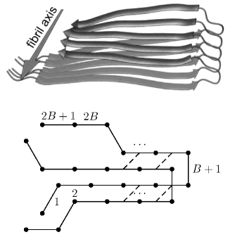
Our model is an extension of the WSME–model, suitable for the thermodynamics of -amyloid aggregation. The basic idea is that peptide monomers can either fold into their native structure or partially lose this feature before aggregating in fibrils. Here we focus, for simplicity, on -helices while the aggregation is assumed to require a hairpin shape and proceed by parallel in–register arrangement as in Fig. 1, thus mimicking real fibrils. Other in–register amyloid structures with more than two -layers Kajava2004 could be also implemented.
We will define the model in a bottom-up approach. Let us begin introducing the free energy of isolated monomers, which can fold into -helix native structure. In such a structure an hydrogen bond is formed between peptide units and so that . Then, for a homogeneous molecule with an odd number of peptide bonds, , following Eq. (1) we choose the free energy as
| (2) |
because if and 0 otherwise. The dimensionless parameters and account respectively for the energy strength of each contact and the entropic cost of ordering each bond.
As far as the interaction between different peptides is concerned, we
assume that aggregation involves and requires a partial
-hairpin shape, which is obtained by removing some helical
contacts. In such a view, the small loop region of the hairpin formed
by a monomer with peptide units is identified with the peptide
bond , from which two strands depart as shown in
Fig. 1.
Fibril formation is triggered by pairing a
part of the ordered fragments of the two strands from one molecule
with the same part of another. A measure of the “-order”
extent associated to a pair of consecutive -hairpins, with
WSME–variables and , is provided by
and
vanishes if loop regions are not both ordered. Otherwise, it is the
common number of ordered peptide units facing each other beginning
from loops. For the case shown in Fig. 1, we have
.
We can then interpret the aggregation phenomenon, which is driven and stabilized by hydrogen bonds, as the formation of contacts between the -portions of the two different monomers, where has thus to be in between 0 and . We assume that the pairing between different peptides starts from loops and go on sequentially along the strands, suggesting the idea that these regions, having the same shape, are the most suitable to initiate the aggregation. In equilibrium conditions this mechanism corresponds to assume that there is only one way to form the above contacts. In Fig. 1 all available interactions of this kind are present.
A segment can gain energy being either in a helical state and unbounded by other peptides or in a -hairpin state and bounded to another hairpin. We assume that, if a molecule binds another one with hydrogen bonds, then the helical contacts including peptide bonds participating to the pairing, that is in the stretch going from to , are suppressed. Hence, the free energy of that monomer becomes
| (3) |
being the free energy of the isolated -helix defined by Eq. (2). In turn, we shall denote by the energetic gain, in unit of , of one contact between different monomers.
Now we take into account the translational and rotational entropy loss due to the aggregation of different peptides with the formation of hydrogen bonds. We will choose so that . At last, the free energy for a system of two close molecules that can aggregate takes the form , with the constraint .
We want to stress that the -hairpin shape is not needed a priori in order to have aggregation between different monomers, but it is rather considered as a concomitant event to the matching process. Moreover, the requirement of ordered stretches of peptide units to form contacts between molecules is just a way to express that only few chain conformations are suitable for aggregation. Intra–helix contacts represent general native interactions protecting isolated conformers from the aggregation–prone states ChitiDobson2 .
Finally, we model the formation of an aggregate as a growth of a “one–dimensional structure”. To this aim, we describe a system of many peptides by placing them on distinct sites , , of a one–dimensional lattice and including in the model only interactions between nearest–neighbor molecules. The occupation number of site is 1 if a monomer is present in that position and 0 otherwise. Furthermore, to each site we associate WSME–variables describing the conformation of the peptide chain placed there and thus will give the state of the -th peptide unit of the molecule at . In order to avoid an unphysical entropic contribution, we set at 0 for any if . For simplicity, the symbol will be used for the array of binary variables related to the node , for all these variables and for the collection of occupation numbers. Finally, we need the variable keeping count of the contacts between close molecules residing at nodes and . This variable ranges from 0 to and, as expected, no interaction is possible between sites and when they are not both occupied.
The free energy of the full model is then a generalization of the one introduced above for two monomers. Using the dummy variables and and noticing that the number of peptide units of the molecule at site involved in contacts with other molecules is properly related to , this free energy reads
| (4) | |||||
The contribution of the chemical potential , which will be determined by imposing a given value to the monomer density, has been here included.
The Boltzmann distribution with the free energy of Eq. (4) provides the possibility to evaluate equilibrium expectation values of physical observables. The present model can be solved exactly by means of a transfer matrix method, because of the presence of short–range interactions in a 1-dimensional system and the possibility of exactly tracing on the WSME–variables. Details are shown in the supplementary material EPAPS . Here we restrict to some results on the behavior of two order parameters related to the fraction of isolated helices and aggregated molecules. The former, EPAPS , measures the global order of peptides when they do not interact at all and is defined as the equilibrium average of the fraction of native bonds per site, normalized to the density , considering only microscopic configurations which exclude aggregation phenomena. The latter, EPAPS , accounts for bonds between different monomers and is given by the fraction of formed contacts between two consecutive lattice nodes, again normalized with respect to .
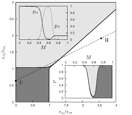
Fig. 2 shows the phase diagram of the model in the thermodynamic limit , where different phases are separated by the conditions and . Here we choose and but other values of only marginally affect this diagram. Moreover, we consider the case and independent of if , assuming that most of the entropy loss in aggregation is due to the formation of just one contact between monomers. Parameters and are referred to the midpoint , depending on both and , of an helix in the pure WSME–model. The energy scale may be obtained by imposing when the density approaches 0.
Three regions are recognized. The first is the region of unfolded isolated peptides where both and are less than 1/2. For small the order parameter is closed to the value of a completely unfolded structure in the WSME–model. The second region, where and , corresponds to native isolated helical peptides. The boundary between the unfolded region and the -helix region is weakly depending on both and and almost coincides with the one obtainable in the plain WSME–model for the same helices. Finally, there is the fibril region where and . Increasing or decreasing favors the aggregation by lowering the boundary between this region and the denatured one.
Since the energetic parameters and are effective parameters mediated by solvent, we may expect them to vary in a non trivial way as external conditions, such as temperature, different denaturant concentrations, solution ionic strength and pH, are changed. The denaturant agent TFE is commonly used in fibril formation assays because, at moderate concentrations, it disrupts the native structures of isolated proteins, without preventing the formation of inter–molecular contacts TFE . At high concentrations, TFE addition results in the stabilization of isolated unfolded proteins TFE .
We can mimic the TFE effect by assuming that both and are simple linear decreasing functions of its concentration , with decreasing more than . For example, by moving along the straight line in Fig. 2 from H at to U at , the observed native–fibril–unfolded pattern TFE can be qualitatively reproduced. Given such a dependence of and on , the top inset in Fig. 2 depicts the profile of and as a function of the TFE concentration whereas the bottom one reports the phase diagram of the model in the plane . The pattern discussed above is present for high values of peptide density, with the fibril stability interval in TFE concentration narrowing with decreasing peptide density. At low density the fibril phase is not present anymore and the peptides remain always isolated going directly from the native to the unfolded state, with increasing TFE concentration.
In summary, in this Letter we have proposed a highly simplified equilibrium model to describe the aggregation of identical monomers and the consequent formation of fibrillar structures. Despite its simplicity, the model has been shown to explain different phases of the system, such as unfolded and aggregated states, and to reproduce qualitatively the observed trend of fibril formation experiment as a function of trifluoroethanol concentration. Moreover, we argue that a kinetic version of the model could shed new light on the protein aggregation dynamics and work is in progress along this line.
This work has been supported by the Italian Ministry of Education, University and Research via the PRIN 2007B57EAB and by University of Padua via Progetto di Ateneo CPDA083702.
References
- (1) F. Chiti and C. M. Dobson, Ann. Rev. Biochem. 75, 333 (2006).
- (2) M. Fandrich and C. M. Dobson, EMBO J. 21, 5682 (2002).
- (3) A. T. Petkova, et. al., Proc. Natl. Acad. Sci. U.S.A. 99, 16742 (2002).
- (4) N. Ferguson, et. al., Proc. Natl. Acad. Sci. U.S.A. 103, 16248 (2006).
- (5) M. R. Sawaya, et. al., Nature 447, 453 (2007).
- (6) A. Trovato, F. Chiti, A. Maritan, and F. Seno, PLoS Comput. Biol. 2, 1608 (2006).
- (7) F. Chiti and C. M. Dobson, Nat. Chem. Biol. 5, 15 (2009).
- (8) R. Gaspari, A. Gliozzi, and R. Ferrando, Phys. Rev. E 76, 041604 (2007).
- (9) M. Nicodemi, A. de Candia, and A. Coniglio, Phys. Rev. E 80, 041914 (2009).
- (10) Chiu Fan Lee, Phys. Rev. E 80, 031922 (2009).
- (11) H. Wako and N. Saitô, J. Phys. Soc. Jpn 44, 1931 (1978).
- (12) H. Wako and N. Saitô, J. Phys. Soc. Jpn 44, 1939 (1978).
- (13) V. Muñoz, P. A. Thompson, J. Hofrichter, and W. A. Eaton, Nature (London) 390, 196 (1997)
- (14) V. Muñoz, E. R. Henry, J. Hofrichter, and W. A. Eaton, Proc. Natl. Acad. Sci. U.S.A. 95, 5872 (1998).
- (15) V. Muñoz and W. A. Eaton, Proc. Natl. Acad. Sci. U.S.A. 96, 11311 (1999).
- (16) P. Bruscolini and A. Pelizzola, Phys. Rev. Lett. 88, 258101 (2002).
- (17) K. Itoh and M. Sasai, Proc. Natl. Acad. Sci. U.S.A 101, 14736 (2004).
- (18) H. Abe and H. Wako, Phys. Rev. E 74, 011913 (2006).
- (19) M. Zamparo and A. Pelizzola, Phys. Rev. Lett. 97, 068106 (2006).
- (20) P. Bruscolini, A. Pelizzola, and M. Zamparo, Phys. Rev. Lett. 99, 038103 (2007).
- (21) A. Imparato, A. Pelizzola, and M. Zamparo, Phys. Rev. Lett.98, 148102 (2007).
- (22) A. Imparato and A. Pelizzola, Phys. Rev. Lett. 100, 158104 (2008).
- (23) N. Gō and H. Taketomi, Proc. Natl. Acad. Sci. U.S.A. 75, 559 (1978).
- (24) A. V. Kajava, U. Baxa, R. B. Wickner, and A. C. Steven, Proc. Natl. Acad. Sci. U.S.A. 101, 7885 (2004).
- (25) Supplementary material. In this appendix we show some analytical properties of our model, focusing in particular on the order parameters and .
- (26) F. Chiti, N. Taddei, M. Bucciantini, P. White, G. Ramponi, and C. M. Dobson, EMBO J. 19, 1441 (2000).