Observation of phonons with resonant inelastic x-ray scattering
Abstract
Phonons, the quantum mechanical representation of lattice vibrations, and their coupling to the electronic degrees of freedom are important for understanding thermal and electric properties of materials. For the first time, phonons have been measured using resonant inelastic x-ray scattering (RIXS) across the Cu -edge in cupric oxide (CuO). Analyzing these spectra using an ultra-short core-hole lifetime approximation and exact diagonalization techniques, we can explain the essential inelastic features. The relative spectral intensities are related to the electron-phonon coupling strengths.
1 Introduction
It is generally accepted that the retardation of the Coulomb interaction due to the coupling between electrons and phonons is the mechanism for conventional BCS superconductivity. On the other hand, despite over two decades of unceasing research efforts, the mechanism for high- superconductivity in cuprates remains evasive. Although it has been suggested that electron-phonon coupling could be a key towards understanding this intriguing phenomenon [1, 2, 3, 4], the topic is still being heavily debated [5, 6, 7, 8]. Whereas information about the phonon states can be obtained through inelastic neutron and x-ray scattering or Raman spectroscopy, the study of the coupling between electrons and phonons is more elusive. A number of experimental techniques provide an indirect measure of the electron-phonon coupling. For example, in angle-resolved photo-emission spectra of high- cuprates, kinks observed in the dispersion of the electron bands have been attributed to strong electron-phonon coupling. However, since the observed spectral features are dominated by electronic excitations, it is difficult to unambiguously assign the origin of these kinks to phonons rather than, for example, spin excitations [5, 6, 7]. Point-contact spectroscopy [10], on the other hand, is sensitive to interaction of electrons with elementary excitations including phonons; however, this technique is not momentum resolved. In addition, the spectra strongly depend on the quality of the tunnel barrier, transmission matrix elements, and inelastic scattering in the barrier region. Resonant Inelastic X-ray Scattering (RIXS) has been shown to be sensitive to phonon excitations, but until now they have only been observed in combination with electronic excitations [9].
Here we demonstrate that RIXS can probe pure phonon excitations through the core-electron excitation, that provide element-specific and momentum-resolved information on the coupling between phonons and electrons, but that separates the phonon and the electron degrees of freedom in the final states. To achieve this, we employed RIXS at the transition-metal -edge. The underlying physical mechanism is as follows. The incoming x-ray excites an electron from the deep-lying core level into the valence band consisting of and states through quadrupolar and dipolar transitions, respectively. At the site where the absorption takes place, this excitation creates a sudden change in charge density, to which the lattice responds through the electron-phonon coupling. Since the deexcitation removes the electronic excitation created in the absorption, final states with only phonon excitations can be reached. Additionally, the very short core-hole lifetime prevents a complete relaxation of the lattice within the time scale of the resonant scattering process.
2 Experiment
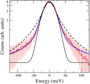
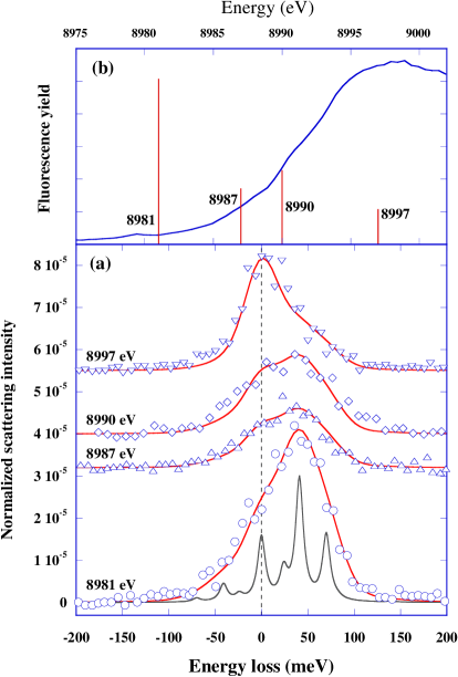
In order to demonstrate the technique, cupric oxide (CuO) was used as a model system. CuO is of particular importance since it is the simplest member of the family that shares the same integral CuO4 plackets with the high- superconducting cuprates [11]. The CuO single crystal was grown and oriented along the direction [12] and measured at the 3-ID beamline of the Advanced Photon Source using a RIXS spectrometer based on a sapphire crystal analyzer at a back-scattering geometry [13]. The monochromatic beam of 22 meV resolution was obtained by the four-bounce asymmetrically cut Si (4 4 4) monochromator. The sample was in vacuum to reduce the background due to air scattering, and the measurements were performed at room temperature. The overall energy resolution of the spectrometer was meV (figure 1) around the Cu -edge as the test measurements of this new spectrometer reported in Ref. [13]. The resolution measurements were repeated for each incident energy before and after the inelastic measurements to assure the observed effect was not due to a possible instrumental glitch. Spectra were collected at four incident photon energies ranging from the (quadruple transition) to (dipole transition). The incident energy extrema of 8981 eV and 8997 eV were the limits of the instrument as the analyzer was designed to operate at a fixed Bragg angle of around 89.8∘ (very close to back-scattering), and analyzer energy was changed by thermal expansion of the scattering crystal, not the Bragg angle. The temperature of the analyzer crystal, ranging from 100 K to 500 K defines the energy, ranging from 8997 eV to 8981 eV, respectively.
3 Results and Discussion
Since there is evidence that electron-lattice coupling is strongest at the zone boundary in cuprates [14], the measurements were taken in the first Brillouin zone such that the momentum transfer vector was along the direction. This geometry corresponds to a scattering angle 2 of 16.93∘ with momentum transfer of 13.4 nm-1 (1.8 nm-1 resolution). Additionally, the phonon branches under investigation have their maximum energies at the zone boundary as reported earlier [15], which makes them relatively easier to detect with the current instrumental resolution.
The RIXS data are shown in figure 2 for several incoming photon energies. In the pre-edge region ( eV), which is dominated by excitations, we observe a clear asymmetry in the spectral features with a maximum intensity around an energy loss of meV. For larger incoming x-ray energies, the asymmetry remains but the intensity decreases. At an incoming x-ray energy of eV, the maximum coincides with the zero-loss peak with some asymmetry still present. It is known that the elastic signal also enhances as a result of -edge resonance and one can argue that the observed spectral change may be due to this resonant behaviour of the zero-loss peak. However, it was not possible to fit the spectra according to this explanation. The effect of photon absorption coefficients cannot explain the reported anomaly either. The energy scan range is 200 meV and the inelastic signal is observed within 100 meV. The absorption coefficients are virtually the same for the short energy scanning range compared to the width of the absorption edge of 20 eV. Therefore, the change in the spectra cannot solely be associated with energy dependence of the absorption coefficients, since photon absorption would only affect the overall intensity, not the line shape.
It is evident that the inelastic features show a clear resonant behaviour. In RIXS measurements, it is a common practice to take a spectrum away from the transition metal edge to prove the resonant behaviour [16, 17]. However, in this case where the non-resonant measurements away from the Cu -edge are not possible (see above for the discussion about the instrument and ref. [13]), the noticeable change in the spectra as a function of incident photon energy should be accepted as the necessary proof.
There are several possible explanations for the origin of these inelastic features. We can readily discount crystal-field excitations, since the lowest transition is expected to be on the order of 1-1.5 eV [18]. Additionally, -edge RIXS is known to be sensitive to magnon excitations [19, 20]. Since the RIXS process conserves spin, only two-magnon excitations are allowed with an expected combined energy of 0.3-0.5 eV [19, 20], which is an order of magnitude larger than the features in the present RIXS data. Therefore, we attribute these loss features to the excitations of lattice vibrations. The closeness of the energy loss of the RIXS spectra to the experimentally observed phonon modes supports our assertion [15, 21].
As a result, it can be safely deduced that the observed variation in the spectral shape as a function of incident photon energy can be explained by the change in the relative intensities of the phonon modes against each other. At this geometry, three phonon modes are detectable with energies of 24 meV, 41 meV, and 70 meV [15]. We repeated these measurements along the direction to make sure which branches are allowed for this geometry. The rough measurements were done using a 2.2 meV-resolution spectrometer that operates at 21 keV [22]. The experimental data demonstrate the coupling between the lattice and the electronic excitations created by the absorption process and this coupling leads to modulation in the phonon intensities. The electron-phonon interaction can be described by
| (1) |
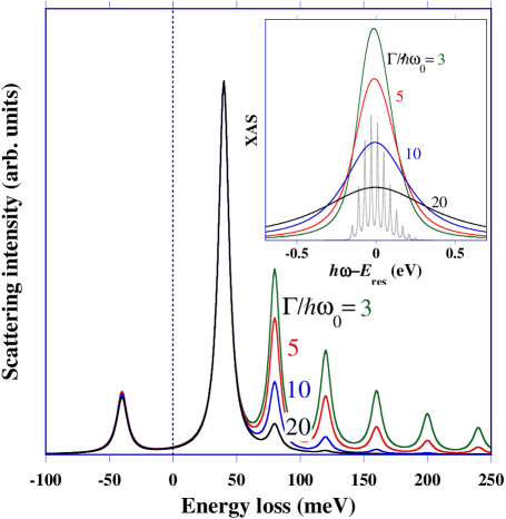
where creates a phonon with mode , wavevector , and energy ; after the absorption process, the coupling strength to the transient change in charge density with respect to the ground state is determined by . In the ground and final states this coupling is zero. This Hamiltonian corresponds to a displaced oscillator, and its solutions are well known. The sudden change in charge density due to the absorption process causes the lattice to respond through the excitation of multiple phonons. To understand the underlying physics, a single dispersionless optical phonon mode with energy and coupling can be taken as an example. In the absorption process, phonons of this mode are excited following a Poisson distribution with the maximum given by , see the inset in Fig. 3. For a typical coupling on the order of meV, and it is expected that several phonons are excited in the intermediate state. One therefore expects that several phonons should be visible in the final state. To verify this, the RIXS cross section is calculated using the Kramers-Heisenberg equation
| (2) |
where , , and are the initial, intermediate, and final states, respectively; and are the energies of the incoming and outgoing x-rays; is the transition operator; and is the full width at half maximum of the lifetime broadening of the core-hole. Figure 3 shows that through the Kramers-Heisenberg equation multiple phonon excitations are expected (see the calculations for and 5). Contrary to the calculations, the experimental spectra do not show multiple phonon excitations. This peculiar effect can be explained through numerical calculations by increasing . Even though the intermediate states are entirely equivalent in all calculations, the large lifetime broadening causes a destructive interference between the different intermediate states leading to the same final state. To understand this, the RIXS cross section must be more closely examined. The electron-phonon contribution to the intermediate state can be solved by introducing the displaced operators , giving a Hamiltonian . Applying an ultra-short core-hole lifetime approximation [23, 24, 25] to the intermediate-state propagator by expanding the denominator around the resonance energy results in
| (3) |
with . On resonance, where and therefore , the above expression is only valid when . At first, this does not appear to be justified since the eigenstates of go to infinity. However, only the eigenstates with a finite spectral weight, which are spread in an energy region of approximately around the resonance frequency, are pertinent to the RIXS cross section. Limiting the calculations to the lowest order, an effective scattering amplitude can be obtained as
| (4) |
by omitting the terms that do not give rise to inelastic x-ray scattering. The transition operators causing the electronic excitation in the intermediate state effectively cancel due to the unilateral consideration of the phonon excitations in the final state. Therefore in the limit of a very short core-hole lifetime (the large limit), only single phonons will be excited, and the probability will be proportional to the coupling constant. The numerical calculation of the Hamiltonian supports the idea behind the expansion as shown in figure 3. For and 5, the lifetime broadening is comparable to the width of the x-ray absorption spectral features (inset in figure 3); the RIXS spectra clearly show multiple phonon excitations (figure 3) and the restriction to lowest order in the ultrafast core-hole lifetime expansion is invalid. For and 20, the multiphonon features are strongly reduced by the destructive interference, and the spectra are dominated by the single-phonon excitations, described by the amplitude in Equation (4). An alternative way to understand the ultra-short core-hole lifetime approximation is by noting that the decay of the core-hole is so fast that the lattice has insufficient time to fully respond to the change in charge distribution caused by the resonant process. At the -edge in transition metal compounds, where eV and , the ultra-short core-hole lifetime approximation is well justified. Note that the ratio between the Stokes and anti-Stokes single-phonon features remains almost constant for all . In figure 2, the experimental spectra are compared with a numerical calculation where the intermediate states are exactly diagonalized. The intermediate-state lifetime broadening is taken to be eV. Some weight has been added in the zero-loss peak to account for elastic diffuse scattering. The final spectra are further broadened with a Gaussian with a width of meV to account for experimental uncertainties, including the instrumental resolution. The relative RIXS intensities of the phonon peaks are proportional to (Equation (1)) and therefore are indicative of the coupling strength between the phonons and the transient change in charge density with respect to the ground state.
From the intensities of the inelastic phonon peaks, see figure 2(b), we observe a clear resonance behaviour. The largest intensity is observed in the pre-edge region (8981 eV), where direct excitations from to are made. This causes a change in the local charge distribution that directly affects the surrounding ligands. The intensity then drops with the main edge ( eV), which is dominated by excitations into the delocalized band that are less effective in coupling to the phonon modes. The multiphonon excitations are strongly suppressed due to the short core-hole lifetime. The best agreement between theory and experiment is obtained when for the 24 and 70 meV features are reduced a factor 0.7 compared to the 41 meV phonon excitations. Noting the relative intensities are directly related to the electron-phonon coupling strength, the spectra reveals direct information of the relative corresponding coupling strengths. High-quality data measured on resonance can therefore provide insight into the electron-phonon coupling. The relative ratio between the Stokes and anti-Stokes is given by the Boltzmann factor at room temperature, a further confirmation that the features are phonon related.
In conclusion, we have for the first time demonstrated the resonant enhancement of phonon excitations at the -edge of copper. The experimental features can be explained by the coupling between the phonons and the transient change in the charge distribution on the site where the resonant scattering process occurs. Due to the fast decay of the core-hole, the system has no time to fully respond to the change in charge density, and multiple phonon excitations are strongly suppressed. Using an ultra-fast core-hole expansion, we demonstrate that the resonant inelastic scattering intensity is directly proportional to the electron-phonon coupling strength. Future experiments should include high- superconductors where knowledge of the relative coupling strengths of different phonon modes might shed additional light on their role in superconductivity. We would like to stress that this technique studies the coupling of a specific element (Cu in this case) to the lattice. One can also perform experiments at different edges to obtain element-specific couplings to the phonon modes. Another aspect not explored in this paper is the momentum dependence. The study of the dependence of the intensity of the inelastic phonon peaks as a function of transferred momentum would provide unique insights into the -dependence of the electron-phonon coupling.
References
References
- [1] Lanzara A et al. 2001 Nature 412 510
- [2] Cuk T et al. 2004 Phys. Rev. Lett. 93 117003
- [3] Devereaux T P, Cuk T, Shen Z-X and Nagaosa N 2004 Phys. Rev. Lett. 93 117004
- [4] Reznik D, Pintschovius L, Ito M, Iikubo S, Sato M, Goka H, Fujita M, Yamada K, Gu G D and Tranquada J M 2006 Nature 440 1170
- [5] Johnson P D et al. 2001 Phys. Rev. Lett. 87 177007
- [6] Giustino F, Cohen M L and Louie S G 2008 Nature 452 975
- [7] Dahm T, Hinkov V, Borisenko S V, Kordyuk A A, Zabolotnyy V B, Fink J, Buchner B, Scalapino D J, Hanke W and Keimer B 2009 Nat. Phys. 5 217
- [8] Reznik D, Sangiovanni G, Gunnarsson O and Devereaux T P 2008 Nature 455 E6
- [9] Hancock J N, Chabot-Couture G and Greven M 2010 New J. Phys. 12 033001
- [10] Jansen A G M, van Gelder A P and Wyder P 1980 Journal of Physics C: Solid State Physics 13 6073
- [11] Asbrink B S and Norrby L J 1970 Acta. Crystallogr. B26 8
- [12] Souptel D, Behr G and Balbashov A 2002 Journal of Crystal Growth 236 583
- [13] Yavas H et al. 2007 Nucl. Instrum. Methods A 582 149
- [14] Egami T 1996 Journal of Low Temperature Physics 105 791
- [15] Reichardt W, Gompf F, Aïn M and Wanklyn B M 1990 Zeitschrift für Physik B Condensed Matter 81 19
- [16] Kao C-C, Caliebe W A L, Hastings J B and Gillet J-M 1996 Phys. Rev. B 54 16361
- [17] Hill J P, Kao C-C, Caliebe W A L, Matsubara M, Kotani A, Peng J L and Greene R L 1998 Phys. Rev. Lett. 80 4967
- [18] Eskes H, Tjeng L H, and Sawatzky G A 1990 Phys. Rev. B 41 288
- [19] Hill J P et al. 2008 Phys. Rev. Lett. 100 097001
- [20] van den Brink J 2007 Europhysics Letters 80 47003
- [21] Guha S, Peebles D and Wieting T J 1991 Phys. Rev. B 43 13092
- [22] Sinn H et al. 2001 Nucl. Instrum. Methods A 467-468 1545
- [23] van Veenendaal M, Carra P and Thole B T 1996 Phys. Rev. B 54 16010
- [24] van den Brink J and van Veenendaal M 2006 Europhysics Letters 73 121
- [25] Ament L J P, Forte F and van den Brink J 2007 Phys. Rev. B 75 115118