DNA-psoralen: single-molecule experiments and first principles calculations
Abstract
The authors measure the persistence and contour lengths of DNA-psoralen complexes, as a function of psoralen concentration, for intercalated and crosslinked complexes. In both cases, the persistence length monotonically increases until a certain critical concentration is reached, above which it abruptly decreases and remains approximately constant. The contour length of the complexes exhibits no such discontinuous behavior. By fitting the relative increase of the contour length to the neighbor exclusion model, we obtain the exclusion number and the intrinsic intercalating constant of the interaction. Ab initio calculations are employed in order to provide an atomistic picture of these experimental findings.
pacs:
87.80.Cc, 87.14.gk, 87.15.LaDNA interactions with ligands such as drugs or proteins have been extensively studied and reviewed by many authors in the past years Fritzsche ; Tessmer ; Sischka ; Sinden ; Rocha ; RochaJCP2 ; Cimino ; McCauley2 . Some of these drugs, like daunomycin and ethidium bromide (EtBr), exhibit intercalative binding RochaJCP2 ; Fritzsche ; Sischka . Psoralen, by contrast, presents other forms of linkage to the DNA bases. In fact, it is well known that this drug can absorb photons and form covalent bonds with the DNA bases, when the sample is illuminated with ultraviolet-A (UVA) light ( = 320-400 nm) Sinden . Psoralens are compounds from the family of furocoumarins, broadly used in the medical treatment of various skin diseases like psoriasis, vitiligo, and some other kinds of dermatitis McNeely .
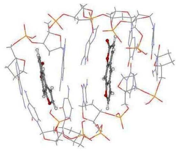
Although psoralens have been extensively used in medicine for many years, the interaction of the drug with DNA is not yet completely understood. In this work, we study the intercalative binding and crosslink formation in DNA-psoralen complexes (Psol-DNA), in order to clarify the quantitative aspects of the interaction between psoralen and DNA. We are interested in probing the modifications of the mechanical properties of DNA molecules when interacting with psoralen. The elasticity and size changes of these molecules, as a function of the total psoralen concentration (), are measured by performing single-molecule stretching experiments, which give information about persistence () and contour () lengths of Psol-DNA. The total psoralen concentration is the sum of the intercalated concentration () and the free concentration in solution of psoralen. We also define the intercalated psoralen fraction =/, where is the DNA base-pair concentration, fixed in all experiments at 11 M. In order to elucidate the microscopic mechanisms behind the psolaren-DNA interaction, we also perform first principles calculations for Psol-DNA fragments (an example is shown in Fig. 1) to determine the behavior of the Psol-DNA stiffness (which is related to the Young modulus) as a function of , and also to obtain the maximum fraction of intercalated psoralen into DNA, i.e., the limiting value of at which a Psol-DNA complex is still structurally stable.
Experimentally, we observe that as increases the Psol-DNA persistence length initially increases and then undergoes an abrupt transition to a lower value at a critical total psoralen concentration (), a behavior we also observe in UV-light illuminated complexes. In the latter case, the Psol-DNA complex attains higher persistence length values for , which is indicative of crosslinking, where the psoralen binds covalently preferably to thymine bases in opposite DNA strands, upon illumination. As discussed below, the ab initio calculations are in qualitative agreement with these experimental findings, enabling us to analyze the changes in the molecular structure that are responsible for the experimentally observed behavior.
The intercalated complexes are obtained by simply waiting for the psoralen to intercalate the -DNA in the sample. The crosslinked complexes are obtained by illuminating the sample with a UVA mercury lamp. We use an optical tweezers to trap a polystyrene bead attached to the DNA molecule, while pulling the microscope stage with a controlled velocity. We then make force extension curves, and use the approximate expression derived by Marko and Siggia Marko to obtain the persistence and contour lengths of the bare DNA and Psol-DNA complexes. The persistence length ( is the Young modulus, is the geometrical moment of inertia of the polymer, and is the thermal energy) is a mesoscopic quantity which depends on the local elasticity of DNA. Its value is 50 nm for bare DNA under physiological conditions. In our experiments, the forces applied to stretch the DNA are within the entropic force regime (forces 3 pN), thus avoiding externally distorting the structure and shifting the DNA/psoralen chemical equilibrium (enthalpic effects). The details about our experimental setup, experimental procedure, and optical-tweezers calibration can be found in Refs. Rocha ; Nathan1 ; RochaJCP2 .
In an attempt to understand the behavior of as a function of , we perform first principles calculations of the structural stability and stiffness changes produced by intercalation of psoralen into fragments of dry poly(dG)-poly(dC) in its acid form. We investigate DNA models with fragments consisting of four and five guanine-citosine (CG) pairs. A five-base-pair fragment with two intercalated psoralens is shown in Fig. 1. Our first principles density functional theory (DFT) simulations use the SIESTA code soler . Exchange and correlation effects are described within the generalized gradient approximation perdew . A double- basis set of atomic orbitals of finite range is used, with polarization functions added for phosphorus and for the atoms involved in the hydrogen bridges. This methodology has been shown to provide a good description of the DNA structure pablo ; parrinelo ; mdna . We treat isolated finite fragments in a periodic supercell with large vacuum regions. For the DNA bases at the edges of the fragments, one atom was held fixed in the same position as in the infinite periodic DNA pablo . Broken bonds at the edges are saturated with hydrogen atoms. Full geometry relaxations were performed (forces eV/Å). The stability of Psol-DNA complexes is addressed by computing their formation energies defined as the difference between the total energy of the Psol-DNA complex and the sum of the total energies of the bare-DNA fragment and the isolated psoralen molecule, after full relaxation of the initial geometries.
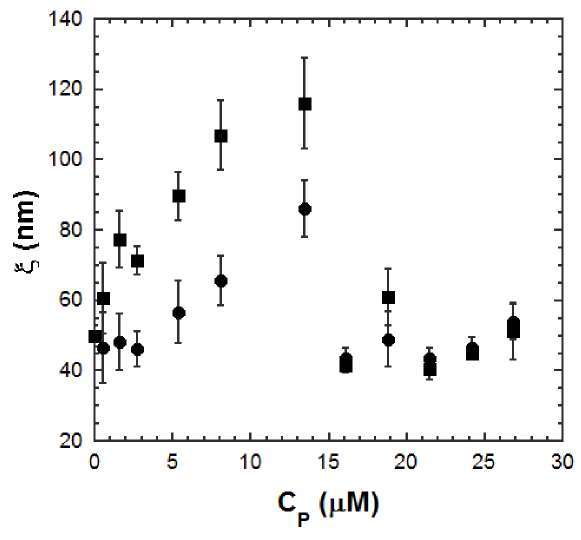
In Fig. 2, we plot the measured values of as a function of , for both intercalated and crosslinked Psol-DNA complexes. The figure shows that increases monotonically until the critical concentration M is reached. For , decays abruptly to around 50 nm, and remains approximately constant at this value, at least for the concentration range in our experiments. Note that is the same for both intercalated and crosslinked complexes. A similar abrupt transition of the persistence length was recently reported by two of us for other intercalating drugs (daunomycin and ethidium bromide) RochaJCP2 . The fact that Psol-DNA complexes exhibit this same behavior is a strong evidence that this transition is of general character for intercalating molecules, at least in the low-force regime ( 3 pN) used in our experiments. This abrupt transition may be caused by local formation of denaturing bubbles, as proposed in Refs. Harris and RochaPB , which softens the structure of the Psol-DNA complex. In Ref. Harris large scale molecular dynamics calculations indicate the formation of denaturing bubbles when pure DNA is stretched above 65 pN forces. Since we use very small stretching forces it is likely that the abrupt transition we observe is caused by some intrinsic DNA structural changes induced by the intercalating drug.
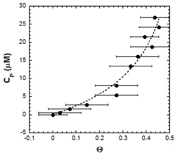
In Fig. 3 we show the relative increase of the contour length, , as a function of for the same intercalated complexes shown in Fig. 2, where is the contour length for . Note that the contour length does not exhibit any abrupt changes. The dashed line in this figure is a fit using Eq.1 below, derived from the neighbor exclusion model (NEM) McGhee (see Ref. RochaJCP2 for details). Considering that each intercalated psoralen molecule increases the length of DNA by the base-pair distance 0.34 nm Sinden , is equal to the intercalated psoralen fraction . In this case the NEM gives
| (1) |
where is the exclusion number and is the intrinsic intercalating constant. From the fitting we determine the parameters and M-1, for intercalative binding of psoralen. The large error bars for are due to the fact that different DNAs were used, and there is a natural length distribution for -DNA. The abrupt transition for occurs around 0.38, while the maximum psoralen intercalated fraction () is between 0.64 and 0.77.
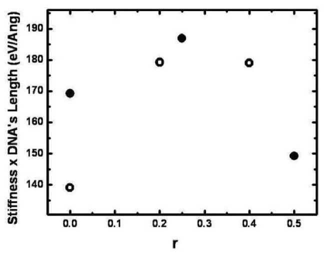
Figure 4 shows the behavior of the stiffness, which is proportional to the Young modulus (for fixed DNA length), as a function of , computed using the DFT methodology. Note that the stiffness increases as increases, and an abrupt decrease is observed for between 0.40 and 0.50, in reasonably good agreement with the data for the persistence length of Fig 2, where this transition occurs around 0.38. Our calculations indicate that before the transition two psoralens are well intercalated into the DNA structure, while after the transition one of the psoralens binds with the citosine, and the hydrogen bond between the two bases is broken, indicating that local denaturation may indeed be the mechanism behind the abrupt change in stiffness and consequently in the persistence length. Moreover, our calculations also indicate that when increases above 0.60, the intercalated structure is unstable, which sets an upper bound on the maximum fraction of intercalated psoralen within this theoretical model, in good agreement with the lower-limit value of 0.64 measured experimentally. Our interpretation for the microscopic nature of the transition rests on the good qualitative and quantitative agreement between theory and experiment. We must, however, not refrain from stating the limitations of our model system, since calculations in such small fragments are strongly affected by boundary effects. This is reflected in the large values we obtain for the theoretical stiffness, which are also due to the fact that such small fragments are much stiffer than the much longer DNA chains in the experiments (The stiffness length we compute for bare DNA using the four-base-pair model is about 20% higher than that of the five-base-pair model.)
In conclusion, the authors observe experimentally that the persistence length of DNA-psoralen complexes increases with psoralen concentration up to a critical concentration, decreasing abruptly and remaining approximately constant above this concentration, a behavior also observed in UV-light illuminated crosslinked complexes. Ab initio calculations show a similar behavior for the stiffness of psoralen-DNA complexes, and also indicate that local denaturation and binding between psoralen and a DNA base may be the mechanism behind this persistence length transition.
The authors acknowledge support from the European Grant EU
(FP6-029192), and from the Brazilian agencies: FAPEMIG, CAPES, CNPq,
Instituto do Milênio de Nanotecnologia/MCT and Instituto do
Milênio de Óptica Não-linear, Fotônica e Biofotônica/MCT.
References
- (1) H. Fritzsche, H. Triebel, J. B. Chaires, N. Dattagupta, and D. M. Crothers, Biochemistry 21 (17), 3940 (1982).
- (2) I. Tessmer, C. G. Baumann, G. M. Skinner, J. E. Molloy, J. G. Hoggett, S. J. B. Tendler, and S. Allen, J. Mod. Optic. 50 (10), 1627 (2003).
- (3) A. Sischka, K. Toensing, R. Eckel, S. D. Wilking, N. Sewald, R. Ros, and D. Anselmetti, Biophys. J. 88 (1), 404 (2005).
- (4) D. W. Ussery, R. W. Hoepfner, and R. R. Sinden, Method. Enzymol. 212, 242 (1992).
- (5) M. S. Rocha, N. B. Viana, and O. N. Mesquita, J. Chem. Phys. 121 (19), 9679 (2004).
- (6) M. S. Rocha, M. C. Ferreira, and O. N. Mesquita, J. Chem. Phys. 127 (10), 105108 (2007).
- (7) G. D. Cimino, H. B. Gamper, S. T. Isaacs, and J. E. Hearst, Annu. Rev. Biochem. 54, 1151 (1985).
- (8) M. J. McCauley and M. C. Williams, Biopolymers 91 (4), 265 (2009).
- (9) W. McNeely and K. L. Goa, Drugs 56 (4), 667 (1998).
- (10) J. F. Marko and E. D. Siggia, Macromolecules 28 (26), 8759 (1995).
- (11) N. B. Viana, R. T. S. Freire, and O. N. Mesquita, Phys. Rev. E 65 (4), 041921 (2002).
- (12) S. A. Harris, Z. A. Sands, and C. A. Laughton, Biophys. J. 88 (3), 1684 (2005).
- (13) M. S. Rocha, Phys. Biol. 6, 036013 (2009).
- (14) J. D. McGhee and P. H. von Hippel, J. Mol. Biol. 86 (2), 469 (1974).
- (15) J. M. Soler, E. Artacho, J. D. Gale, A. Garcia, J. Junquera, P. Ordejon, and D. Sanchez-Portal, J. Phys-Condens. Mat. 14, 2745 (2002).
- (16) J. P. Perdew, K. Burke and M. Ernzerhof, Phys. Rev. Lett. 77, 3865 (1996).
- (17) P. J. de Pablo, F. Moreno-Herrero, J. Colchero, J. Gómez Herrero, P. Herrero, A. M. Baró, P. Ordejón, J. M. Soler, and E. Artacho, Phys. Rev. Lett. 85, 4992 (2000).
- (18) F. L. Gervasio, P. Carloni, and M. Parrinello, Phys. Rev. Lett. 89, 108102 (2002).
- (19) S. S. Alexandre, J. M. Soler, L. Seijo, and F. Zamora, Phys. Rev. B 73, 205112 (2006).