kxl014 \gridframeN\cropmarkN
Size-dependent rheology of type-I collagen networks
Abstract
We investigate the system size dependent rheological response of branched type I collagen gels. When subjected to a shear strain, the highly interconnected mesh dynamically reorients, resulting in overall stiffening of the network. When a continuous shear strain is applied to a collagen network, we observe that the local apparent modulus, in the strain-stiffening regime, is strongly dependent on the gel thickness. In addition, we demonstrate that the overall network failure is determined by the ratio of the gel thickness to the mesh size. These findings have broad implications for cell-matrix interactions, the interpretation of rheological tissue data, and the engineering of biomimetic scaffolds.Received for publication XX and in final form XX. Address reprint requests and inquiries to D. L. Blair, E-mail: blair@physics.georgetown.edu.
doi:
doi:The increasing variety and availability of purified extra- and intra-cellular matrix proteins provides an unprecedented opportunity to quantify the mechanical properties of the cellular environment. Reconstituted biopolymer fiber networks can exhibit nonlinear rheology that is dramatically different than synthetic polymer gels [1, 2, 3, 4, 5]. Tying the microscopic physical behavior of the network constituents to the meso- and macro-scale rheology is critical for determining the mechanosensory mechanisms that underlie such diverse processes as cell motility, cancer metastasis and tumor proliferation. In vivo, cells sense, and respond to forces generated within or transmitted through the extracellular matrix (ECM). In turn, the regulation of sensory cues can be highly susceptible to changes in the ECM stiffness on length scales comparable to the cell size. However, in spite of the increasing number of experimental and theoretical investigations of the nonlinear rheological behavior of semi-flexible and stiff polymer networks [6, 7, 8, 9], the effects of system size on strain-stiffening remains essentially unexplored. Most theoretical models focus on the global response of biopolymer networks using three distinct microscopic mechanisms; non-affine deformations of crosslinked rigid rods [9], entropic penalties of fiber stretching in entangled networks [3], or changes to the network geometry [10]. There is little experimental data to directly verify these microscopic mechanisms, in part because of the difficulty in extracting the relevant quantities experimentally [11, 12]. In addition, the nonlinear rheology of stiff networks can only be investigated with bulk techniques, so the behavior at biologically relevant microscopic and mesoscopic scales is largely unknown.
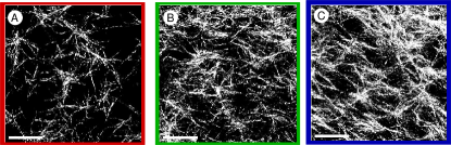
Collagen is the most abundant ECM protein, and a common component of in vitro cell cultures and bioengineered scaffolds. Under appropriate conditions, type I collagen self-assembles in vitro to form percolated networks (gels) [13] with mesh sizes that depend on the concentration and polymerization conditions (Fig. 1A-C) [14, 12]. These branched networks substantially strain-stiffen over a broad range of concentrations [15, 16, 17]; one interpretation of the biomechanical function associated with nonlinear stiffening is a passive protection for soft tissues when shear deformations become large. Collagen networks are not only excellent ECM model systems, but provide a platform for investigating cellular motility within three-dimensional microenvironments [18], and provide bio-compatible scaffolds for tissue growth and organ regeneration [19, 20, 21, 22].
In this work, we investigate strain-stiffening of collagen networks under steady shear and observe that the nonlinear rheological response is strongly dependent on the material thickness. Moreover, the apparent moduli near yield decreases dramatically in thin samples in a process that is controlled by the ratio of the sample size to the network mesh size. The system size dependence is not accounted for in current models of nonlinear strain-stiffening in biopolymer networks.
We apply continuous shear strains to collagen networks using an Anton-Paar MCR-301 bulk rheometer with a 25 mm diameter parallel-plate geometry within a temperature and humidity controlled environment. All continuous shear strain experiments are performed at a strain rate of s-1. Type I rat tail collagen (BD Bioscience, San Mateo, CA, 3.27 mg/mL) is polymerized at 23oC for 45 minutes in 10X PBS at pH 7.0 with ionic strengths for the concentrations mg/ml, respectively.
\doilineThe plate tool provides precise control of the sample thickness through a change of the rheometer gap . At low strains, the measured stress is proportional to the applied strain , for all values of . Intermediate strains reveal that the network undergoes a substantial nonlinear increase of , indicative of strain-stiffening [3]. Specifically, for m, is -independent. However, for smaller gaps, we observe that the maximum of moves to larger values of (Fig. 2). The magnitude of at the peak remains relatively constant. The yield strain in thin samples is more than twice that of thick samples, and the apparent modulus of thin samples can be up to 3 times smaller than the bulk value. To ensure that the changes in are not introducing variability in the rheological measurements, particularly at small , we measure the linear viscoelastic modulus using oscillatory rheology at a frequency of rads s-1 at a strain of . For each concentration, we observe that is constant over the entire range of (Fig. 3A).
Higher strains lead to irreversible deformations and yielding, as evidenced by a reduction in the overall modulus. In addition to this distinctive rheological signature, we observe a dramatic change in the rheological response by varying the sample thickness from m, at a fixed collagen concentration ( mg/ml).
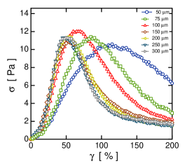
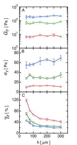
To quantify the -dependence of the nonlinear rheology, we perform a least squares polynomial fit to the peaks of the rheology curves in Figure 2A. We use the peak positions to define the yield values for the stress and strain . We determine as a function of for three different collagen concentrations. For each concentration, exhibits a clear increase for small and approaches a constant value for large (Fig. 3C). Interestingly, for the entire range of and concentration the values of show weak or no gap dependence (Fig. 3B). We speculate that is determined by the strength of fiber-fiber or fiber-boundary junctions, and depends only on the total applied stress, independent of .
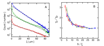
To elucidate the role of the microscopic length scale in the observed size-dependent rheology, we replot the data in Figure 3C with rescaled by . To quantify the mesh size , we analyze the spacing between fibers extracted along horizontal and vertical lines within -resolved planar confocal images [14]. We fit the distribution of the fiber spacing to an exponential decay for each concentration, mg/ml, to obtain the characteristic mesh sizes 14.7, 10.4, 9.4 m (Fig. 4A). To account for the concentration dependent modulus, we also rescale by the -dependent plateau values , determined by fitting to an exponential decay with an offset. The data from the three concentrations collapse onto a single curve (Fig. 4B).
We conclude that the nonlinear rheology of collagen is determined by the interplay between the macroscopic material thickness and the mesh size. We do not claim that the mesh size determines the network rheology; this is most likely determined by some complex interplay of the network structure and topology. In addition, we note that the range of gaps investigated directly corresponds to the radial gap separation in many cone-plate tool geometries, and is equivalent to length scales of many important biological processes.
The observed size dependence of the nonlinear rheology indicates that the material response is non-uniform on scales significantly larger than the mesh size, and is therefore inconsistent with models that attribute strain stiffening to the affine deformation of networks of semi-flexible filaments. Collagen fibers are relatively stiff, and therefore more likely described by athermal models that attribute strain stiffening to non-affine filament stretching [23]. The non-affine length scale can be significantly larger than the mesh size [24], but to our knowledge the implications for system size dependence has not been investigated. Alternatively, heterogeneous localization of strain within the network that is either uniformly distributed through the gel or localized near the boundaries could produce size dependent effects. Reflectance images do not show any measurable variation in collagen structure near the boundary, but more subtle boundary effects may also play a role. Finally, the long persistence length of the collagen fibers may directly contribute to a finite size effect. Whatever the physical mechanism, the size-dependent rheology extends to important length scales for a range of fundamental studies and applications.
Acknowledgements
We thank P. Janmey, F. MacKintosh and Andreas Bausch for helpful discussions and W. Rosoff and D. Koch for the collagen preparation. This was funded by the NSF through grant DMR-0804782 and from the AFOSR through grant FA9550-07-1-0130.
References
- [1] Djabourov, M., Lechaire, J.-P., and Gaill, F. 1993. Structure and rheology of gelatin and collagen gels. Biorheology. 30:191–205.
- [2] Gardel, M., Shin, J., MacKintosh, F., Mahadevan, L., Matsudaira, P., and Weitz, D. 2004. Elastic behavior of cross-linked and bundled actin networks. Science. 304:1301–1305.
- [3] Storm, C., Pastore, J., MacKintosh, F., Lubensky, T., and Janmey, P. 2005. Nonlinear elasticity in biological gels. Nature. 435:191–194.
- [4] Liu, J., Koenderink, G. H., Kasza, K. E., MacKintosh, F. C., and Weitz, D. A. 2007. Visualizing the strain field in semiflexible polymer networks: Strain fluctuations and nonlinear rheology of -actin gels. Phys. Rev. Lett. 98:198304.
- [5] Chaudhuri, O., Parekh, S. H., and Fletcher, D. A. 2007. Reversible stress softening of actin networks. Nature. 445:295–298.
- [6] MacKintosh, F. C., Käs, J., and Janmey, P. A. 1995. Elasticity of semiflexible biopolymer networks. Phys. Rev. Lett. 75:4425–4428.
- [7] Wilhelm, J. and Frey, E. 2003. Elasticity of stiff polymer networks. Phys. Rev. Lett. 91:108103.
- [8] Head, D. A., Levine, A. J., and MacKintosh, F. C. 2003. Distinct regimes of elastic response and deformation modes of cross-linked cytoskeletal and semiflexible polymer networks. Phys. Rev. E. 68:061907.
- [9] Heussinger, C., Schaefer, B., and Frey, E. 2007. Nonaffine rubber elasticity for stiff polymer networks. Phys. Rev. E. 76:031906.
- [10] Onck, P. R., Koeman, T., van Dillen, T., and van der Giessen, E. 2005. Alternative explanation of stiffening in cross-linked semiflexible networks. Phys. Rev. Lett. 95:178102.
- [11] Stein, A. M., Vader, D. A., Jawerth, L. M., Weitz, D. A., and Sander, L. M. 2008. An algorithm for extracting the network geometry of three-dimensional collagen gels. Journal of Microscopy. 232:463 – 475.
- [12] Yang, Y.-L., Leone, L., and Kaufman, L. 2009. Elastic moduli of collagen gels can be predicted from two-dimensional confocal microscopy. Biophysical Journal. 97:2051–2060.
- [13] Forgacs, G., Newman, S., Hinner, B., Maier, C., and Sackmann, E. 2003. Assembly of collagen matrices as a phase transition revealed by structural and rheologic studies. Biophysical Journal. 84:1272–1280.
- [14] Kaufman, L., Brangwynne, C., Kasza, K., Filippidi, E., Gordon, V., Deisboeck, T., and Weitz, D. 2005. Glioma expansion in collagen i matrices: Analyzing collagen concentration-dependent growth and motility patterns. Biophysical Journal. 89:635–650.
- [15] Janmey, P. A., Mccormick, M. E., Rammensee, S., Leight, J. L., Georges, P. C., and Mackintosh, F. C. 2007. Negative normal stress in semiflexible biopolymer gels. Nature Materials. 6:48–51.
- [16] Kang, H., Wen, Q., Janmey, P., Tang, J., Conti, E., and MacKintosh, F. 2009. Nonlinear elasticity of stiff filament networks: Strain stiffening, negative normal stress, and filament alignment in fibrin gels. Journal of Physical Chemistry B. 113:3799–3805.
- [17] Vader, D., Kabla, A., Weitz, D., and Mahadevan, L. 2009. Strain-induced alignment in collagen gels. PLoS ONE. 4:e5902.
- [18] Mierke, C. T., Rösel, D., Fabry, B., and Brábek, J. 2008. Contractile forces in tumor cell migration. European Journal of Cell Biology. 87:669–676.
- [19] Ott, H. C., Matthiesen, T. S., Goh, S.-K., Black, L. D., Kren, S. M., Netof, T. I., and Taylor, D. A. 2008. Perfusion-decellularized matrix: using nature’s platform to engineer a bioartificial heart. Nature Medicine. 14:213–221.
- [20] Sabass, B., Gardel, M. L., Waterman, C. M., and Schwarz, U. S. 2008. High resolution traction force microscopy based on experimental and computational advances. Biophysical Journal. 94:207 – 220.
- [21] Beningo, K., Lo, C.-M., and Wang, Y.-L. 2002. Flexible polyacrylamide substrata for the analysis of mechanical interactions at cell-substratum adhesions. Methods in Cell Biology. 69:325–339.
- [22] Gaudet, C., Marganski, W., Kim, S., Brown, C., Gunderia, V., Dembo, M., and Wong, J. 2003. Influence of type i collagen surface density on fibroblast spreading, motility, and contractility. Biophysical Journal. 85:3329–3335.
- [23] Heussinger, C. and Frey, E. 2007. Role of architecture in the elastic response of semiflexible polymer and fiber networks. Phys. Rev. E. 75:011917.
- [24] Lieleg, O., Claessens, M. M. A. E., Heussinger, C., Frey, E., and Bausch, A. R. 2007. Mechanics of bundled semiflexible polymer networks. Phys. Rev. Lett. 99:088102.