Structure and optical properties of high light output halide scintillators
Abstract
Structural and optical properties of several high light output halide scintillators and closely related materials are presented based on first principles calculations. The optical properties are based on the Engel-Vosko generalized gradient approximation and the recently developed density functional of Tran and Blaha. The materials investigated are BaBr2, BaIBr, BaCl2, BaF2, BaI2, BiI3, CaI2, Cs2LiYCl6, CsBa2Br5, CsBa2I5, K2LaBr5, K2LaCl5,K2LaI5, LaBr3, LaCl3, SrBr2, and YI3. For comparison results are presented for the oxide CdWO4. We find that the Tran Blaha functional gives greatly improved band gaps and optical properties in this class of materials. Furthermore, we find that unlike CdWO4, most of these halides are highly isotropic from an optical point of view even though in many cases the crystal structures and other properties are not. This general result is rationalized in terms of halide chemistry. Implications for the development of ceramic halide scintillators are discussed.
pacs:
78.20.Ci,78.20.Bh,61.66.FnI introduction
Scintillators are widely used in radiation detection applications including medical imaging, oil well drilling, nuclear security and high energy physics experiments. These materials function by emitting light when excited by ionizing radiation, such as gamma rays. This light is then coupled to a photomultiplier or other light detector to produce electrical signals. The performance of scintillators is characterized by their light output, normally given in terms of photons per MeV of excitation energy, proportionality (how linear the light output is as a function of excitation energy), density (related to stopping power), decay time and energy resolution. Knoll (2000)
Scintillator performance depends on energy transport between the energy absorption events and the scintillation centers, suppression of non-radiative recombination channels and fast efficient light emission from the scintillation center. These scintillation centers can be intrinsic, via a mechanism such as decay of a self trapped exciton, or, as is commonly the case, at an activation center (e.g. Ce3+ ions substituting for La in LaBr3) where electron hole pairs generated by the radiation recombine. These processes are sensitive to the details of the material and in the case of materials with activators the environment of the activator ions. Halides generally have soft lattices that disfavor non-radiative recombination and can typically incorporate rare earth and other activator ions. More importantly, halide chemistry is very rich, with a wide variety of crystal structures that provide different bonding topologies for energy transfer and various environments for activator ions. Pauling (1960); Meyer (1982) In fact, one of the best known scintillators is the halide, NaI activated with Tl+. This material has high light output, but is slow, non-proportional and has poor energy resolution. de Haas and Dorenbos (2008) The finding that Ce3+ activated LaBr3 is a very high light output proportional scintillator with energy resolution better than 3% at 662 KeV van Loef et al. (2001) has led to renewed interest in halides as scintillator hosts, especially for spectroscopic gamma ray detection. This interest has resulted in the discovery of several other interesting halide scintillators, including heavily Eu2 activated SrI2, which is very proportional and has a light output exceeding that of LaBr3, Cherepy et al. (2008) and Ce3+ activated YI3, which is another very high light output material. Glodo et al. (2008)
Full characterization of the optical properties of scintillators is very useful both from the point of view of improving the design of systems as regards light coupling and also importantly in selecting candidate materials for ceramic scintillators. However, full optical characterization of these halides is complicated by sensitivity to moisture and other experimental difficulties. As a result only limited data is available.
The key requirements for a ceramic scintillator are sinterability and optical isotropy. This latter requirement comes from the need to avoid light scattering at misoriented grain boundaries. It is commonly thought that because of this requirement cubic material is needed for a true transparent ceramic. However, radiation detection is not an optical imaging application. Therefore weak scattering and image distortion are not as detrimental as they would be in an optical application such as a ceramic lens. In fact ceramic scintillators based on monoclinic Lu2SiO5 (LSO) have been demonstrated. Lempicki et al. (22 November 2000); Wisniewski et al. (2008) We showed in previous work that the high light output halide scintillator, SrI2 is in fact very nearly optically isotropic in spite of its orthorhombic () crystal structure. Singh (2008) This was an expected result considering the strongly orthorhombic lattice. It is of interest to determine whether this is the case for other high light output halide scintillators. Here we present a consistent set of first principles data for structural and optical properties of a number of high light output halide scintillators and closely related materials. We find, quite unexpectedly, that the halides we investigate have remarkably little optical anisotropy, even though a number of them are very anisotropic from other points of view.
II approach
The density functional calculations reported here were performed using the general potential linearized augmented planewave (LAPW) method as implemented in the WIEN2K code. Blaha et al. (2001) We used well converged Brillouin zone samples and basis sets, with the standard LAPW augmentation plus local orbitals. Singh (1991) Relativity was included at the scalar relativistic level except for BiI3, where spin-orbit was included for the electronic structure.
The crystal structure plays a fundamental role in determining the electronic and optical properties of a material. We began our calculations by fully relaxing all free internal atomic positions consistent with the crystal symmetry for each material. The lattice parameters were held fixed at their experimental values, which are no doubt more precise than the values that can be obtained using density functional calculations. This relaxation was done using the generalized gradient approximation (GGA) of Perdew, Burke and Ernzerhof (PBE). Perdew et al. (1996).
The PBE functional, like other standard generalized gradient approximations, is based on the total energy in terms of the coupling constant averaged exchange correlation hole and is designed to reproduce the total energy. Perdew et al. (1992) While these functionals are very useful in obtaining structures and other properties related to total energies, they underestimate, often strongly, the band gaps of most semiconductors and simple insulators. Accordingly, for the optical properties we use two other functionals. The first is the Engel-Vosko GGA (EV). Engel and Vosko (1993) This functional was designed to reproduce the exchange-correlation potential rather than the total energy, and gives improved band gaps. Dufek et al. (1994) We used it in our prior studies on transport in PbTe and optical properties of SrI2 and Bi4Ge3O12 scintillators. Singh (2010, 2008); Jellison, Jr. et al. (2010) The second is the semi-local functional of Tran and Blaha (TB-mBJ). Tran and Blaha (2009) This relatively recent functional is more sophisticated than the Engel-Vosko GGA and has been shown to give very much improved band gaps for a variety of semiconductors and insulators. We find that where comparison with experiment is possible this functional also gives very much improved band gaps as well as optical properties for these halides. As such, we focus on the results obtained using the TB-mBJ functional. The electronic structures were calculated with these two functionals based on the relaxed crystal structures from the PBE calculations. Optical properties were then obtained using the dipole matrix elements with the WIEN2K optical package. Blaha et al. (2001) A 0.1 eV broadening was applied to the spectra.
III materials and structures
We begin with the calculated structural parameters and brief introductions to the materials that we study.
BaF2 is a very well characterized material. It has been applied as a scintillator, both in pure form and with activation. Laval et al. (1983); Woody et al. (1989); Visser et al. (1991); Wojtowicz et al. (2000); Dinca et al. (2002) In pure form it has a very fast component (0.8 ns), as well as a slow component, which can be at least partially suppressed by La doping. Fast response is of importance in applications with very high count rates or where timing is critical. It occurs in the cubic () CaF2 structure, Ba on Wyckoff site (0,0,0) and F on (1/4,1/4,1/4). In our calculations we used the experimental lattice parameter,Swanson and Tatge (1953) =6.2001 Å.
Several of the alkaline earth di-halides, , are high light output proportional scintillators when activated with Eu2+. Cherepy et al. (2008); Hofstadter et al. (1964a, b); Jestin Lenus et al. (2002); Selling et al. (2007a) As with BaF2, activation with Ce3+ is also possible in some cases. Selling et al. (2007b) Undoped BaCl2 has a very fast response, with a short component lifetime of 1.6 ns. Koshimizu et al. (2009) SrI2:Eu is equal to or superior to LaBr3:Ce3+ as regards light output and proportionality, although it suffers from a slower response. Cherepy et al. (2008) Even though the material is orthorhombic, we found it to be optically nearly isotropic. Singh (2008) CaI2:Eu2+ is another material with very high light output, photons/MeV. Hofstadter et al. (1964b); Cherepy et al. (2008) BaIBr:Eu2+ crystals have been shown to have a light output of 81,0003,000 photons/MeV with a 662 KeV energy resolution better than 5%. Bourret-Courchesne et al. (2010) Regarding structure, BaCl2, BaBr2, BaI2 and BaIBr occur in an orthorhombic , PbCl2 type structure with four formula units per cell. The structural parameters as determined from relaxation are given in Table 1. SrBr2 has a large tetragonal unit cell () with ten formula units per cell. Smeggil and Eick (1971) The calculated internal parameters are as given in Table 2. CaI2 is hexagonal (), with Ca on site (0,0,0) and I on (1/2,2/3,). In our optical calculations we used the experimental lattice parameters, =4.49 Å, =6.975 Å, with the calculated =0.2535 from total energy minimization with the PBE GGA (the reported experimental value is 0.25). Blum (1933)
LaCl3 and LaBr3 activated with Ce3+ are very high light output, proportional scintillators with excellent energy resolution. van Loef et al. (2001) They are hexagonal () with the UCl3 structure type. There are two formula units per cell, with La on site (1/3,2/3,1/4) and the halogen on (,,1/4). We used the experimental lattice parameters, =7.4779 Å, =4.3745 Å, for LaCl3 (Ref. Morosin, 1968) and =7.9648 Å, =4.5119 Å, for LaBr3 (Ref. Kraemer et al., 1989). The calculated internal parameters were =0.9145, =0.6132 for LaCl3 and =0.9144, =0.6159 for LaBr3.
Cs2LiYCl6 is a member of the elpasolite family. The elpasolites are cubic halides, with general formula , with and alkali metals, a halogen and a rare earth or other trivalent element. is on site (1/4,1/4,1/4), is on (1/2,1/2,1/2), is on (0,0,0) and is on (,0,0). A large number of such compounds are known. Meyer (1982) However, only a fraction have been studied as potential scintillator materials. van Eijk et al. (2005); van Loef et al. (2002); Birowosuto et al. (2006, 2008); Bessiere et al. (2004); Grimm et al. (2007); Higgins et al. (2010) Cs2LiYCl6 is of particular interest because of its high Li content, which makes it useful as a neutron detector. We used the experimental lattice parameter for Cs2LiYCl6 from Reber and co-workers, Reber et al. (1989) =10.4857 Å, with the calculated internal parameter =0.2517 (an experimental value of 0.25046 was reported by Reber and co-workers).
The scintillation properties of Ce3+ activated K2LaCl5, K2LaBr5 and K2LaI5 were investigated by van Loef and co-workers. van Loef et al. (2003, 2005) These isostructural orthorhombic compounds showed high light output, up to 55,000 photons / MeV (for K2LaI5:Ce3+). The iodide also showed a reasonable decay time of 245 ns and 662 KeV energy resolution of 4.50.5%, although use of this scintillator is complicated because of self-activity associated with K. Our calculated structural properties are give in Table 3.
| BaCl2 | BaBr2 | BaI2 | BaIBr | |
|---|---|---|---|---|
| (Å) | 7.878 | 8.276 | 8.922 | 8.6 |
| (Å) | 4.731 | 4.956 | 5.304 | 5.12 |
| (Å) | 9.415 | 9.919 | 10.695 | 10.31 |
| Ba | 0.2481 | 0.2470 | 0.2466 | 0.2302 |
| Ba | 0.3838 | 0.1163 | 0.1166 | 0.1228 |
| 1 | 0.1423 | 0.6426 | 0.6424 | 0.9722 |
| 1 | 0.0708 | 0.0714 | 0.0721 | 0.6701 |
| 2 | 0.5274 | 0.5266 | 0.5246 | 0.3538 |
| 2 | 0.8313 | 0.6686 | 0.6665 | 0.4325 |
| Sr1 | Sr2 | Br1 | Br2 | Br3 | Br4 | |
|---|---|---|---|---|---|---|
| 0.4141 | 0.7500 | 0.5402 | 0.5426 | 0.2500 | 0.2500 | |
| 0.6035 | 0.7500 | 0.6527 | 0.8386 | 0.7500 | 0.7500 | |
| 0.7520 | 0.1487 | 0.3761 | 0.9004 | 0.0000 | 0.5000 |
| K2LaCl5 | K2LaBr5 | K2LaI5 | |
|---|---|---|---|
| (Å) | 12.742 | 13.36 | 14.332 |
| (Å) | 8.868 | 9.272 | 9.912 |
| (Å) | 8.022 | 8.462 | 9.132 |
| K | 0.3279 | 0.3277 | 0.3264 |
| K | 0.9952 | 0.5049 | 0.5035 |
| K | 0.0475 | 0.4465 | 0.4419 |
| La | 0.0066 | 0.0070 | 0.0064 |
| La | 0.0774 | 0.4202 | 0.4182 |
| 1 | 0.1818 | 0.0056 | 0.0053 |
| 1 | 0.8644 | 0.0646 | 0.0609 |
| 2 | 0.0065 | 0.1841 | 0.1847 |
| 2 | 0.4311 | 0.6329 | 0.6327 |
| 3 | 0.2912 | 0.2898 | 0.2879 |
| 3 | 0.3298 | 0.1674 | 0.1654 |
| 4 | 0.0816 | 0.0799 | 0.0792 |
| 4 | 0.5435 | 0.5459 | 0.5471 |
| 4 | 0.1659 | 0.3298 | 0.3282 |
| CsBa2Br5 | CsBa2I5 | |||||
|---|---|---|---|---|---|---|
| (Å) | 13.816 | 9.987 | 8.665 | 14.637 | 10.541 | 9.256 |
| 90.2 | 90.194 | |||||
| Cs | 0.1665 | 0.9897 | 0.5586 | 0.1675 | 0.9911 | 0.5594 |
| Ba1 | 0.1768 | 0.5002 | 0.5300 | 0.1782 | 0.4999 | 0.5332 |
| Ba2 | 0.4921 | 0.2511 | 0.5704 | 0.4913 | 0.2500 | 0.5772 |
| 1 | 0.4211 | 0.9614 | 0.6782 | 0.4205 | 0.9562 | 0.6810 |
| 2 | 0.0980 | 0.4602 | 0.1643 | 0.0956 | 0.4565 | 0.1669 |
| 3 | 0.4980 | 0.2849 | 0.9535 | 0.4985 | 0.2813 | 0.9592 |
| 4 | 0.3122 | 0.2868 | 0.3356 | 0.3122 | 0.2819 | 0.3375 |
| 5 | 0.2192 | 0.2732 | 0.8099 | 0.2216 | 0.2694 | 0.8110 |
CsBa2Br5 and CsBa2I5 occur in a monoclinic (No. 14) structure. Schilling and Meyer (1996); Bourret-Courchesne et al. (2009) Eu2+ doped CsBa2I5 was reported to have a light yield of approximately 97,000 photons per MeV and to be less hygroscopic than LaBr3 or SrI2. Bourret-Courchesne et al. (2009) The calculated structural parameters are as given in Table 4.
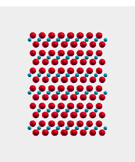
YI3 and BiI3 are related materials. Ce3+ activated YI3 was recently reported to have a very high light yield of almost 100,000 photons per MeV. Glodo et al. (2008); van Loef et al. (2008) The related compound BiI3 is of interest mainly as a semiconductor for radiation detection, Nason and Keller (1995) rather than as a scintillator. We include it here for comparison with YI3. Unlike YI3, BiI3 has substantial covalency between Bi and halogen states, which leads to enhanced Born charges and may be expected to affect the optical properties. Du and Singh (2010) These compounds occur in a rhombohedral structure, with the cations on site (0,0,) and I on (,,). With the hexagonal setting, the experimental lattice parameters are =7.4864 Å, =20.880 Å, for YI3 (Ref. Jongen and Meyer, 2005) and =7.516 Å, =20.7180 Å, for BiI3 (Ref. Trotter and Zobel, 1947). The calculated internal coordinates are =0.1664, =0.6568, =0.9993, =0.4156, for YI3 and =0.1664, =0.0022, =0.6685, =0.4123, for BiI3.
IV Electronic Structure and Optical Properties
As mentioned, three functionals were used in the present study, the PBE GGA for the structural properties, and the Engel-Vosko and TB-mBJ functionals for the optical properties. The recently developed TB-mBJ has been shown to give much more accurate band gaps than other semilocal functionals in a wide variety of materials. Tran and Blaha (2009) As such, we emphasize results obtained using that functional. For comparison, the fundamental band gaps of the compounds studied are listed in Table 5. The EV gaps are invariably intermediate between those of the standard PBE and the TB-mBJ functionals with the exception of the La containing compounds. In those compounds, the -resonance is found inside the insulating gap for all three functionals. The position of the -resonance relative to the valence band edge depends on the compound, but for a given material is practically unchanged between the different functionals. For these compounds we show both the gap between the valence band maximum and the bottom of the -bands making up the resonance, as well as the gap between the valence bands and the non- conduction bands. Experimental band gaps are unavailable for most of these materials.
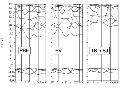
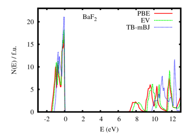
We start with BaF2, which as mentioned is a relatively well characterized material. The calculated band structure with the PBE, EV and TB-mBJ functionals is as shown in Fig. 1, while the corresponding electronic density of states (DOS) is given in Fig. 2. The PBE band structure has a direct gap at that is clearly underestimated with respect to experiment. Rubloff (1972) The valence bands, of F character, are narrow with a width of less that 2 eV, while the metal derived conduction bands are more dispersive. The EV band structure is similar to the PBE result but with a somewhat larger band gap. The mBJ band structure has a considerably enhanced gap of 9.8 eV. Furthermore, the conduction bands are distorted relative to the PBE result, with the conduction band minimum now at the point, although it should be noted that the distinction between direct and indirect is not so important for the properties of this compound because of the very flat valence bands.
| PBE | EV | TB-mBJ | |
|---|---|---|---|
| BaF2 | 6.87 | 7.46 | 9.77 |
| BaCl2 | 5.23 | 5.66 | 6.45 |
| BaBr2 | 4.45 | 4.85 | 5.39 |
| BaIBr | 3.79 | 4.13 | 4.45 |
| BaI2 | 3.50 | 3.87 | 4.14 |
| CaI2 | 3.49 | 3.92 | 4.26 |
| SrBr2 | 4.99 | 5.25 | 5.91 |
| YI3 | 2.81 | 3.02 | 3.31 |
| BiI3 | 1.43 | 1.67 | 1.82 |
| Cs2LiYCl6 | 4.90 | 5.30 | 5.97 |
| CsBa2Br5 | 4.53 | 5.13 | 5.77 |
| CsBa2I5 | 3.69 | 4.14 | 4.49 |
| K2LaCl | 4.46 | 4.46 | 4.59 |
| K2LaCl5 cb | 5.23 | 5.49 | 6.32 |
| K2LaBr | 3.69 | 3.76 | 3.74 |
| K2LaBr5 cb | 4.33 | 4.66 | 5.26 |
| K2LaI | 2.79 | 2.95 | 2.80 |
| K2LaI5 cb | 3.38∗ | 3.61 | 3.99 |
| LaCl | 3.97 | 4.04 | 4.15 |
| LaCl3 cb | 4.95 | 5.26 | 5.82 |
| LaBr | 3.20 | 3.28 | 3.24 |
| LaBr3 cb | 3.98 | 4.24 | 4.60 |
-resonance overlaps bottom of conduction band.
The experimental band gap of BaF2 is usually quoted as 11 eV based on the measured ultraviolet reflectance spectrum. Rubloff (1972) This spectrum shows several features near the gap: a prominent feature at 10.00 eV, assigned as an exciton, a weaker feature at 10.57, also assigned as an exciton, and a direct band edge at 11.0 eV. Subsequent two photon absorption experiments assigned the gap as 10.6 eV. Tsujibayashi et al. (2002) The TB-mBJ direct gap at of 9.90 eV is 1 eV lower. Importantly, the experimental spectrum has three large peaks in the valence band region below 16 eV. The calculated TB-mBJ reflectance spectrum is shown in Fig. 3. There are two prominent peaks that can be matched with the upper two peaks in the experimental spectrum (Ref. Rubloff, 1972) using a band shift of 1 eV. This is consistent with the assignment of the lower experimental peak as excitonic and the conclusion that the TB-mBJ band gap is underestimated by 1 eV.

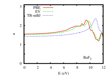
The calculated refractive index is shown as a function of energy for the three functionals in Fig. 4. The refractive indices follow the trend in the band gaps, with the result of the TB-mBJ functional lowest. Experimentally, the refractive index of BaF2 is practically constant, rising from =1.465 at low energy to =1.474 at 589 nm (2.107 eV). Shannon et al. (2002) This is in close agreement with the results using the TB-mBJ functional. We obtain a low frequency value of =1.42 with a weak dispersion as in experiment. The low frequency PBE value is higher at =1.56 and shows a stronger energy dependence consistent with the smaller gap, while the EV value of =1.52 is intermediate.
This result shows that the TB-mBJ functional not only improves the band gap, but also improves the optical response of this halide relative to the PBE functional, which is regarded as state of the art for calculations of total energies. This is important because the response depends not only on band energies but also on the wavefunctions, specifically through the dipole matrix elements. The charge density and total energy are fundamental quantities within density functional theory while the single particle energies are not. It is strongly thought that the Kohn-Sham eigenvalues of the exact density functional will not produce correct band gaps in insulators, even though its charge density and static response will be exact. Jones and Gunnarson (1989) Therefore it is of interest to note that the improvement in the band gap of semi-local functionals, particularly going to the EV and then the TB-mBJ functional improve the static response at the same time as they improve the band gap. In the following we focus on results obtained with the TB-mBJ functional.
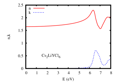
The calculated TB-mBJ band gap of 5.97 eV for the high light output elpasolite scintillator, Cs2LiYCl6 agrees well with the experimental onset of optical absorption at 5.9 eV. The refractive index is shown in Fig. 5. van Loef et al. (2002) The low frequency limit is =1.65. There is more dispersion than in BaF2, but the spectrum is otherwise featureless almost up to the band edge. We are not aware of experimental measurements of the optical properties of this material.
The isostructural tri-iodides, YI3 and BiI3 offer an interesting contrast. As mentioned, when activated with Ce3+, YI3 is a very high light output scintillator, while BiI3 is not. The emission of YI3:Ce3+ starts at 2.9 eV and peaks at 2.26 eV. Glodo et al. (2008) The high energy onset is below our calculated TB-mBJ gap of 3.31 eV, but is higher than the gap obtained with the standard PBE GGA and prior calculations done with the local density approximation (LDA). Boutchko et al. (2009) The difference between YI3 and BiI3 can be understood in terms of the electronic structure, specifically that the band gap of BiI3 is too small and that this is due to low lying Bi states.
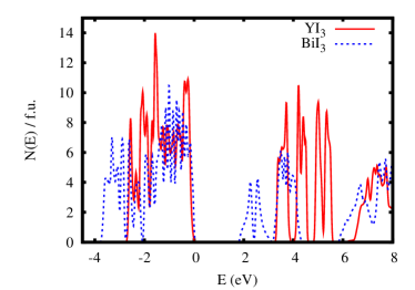

In order for scintillation to take place an electron hole pair must recombine at the Ce3+ site. For this to be efficient both the upper and lower states for the activator ion should be in the band gap. This is evidently not possible in BiI3. As mentioned, the Ce3+ emission in YI3 has a high energy onset of 2.9 eV, which is similar to that in other iodides. This is a lower bound on the energy difference between the upper and lower states of the activator ion. A comparison of the calculated TB-mBJ electronic DOS of the two materials is given in Fig. 6. The lowest conduction bands in YI3 are formed from Y states. In contrast, there is a manifold of Bi states making up the conduction bands in BiI3, and these occur at lower energy. This leads to a 1.49 eV lower band gap in the Bi compound. Our TB-mBJ gap for BiI3 is substantially higher than that obtained in prior calculations using other functionals, Schluter et al. (1976); Yorikawa and Muramatsu (2008) but it is still only 1.82 eV. The experimental optical gap is 2.0 eV. Jellison, Jr. et al. (1999) This does not leave a sufficient energy range for both the upper and lower states to be in the gap. This is similar to the problem in activating Pb phosphate glasses with rare earths. Singh et al. (2006) In fact, considering the fact that, all things being equal, lower gaps favor higher light output, but that too low a gap prevents activation, the gap of YI3 is probably close to optimum for a Ce3+ activated material.
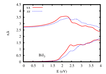
The refractive indices of YI3 and BiI3 are presented in Fig. 7. Surprisingly, the refractive index of YI3 is almost isotropic, even though the material itself is structurally very anisotropic as shown in Fig. 8. The low energy limits of the refractive indices of YI3 are =2.08 and =2.06, which is an anisotropy of 1%. BiI3 is slightly less isotropic, but is still remarkably so for such an anisotropic layered crystal structure. The low energy refractive indices are =2.76 and =2.71 for BiI3. Jellison and co-workers Jellison, Jr. et al. (1999) reported a value of 3.1 at 1.6 eV. For comparison, our calculated values at 1.6 eV are 3.16 in-plane () and 3.02 out-of-plane (), in excellent agreement with the experiment.
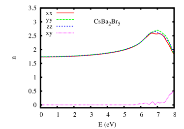
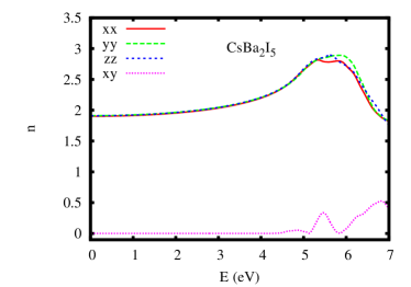
We find a similar remarkable near isotropy of the optical properties in several other halides as well. This is the case even for monoclinic CsBa2Br5 and CsBa2I5 as shown in Fig. 9. The three diagonal components of the refractive index are practically the same almost up to the band edge, while the off-diagonal component associated with the monoclinic symmetry is practically zero over this energy range.
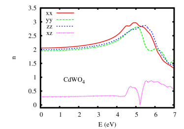
For comparison, we also performed calculations for monoclinic CdWO4. We followed the same procedure relaxing the internal coordinates using the PBE functional and then performing optical calculations using the TB-mBJ functional. The calculated band gaps are 2.99 eV with the PBE functional and 4.16 eV with the TB-mBJ form. The PBE value is similar to that obtained previously by Abraham and co-workers. Abraham et al. (2000) The TB-mBJ value is close to the experimental value of 3.8 – 4.1 eV (see Ref. Abraham et al., 2000). The refractive index is shown in Fig. 10. As may be seen, in contrast to the halides, it is quite anisotropic with a substantial off-diagonal component.
Returning to halides, we next discuss La containing materials. The band structure of these materials may be described as that of an equivalent material based on a simple trivalent ion, e.g. Y, plus additional unoccupied derived bands that occur in the band gap. The bands comprise the so-called -resonance. In Table 5 we give both the value of the fundamental gap, which is from the top of the halogen derived valence bands to the bottom of the -resonance as well as a larger gap, denoted “cb”, which is from the top of the valence bands to the bottom of the conduction bands, excluding the -resonance.
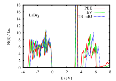
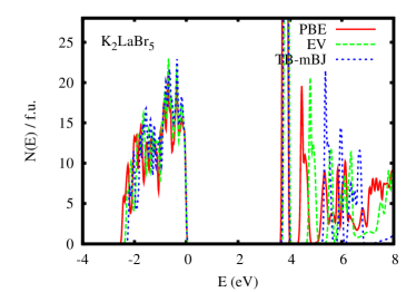
We find that in contrast to the band gaps of the other materials studied, the fundamental gap of these materials is practically unchanged upon going from the PBE to the TB-mBJ functional. That is the position of the bands relative to the valence band maximum is almost the same with these three functionals. This is illustrated for LaBr3 and K2LaBr5 in Fig. 11. The position of the higher lying non- conduction bands is, however, increased with the TB-mBJ functional. This does lower the optical refractive index, even though the fundamental band gap is unchanged.
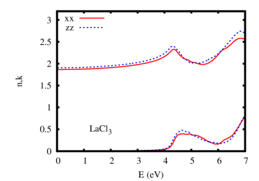
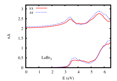
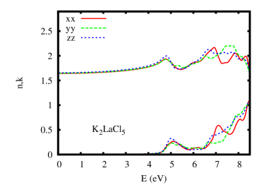
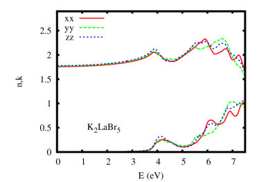
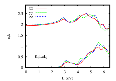
The calculated refractive indices of LaCl3 and LaBr3 are shown in Fig. 12. The low energy values are =1.87 and =1.91 for LaCl3 and =2.04 and =2.09 for LaBr3. This follows the expected trend where bromides have higher refractive index than chlorides. Again, we find low anisotropy in materials that are structurally anisotropic. The same trend with halogen atomic number and also very low optical anisotropy is found in K2LaCl5, K2LaBr5 and K2LaI5, as shown in Fig. 13. The calculated TB-mBJ band gap to the non- upper conduction band edges (“cb”) for K2LaCl5, K2LaBr5 and K2LaI5 are 6.32 eV, 5.26 eV and 3.99 eV, respectively, as compared to the experimental estimates of 6.6 eV, 5.5 eV and 4.5 eV. van Loef et al. (2005, 2003)
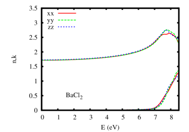
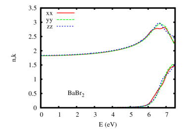
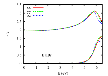
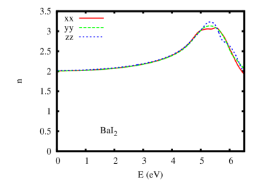
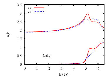
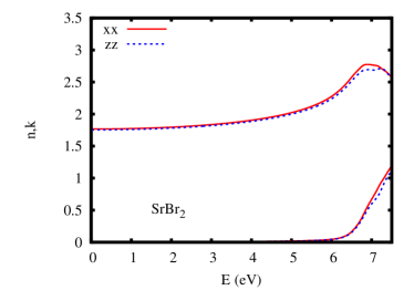
The refractive indices of BaCl2, BaBr2, BaIBr and BaI2 are given in Fig. 14, while those of CaI2 and SrBr2 are given in Figs. 15 and 16, respectively. The calculated TB-mBJ band gap for BaCl2 of 6.45 eV is in reasonable accord with the estimate from Cl- x-ray spectroscopy of 7.0 eV. Sugiura (1974) As mentioned, BaCl2, BaBr2, BaIBr and BaI2 occur in a complex orthorhombic structure. SrBr2 is tetragonal, while CaI2 is rhombohedral. None of these materials is cubic. CaI2 activated with Eu2+ has been known to be an extremely high light output material since the 1960’s. Hofstadter et al. (1964a) However, this hexagonal material has not been used in applications because of crystal growth problems due to its platelet growth habit. Nonetheless, again we find only very small anisotropy in the optical properties for all of these materials, similar to what we found previously for SrI2. Singh (2008)
V discussion
We have two main conclusions, besides the numerical data, which we hope will be useful in improving the design of scintillator systems for better light coupling. First of all, we find that the newly developed TB-mBJ functional greatly improves both the band gaps and the optical properties in a broad class of halide materials, consistent with results reported for other compounds. Tran and Blaha (2009) Considering the computational efficiency of this method, which is similar to standard density functional methods, we expect that this method will enable optical characterization of new complex halide scintillators and perhaps, considering that band gap is a key parameter, more effective theoretical screens for new scintillators.
Secondly, and quite unexpectedly, we find that a wide variety of halide scintillators based on Cl, Br and I are practically isotropic from an optical point of view, even though many of them are highly anisotropic from the point of view of structure and other properties. The broad range of materials in which this occurs implies that it is a general feature of halide chemistry rather than a special coincidence for certain compounds.
Qualitatively, it may be understood from the coordination environments and general band structure features. In particular, these materials have relatively wide band gaps due to the large electronegativity difference between the cations and the halogen atoms. The halogen derived valence bands are narrow compared to typical metal oxides. This narrow band width suggests that one can understand the properties in real space instead of depending essentially on detailed band dispersions. Additionally, the valence band formation comes at least largely from direct hoping between the halogen orbitals on adjacent sites these materials. The structures can be described in anion contact terms. Pauling (1960) From a structural point of view, the anion lattices of these compounds are distortions of high symmetry structures, with most of the anisotropy coming from the cation placement in the intertices of the anion lattice. This places the cations in locally highly symmetric cages based on high nearest neighbor anion coordination numbers. Since the anion bands are relatively non-dispersive, the main crystal structure dependence comes from the conduction bands. Therefore, the small optical anisotropy of these materials can be rationalized in terms of the highly symmetric local environments of the cations as far as nearest neighbor anion coordination is concerned. Further work to elucidate this is clearly needed.
In any case, this result has important implications for the development of halide scintillators. Gamma spectroscopy requires high quality uniform crystals of sufficient size to effectively stop the Gamma rays in the scintillator volume. This typically requires cm sized crystals, and many applications benefit from still larger sizes. As a result, crystal growth is one of the main challenges in the development of new halide scintillators. The near optical isotropy of these materials, however, suggests that ceramic scintillators of sufficient size to be useful for Gamma spectroscopy can be made. While this will require solution of a number of problems, for example, the development of methods to produce dense sintered bodies without contamination in these often hygroscopic, air sensitive materials, it may enable the use of low symmetry difficult to grow halide materials for gamma spectroscopy and other scintillator applications. Furthermore, ceramic materials are generally lower cost than single crystals, especially if large volumes of material are needed.
Acknowledgements.
Work at ORNL was supported by the Department of Energy, Nonproliferation and Verification Research and Development, NA-22.References
- Knoll (2000) G. Knoll, Radiation Detection and Measurement, 3rd Ed. (Wiley, New York, 2000).
- Pauling (1960) L. Pauling, The Nature of the Chemical Bond and the Structure of Molecules and Crystals: An Introduction to Modern Structural Chemistry, 3rd Ed. (Cornell University Press, Ithaca, N.Y., 1960).
- Meyer (1982) G. Meyer, Prog. Solid State Chem. 14, 141 (1982).
- de Haas and Dorenbos (2008) J. T. M. de Haas and P. Dorenbos, IEEE Trans. Nuc. Sci. 55, 1086 (2008).
- van Loef et al. (2001) E. V. D. van Loef, P. Dorenbos, C. W. E. van Eijk, K. Kramer, and H. U. Gudel, Appl. Phys. Lett. 79, 1573 (2001).
- Cherepy et al. (2008) N. J. Cherepy, G. Hull, A. D. Drobshoff, S. A. P. amd E van Loef, C. M. Wilson, K. S. Shah, U. N. Roy, A. Burger, L. A. Boatner, W. S. Choong, et al., Appl. Phys. Lett. 92, 083508 (2008).
- Glodo et al. (2008) J. Glodo, E. V. D. van Loef, W. M. Higgins, and K. S. Shah, IEEE Trans. Nucl. Sci. 55, 1496 (2008).
- Lempicki et al. (22 November 2000) A. Lempicki, C. Brecher, H. Lingertat, and V. K. Sarin, U.S. Patent No. 6,967,330 (22 November 2000).
- Wisniewski et al. (2008) D. J. Wisniewski, L. A. Boatner, J. S. Neal, G. E. Jellison, J. O. Ramey, A. North, M. Wisniewska, A. E. Payzant, J. Y. Howe, A. Lempicki, et al., IEEE Trans. Nucl. Sci. 55, 1501 (2008).
- Singh (2008) D. J. Singh, Appl. Phys. Lett. 92, 201908 (2008).
- Blaha et al. (2001) P. Blaha, K. Schwarz, G. Madsen, D. Kvasnicka, and J. Luitz, WIEN2k, An Augmented Plane Wave + Local Orbitals Program for Calculating Crystal Properties (K. Schwarz, Tech. Univ. Wien, Austria) (2001).
- Singh (1991) D. Singh, Phys. Rev. B 43, 6388 (1991).
- Perdew et al. (1996) J. P. Perdew, K. Burke, and M. Ernzerhof, Phys. Rev. Lett. 77, 3865 (1996).
- Perdew et al. (1992) J. P. Perdew, J. A. Chevary, S. H. Vosko, K. A. Jackson, M. R. Pederson, D. J. Singh, and C. Fiolhais, Phys. Rev. B 46, 6671 (1992).
- Engel and Vosko (1993) E. Engel and S. H. Vosko, Phys. Rev. B 47, 13164 (1993).
- Dufek et al. (1994) P. Dufek, P. Blaha, and K. Schwarz, Phys. Rev. B 50, 7279 (1994).
- Singh (2010) D. J. Singh, Phys. Rev. B 81, 195217 (2010).
- Jellison, Jr. et al. (2010) G. E. Jellison, Jr., S. Auluck, D. J. Singh, and L. A. Boatner, J. Appl. Phys. p. 013514 (2010).
- Tran and Blaha (2009) F. Tran and P. Blaha, Phys. Rev. Lett. 102, 226401 (2009).
- Laval et al. (1983) M. Laval, M. Mosszynski, R. Allemand, E. Cormoreche, P. Guinet, R. Odru, and J. Vacher, Nucl. Inst. Meth. Phys. Res. 206, 169 (1983).
- Woody et al. (1989) C. L. Woody, P. W. Levy, and J. A. Kierstead, IEEE Trans. Nucl. Sci. 36, 536 (1989).
- Visser et al. (1991) R. Visser, P. Dorenbos, C. W. E. van Eijk, R. W. Hollander, and P. Schotanus, IEEE Trans. Nucl. Sci. 38, 178 (1991).
- Wojtowicz et al. (2000) A. J. Wojtowicz, P. Szupryczynski, J. Glodo, W. Drozdowski, and D. Wisniewski, J. Phys. Condens. Matter 12, 4097 (2000).
- Dinca et al. (2002) L. E. Dinca, P. Dorenbos, J. T. M. de Haas, V. R. Bom, and C. W. E. Van Eijk, Nucl. Inst. Meth. Phys. Res. A 486, 141 (2002).
- Swanson and Tatge (1953) A. H. Swanson and E. Tatge, Standard x-ray diffraction powder patterns, National Bureau of Standards Circular 539 (U.S. Gov’t Printing Office, 1953).
- Hofstadter et al. (1964a) R. Hofstadter, E. W. O’Dell, and C. T. Schmidt, IEEE Trans. Nucl. Sci. 11, 12 (1964a).
- Hofstadter et al. (1964b) R. Hofstadter, E. W. O’Dell, and C. T. Schmidt, Rev. Sci. Inst. 35, 246 (1964b).
- Jestin Lenus et al. (2002) A. Jestin Lenus, D. Sornadurai, K. Govinda Rajan, and B. Purniah, Mater. Lett. 57, 635 (2002).
- Selling et al. (2007a) J. Selling, M. D. Birowosuto, P. Dorenbos, and S. Schweizer, J. Appl. Phys. 101 (2007a).
- Selling et al. (2007b) J. Selling, S. Schweizer, M. D. Birowosuto, and P. Dorenbos, J. Appl. Phys. 102, 074915 (2007b).
- Koshimizu et al. (2009) M. Koshimizu, K. Onodera, K. Shibuya, H. Saito, and K. Asai, J. Appl. Phys. 105, 114912 (2009).
- Bourret-Courchesne et al. (2010) E. D. Bourret-Courchesne, G. Bizarri, S. M. Hanrahan, G. Gundiah, Z. Yan, and S. E. Derenzo, Nucl. Inst. Meth. Phys. Res. A 613, 95 (2010).
- Smeggil and Eick (1971) J. G. Smeggil and H. A. Eick, Inorg. Chem. 10, 1458 (1971).
- Blum (1933) H. Blum, Z. Phys. Chem. B 22, 298 (1933).
- Morosin (1968) B. Morosin, J. Chem. Phys. 49, 3007 (1968).
- Kraemer et al. (1989) K. Kraemer, T. Schleid, M. Schulze, W. Urland, and G. Meyer, Z. Anorg. Allg. Chem. 575, 61 (1989).
- van Eijk et al. (2005) C. W. E. van Eijk, J. T. M. de Haas, P. Dorenbos, K. W. Kramer, and H. U. Gudel, IEEE Nucl. Sci. Symp. Conf. Record 1, 239 (2005).
- van Loef et al. (2002) E. V. D. van Loef, P. Dorenbos, C. W. E. van Eijk, K. W. Kramer, and H. U. Gudel, J. Phys. Condens. Matter 14, 8481 (2002).
- Birowosuto et al. (2006) M. D. Birowosuto, P. Dorenbos, C. W. E. van Eijk, K. W. Kramer, and H. U. Gudel, J. Phys. Condens. Matter 18, 6133 (2006).
- Birowosuto et al. (2008) M. D. Birowosuto, P. Dorenbos, J. T. M. de Haas, C. W. E. van Eijk, K. W. Kramer, and H. U. Gudel, IEEE Trans. Nuc. Sci. 55, 1152 (2008).
- Bessiere et al. (2004) A. Bessiere, P. Dorenbos, C. W. E. van Eijk, L. Pidol, K. W. Kramer, and H. U. Gudel, J. Phys. Condens. Matter 16, 1887 (2004).
- Grimm et al. (2007) J. Grimm, J. Fleniken, K. W. Kramer, D. B. andU Happek, and H. U. Gudel, J. Lumin. 122-123, 325 (2007).
- Higgins et al. (2010) W. M. Higgins, J. Glodo, U. Shirwadkar, A. Churilov, E. Van Loef, R. Hawrami, G. Ciampi, C. Hines, and K. S. Shah, J. Cryst. Growth 312, 1216 (2010).
- Reber et al. (1989) C. Reber, H. U. Guedel, G. Meyer, T. Schleid, and C. A. Daul, Inorg. Chem. 28, 3249 (1989).
- van Loef et al. (2003) E. V. D. van Loef, P. Dorenbos, C. W. E. van Eijk, K. W. Kramer, and H. U. Gudel, Phys. Rev. B 68, 045108 (2003).
- van Loef et al. (2005) E. V. D. van Loef, P. Dorenbos, C. W. E. van Eijk, K. W. Kramer, and H. U. Gudel, Nucl. Inst. Meth. Phys. Res. A 537, 232 (2005).
- Sahl (1963) K. Sahl, Beitrage zur Mineralogie und Petrographie 9, 111 (1963).
- Brackett et al. (1939) E. B. Brackett, T. E. Brackett, and R. L. Sass, Z. Anorg. Allg. Chem. 241, 239 (1939).
- Frit et al. (1968) B. Frit, M. Mokail-Chbany, and P. Hagenmuller, Comptes Rendus Acad. Sci. Paris Ser. C 267, 1046 (1968).
- Meyer and Huttl (1983) G. Meyer and E. Huttl, Z. Anorg. Allg. Chem. 497, 191 (1983).
- Meyer et al. (1985) G. Meyer, J. Soose, A. Moritz, V. Vitt, and T. Hollhes, Z. Anorg. Allg. Chem. 521, 161 (1985).
- Schilling and Meyer (1996) G. Schilling and G. Meyer, Z Krist. 211, 255 (1996).
- Bourret-Courchesne et al. (2009) E. D. Bourret-Courchesne, G. Bizarri, R. Borade, Z. Yan, S. M. Hanrahan, G. Gundiah, A. Chaudhry, A. Canning, and S. E. Derenzo, Nucl. Inst. Meth. Phys. Res. A 612, 138 (2009).
- van Loef et al. (2008) E. V. van Loef, W. M. Higgins, J. Glodo, A. V. Churilov, and K. S. Shah, J. Cryst. Growth 310, 2090 (2008).
- Nason and Keller (1995) D. Nason and L. Keller, J. Cryst. Growth 156, 221 (1995).
- Du and Singh (2010) M. H. Du and D. J. Singh, Phys. Rev. B 82, 045203 (2010).
- Jongen and Meyer (2005) L. Jongen and G. Meyer, Acta Cryst. E 61, i51 (2005).
- Trotter and Zobel (1947) J. Trotter and T. Zobel, Skrif. Norske Videnskaps-Akademi Oslo 1947, 73 (1947).
- Rubloff (1972) G. W. Rubloff, Phys. Rev. B 5, 662 (1972).
- Tsujibayashi et al. (2002) T. Tsujibayashi, K. Toyoda, S. Sakuragi, M. Kamada, and M. Itoh, Appl. Phys. Lett. 80, 2883 (2002).
- Shannon et al. (2002) R. D. Shannon, R. C. Shannon, O. Medenbach, and R. X. Fischer, J. Phys. Chem. Ref. Data 31, 931 (2002).
- Jones and Gunnarson (1989) R. O. Jones and O. Gunnarson, Rev. Mod. Phys. 61, 689 (1989).
- Boutchko et al. (2009) R. Boutchko, A. Canning, A. Chaudhry, R. Borade, E. Bourret-Courchesne, and S. E. Derenzo, IEEE Trans. Nucl. Sci. 56, 977 (2009).
- Schluter et al. (1976) M. Schluter, M. L. Cohen, S. E. Kohn, and C. Y. Fong, Phys. Status Solidi b 78, 737 (1976).
- Yorikawa and Muramatsu (2008) H. Yorikawa and S. Muramatsu, J. Phys. Condens. Matter 20, 325220 (2008).
- Jellison, Jr. et al. (1999) G. E. Jellison, Jr., J. O. Ramey, and L. A. Boatner, Phys. Rev. B 59, 9718 (1999).
- Singh et al. (2006) D. J. Singh, G. E. Jellison, Jr., and L. A. Boatner, Phys. Rev. B 74, 155126 (2006).
- Abraham et al. (2000) Y. Abraham, N. A. W. Holzwarth, and R. T. Williams, Phys. Rev. B 62, 1733 (2000).
- Sugiura (1974) C. Sugiura, Phys. Rev. B 9, 2679 (1974).