Membrane morphology induced by anisotropic proteins
Abstract
There are a great many proteins that localize to and collectively generate curvature in biological fluid membranes. We study changes in the topology of fluid membranes due to the presence of highly anisotropic, curvature-inducing proteins. Generically, we find a surprisingly rich phase diagram with phases of both positive and negative Gaussian curvature. As a concrete example modeled on experiments, we find that a lamellar phase in a negative Gaussian curvature regime exhibits a propensity to form screw dislocations of definite burgers scalar but of both chirality. The induced curvature depends strongly on the membrane rigidity, suggesting membrane composition can be a factor regulating membrane sculpting to to curvature-inducing proteins.
To form the structures of many cellular organelles from the Golgi apparatus to the endoplasmic reticulum to the mitochondrial inner membrane, lipid bilayers must be molded and shaped by an army of proteins nature . Among these are proteins known to localize to and, collectively, induce curvature in fluid membranes nature ; BAR ; bax . A flurry of theory fournier ; iglic ; iglic2 ; may , simulations deserno ; voth , and experiments wong ; wong2 has established that proteins can, in principal, induce large-scale shape changes in membranes. Nevertheless, how these proteins control morphology and what other factors are important in determining morphology is still poorly understood.
In this paper, we develop a mean-field model to describe how anisotropic, curvature-inducing proteins induce topological changes in membranes. Using our general model, we show that the magnitude and sign of the induced membrane curvature depends on the protein intrinsic curvatures and concentrations as well as on the rigidities of the underlying membrane. Since the effective membrane rigidity can depend on lipid composition, our model qualitatively explains the observed role of membrane composition in determining curvature wong ; wong2 . For concreteness, we explicitly apply our model to the formation of bicontinuous structures in a lamellar phase doped with saddle-forming proteins. Note only have bicontinuous, cubic phases been observed in model membranes using a variety of proteins wong2 ; wong3 , but they are also seen in vitro in the inner membranes of the mitochondria of starved ameoba dsurface .
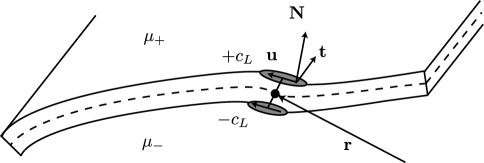
To each bound protein, we associate a position on a two-dimensional surface by projecting the protein center along the normal to the midsurface. A unit vector , tangent to the surface, points along its backbone (see Fig. 1). The second fundamental form of the midsurface can be expressed in terms of two principal curvatures and as , where the are unit vectors along the principal curvature directions. Finally, we define a unit vector , where is the surface normal, that points transverse to the protein backbone. It will prove convenient to define the angle , where the dot product is taken with respect to the surface metric, and dimensionless prescribed curvatures . Assuming Hookean elasticity for the protein-membrane interaction, we obtain the energy
where the sign refers to the sign of . The first term describes the interaction along u while the last term describes the transverse interaction, which induces the curvature along . One can think of the as the energetic cost, in units of , of binding an intrinsically-curved protein to a flat membrane. When do we recover the previously studied models fournier ; iglic .
Rather than fixing the protein density, we calculate in a fixed chemical potential ensemble. Since the proteins on each leaf need not be in equilibrium, we must introduce different chemical potentials, . If a protein, when bound to one leaf of a bilayer, induces a curvature , it must induce a curvature when bound to the opposite leaf (Fig. 1). Therefore, we associate with the positive or negative sign of Eq. (Membrane morphology induced by anisotropic proteins) respectively. Since the proteins only interact through the membrane curvature, the partition function for either membrane leaf is found simply by exponentiating the single protein partition function,
| (2) |
where is the area measure on the membrane midsurface, is a characteristic cross-sectional area of the protein and are the protein fugacities. The average protein concentration and orientation is, therefore, .
Finally, we combine Eq. (2) with the Helfrich energy for a membrane, finding
| (3) |
where , is the mean curvature, the spontaneous curvature, the Gaussian curvature. We have also subtracted off a contribution in , unphysical in a fixed area ensemble, involving the interaction energy between the proteins and a flat membrane. It is straightforward to generalize Eq. (3) to include multiple species of proteins.
We seek to minimize Eq. (3) with respect to the membrane shape. Before proceeding, it is worth noting that a straightforward minimization will not capture the correlated fluctuations of the membrane and, by proxy, the correlated fluctuations of the proteins themselves. Hence, the proteins interact only through the large-scale equilibrium deformations of the membrane. This restriction can be corrected by computing the fluctuation corrections to Eq. (3).
In order to establish our main points with minimal algebraic complication, we will mostly specialize to the extremely anisotropic case . As in the lamellar experiments, we will assume that proteins can bind to either leaf with equal fugacity, so that . Later, we will reintroduce the remaining couplings to see how this basic picture is modified. With this simplification, and in Eq. (3).
As a first step toward a topological phase diagram, we ask what combinations of membrane curvatures and minimize the free energy density. Fig. 2 shows a typical phase diagram as a function of and fugacity . We generically find three morphologies: a flat phase (), a saddle phase (), and a spherical phase (). When , the transition from flat to minimal surface occurs along the line ; therefore, we use in Fig. 2 to highlight the role of .
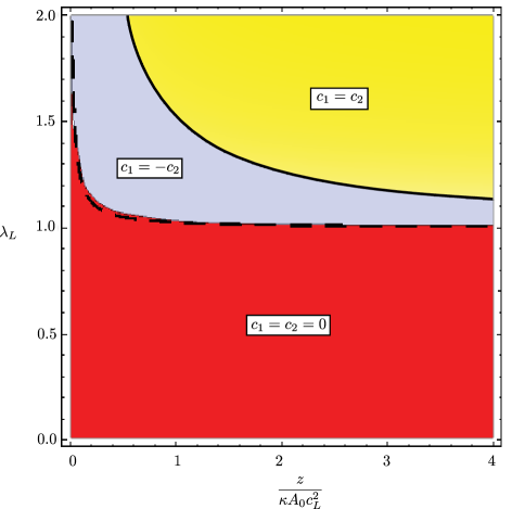
We can understand the transition from the flat to curved phases in terms of the two “topological moduli”, and helfrich ; morse . When , the membrane becomes unstable to topological rearrangements that induce positive Gaussian curvature, for example to spherical vesicles morse . Similarly, when , the membrane becomes unstable toward the formation of negative Gaussian curvature. By expanding Eq. (3) in powers of the curvature, which is valid only in the flat phase, we find effective moduli are shifted by and may so that when
| (4) |
and when
| (5) |
Eq. (5) predicts the boundary between the flat and saddle phases in Fig. 2 quite well. If , the transition from the flat phase proceeds directly to spheres, as can also be seen from Eqs. (4) and (5). In contrast, an instability with respect to continuous perturbations occurs at a critical protein fugacity above which the effective bending modulus . There is also a shift in the spontaneous curvature of which vanishes when the fugacities are balanced.
Why do anisotropic couplings lead to such rich phase diagrams? When , the protein curvature can be accommodated by both and since they need only find a single direction in which the membrane curvature is commensurate with the protein’s intrinsic curvature. However, the orientational entropy of the protein is maximized at umbilics () while the bending energy is minimized at minimal surfaces (). The dependence on the combination arises directly from this competition.
Terms higher than second order in the curvature arising in Eq. (3) are responsible for stabilizing the highly curved phases in Fig. 2. These terms typically compete with potential higher order terms in the Helfrich energy. These Helfrich terms are typically ignored since, in the absence of other physics fournier3 , they are associated with microscopic length scales, . The higher order terms in our model, on the other hand, scale with , the average protein density on a flat membrane. If we denote as the average separation between proteins on the membrane, we obtain that the regime of validity of our model, with respect to higher order corrections, for our model to be valid without these additional corrections.
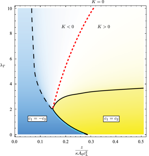
If we introduce the coupling , the spherical phase increases its range of stability. Even when , however, all three phases remain at higher couplings, though at small couplings () the spherical phase preempts the saddle phase entirely. When as well, additional phases can appear. If , for example, a “cylindrical” phase, with and , is introduced. As an example, a cross-section of the phase diagram along the slice is plotted in Fig. 3. The transitions meet at a single critical point at which . For completely isotropic couplings (), there is a “cylindrical” phase (with a small but nonzero ) above a critical fugacity and coupling strength. At small but isotropic couplings, however, protein entropy dominates so that only the flat and spherical phase remain.
Additional complications arise when we make the transition from local phase diagrams, such as those in Figs. 2 and 3, to equilibrium membrane morphologies. We focus on the case that and . In the case of the spherical phase, the lowest energy state is one of monodisperse spherical vesicles of radius . In the saddle phase, however, the lowest energy state occurs when . This is impossible in a three dimensional Euclidean space – the membrane energy is frustrated by the constraints of geometry. As a prototype of a topological transition driven by proteins, we consider the lamellar to bicontinuous phase transition observed experimentally in the presence of certain curvature-inducing peptides wong2 . Generically, lamellar phases that have a tendancy toward negative Gaussian curvature can display a variety of complex morphologies didonna ; in many cases, however, these complex layered structures can be built from a superposition of simple defects schnerk . In fact, screw dislocations are the building blocks of more complex minimal surfaces colding .
Here, we will consider the free energy of inserting a single screw dislocations into the lamellar order. Once it becomes energetically favorable to insert dislocations, we should expect them to proliferate in the ground state. Since screw dislocations of either sign will be degenerate, we should expect the formation of a globally achiral structure such as that explored in Ref. schnerk .
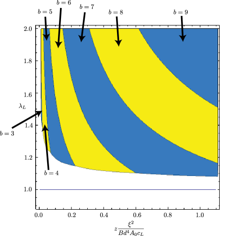
In terms of coordinates on a flat reference layer, a screw dislocation can be described by the multivalued height function of a helicoid, , where is the Burgers scalar, an integer, and the layer spacing. In the absence of proteins, this is an extremum of both the bending and compression energies degennes ; since we are only after the stability of the lamellar phase, we will assume the same height function with proteins present. Screw dislocations have a large elastic contribution to their line tension, arising from the deviation from the equilibrium layer spacing near the core, of the form , where is the bulk modulus and is a microscopic core size degennes . Due to the divergence as , we neglect an additional core energy in comparison to the elastic line tension.
Since , this line tension must be balanced with the contribution from Eq. (3), which we evaluate numerically. We find that a screw dislocation becomes energetically favorable when compared to the flat phase above the critical line shown in Fig. 4. Increasing fugacity prefers an increasing burgers scalar, with both chiralities degenerate. This critical line occurs at larger than predicted by the local phase diagram. The transition also depends strongly on the bulk modulus of the lamellar phase, though we note that the layer spacing is typically set by degennes . If we use for the core size we obtain that , so the horizontal axis of Fig. 4 is roughly comparable to that of our local phase diagrams when .
Finally, the dependence of our phase diagrams on membrane rigidity imply that the induced morphology also depends on lipid composition, since lipids with intrinsic curvature can modify the membrane rigidity may . For an anisotropic lipid with , for example, our results indicate that , suggesting that these intrinsically-curved lipids can drive a membrane coated with proteins from a spherical to saddle phase. Should curvature-inducing proteins take advantage of our mechanism for sculpting membranes, tuning the membrane moduli by adding cosurfacants or changing lipid mixtures will induce a corresponding transition in membrane Gaussian curvature.
In summary, we have studied topological transitions in membranes induced by anisotropic, curvature-inducing proteins. We predict a surprising variety of morphologies having both positive and negative Gaussian curvatures depending on the protein densities and membrane moduli. In a regime of negative Gaussian curvature, this induces a transition from a lamellar phase to one with screw dislocations of both chiralities in the ground state.
This work was funded by the National Science Foundation through DMR-0846582.
References
- (1) H.T. McMahon and J.L. Gallop, Nature 438, 590 (2005).
- (2) B.J. Peter et al., Science 303, 495 (2004).
- (3) R.F. Epand et al., Biomed. Biophys. Res. Comm. 298, 744 (2002); Y. Feng et al., Arch. Biochem. Biophys. 484, 46 (2009).
- (4) J.B. Fournier, Phys. Rev. Lett. 76, 4436 (1996); P.G. Dommersnes and J.P. Fournier, Biophys. J. 83, 2898 (2002).
- (5) M. Fos̆naric̆, A. Igic̆, T. Slivnik and V. Kralj-Iglic̆, Advances in Planar Lipid Bilayers and Liposomes (vol. 8), Ed. A.L. Liu, Chapter 6 (Elsevier, New York, 2008).
- (6) V. Kralj-Iglic̆, V. Heinrich, S. Svetina and B. Zeks, Eur. Phys. J. B 10, 5 (1999).
- (7) M. Fos̆naric̆, A. Igic̆,and S. May, Phys. Rev. E 74, 051503 (2006).
- (8) B.J. Reynwar et al., Nature 447, 461 (2007).
- (9) G.S. Ayton et al., Biophys. J. 97, 1616 (2009).
- (10) Lihua Yang et al., Proc. Nat. Acad. Sci. 105, 20595 (2008); Lihua Yang et al., J. Am. Chem. Soc. 129, 12141 (2007).
- (11) A. Mishra et al., Angew. Chem. Int. Ed. 47, 2986 (2008).
- (12) N. Schmidt, A. Mishra, G.H. Lai and G.C.L. Wong, FEBS Lett. 584, 1806 (2010) and references therein.
- (13) Y. Deng et al., J. Struct. Bio. 127, 231 (1999).
- (14) W. Helfrich, in Physics of Defects, Les Houches Summer School. Balian, R., M. Kleman, J.-P. Poirier, eds. (North Holland, Amsterdam, 1980).
- (15) D.C. Morse, Phys. Rev. E 50, R2423 (1994).
- (16) J.B. Fournier and P. Galatola, EPL 39, 225 (1997).
- (17) B.A. DiDonna and R.D. Kamien, Phys. Rev. Lett. 89, 215504 (2002).
- (18) C.D. Santangelo and R.D. Kamien 96, 137801 (2006).
- (19) T.H. Colding and W.P. Minicozzi II, Proc. Nat. Acad. Sci. 103, 11106 (2006).
- (20) P.G. de Gennes and J. Prost, The physics of liquid crystals (Oxford University Press, Oxford,1995).