Statistical Mechanics Model for Protein Folding
Abstract
We present a novel statistical mechanics formalism for the theoretical description of the process of protein foldingunfolding transition in water environment. The formalism is based on the construction of the partition function of a protein obeying two-stage-like folding kinetics. Using the statistical mechanics model of solvation of hydrophobic hydrocarbons we obtain the partition function of infinitely diluted solution of proteins in water environment. The calculated dependencies of the protein heat capacities upon temperature are compared with the corresponding results of experimental measurements for staphylococcal nuclease and metmyoglobin.
I Introduction
Proteins are biological polymers consisting of elementary structural units, amino acids. Being synthesized at ribosome, proteins are exposed to the cell interior where they fold into their unique three dimensional structure. The process of forming the protein’s three dimensional structure is called the process of protein folding. The correct folding of protein is of crucial importance for the protein’s proper functioning. Despite numerous works devoted to investigation of protein folding this process is still not entirely understood. The current state-of-the-art in experimental and theoretical studies of the protein folding process are described in recent reviews and references therein Munoz07 ; Dill08 ; Onuchic04 ; Shakhnovich06 ; Prabhu06 .
In this paper we develop a novel theoretical method for the description of the protein folding process which is based on the statistical mechanics principles. Considering the process of protein folding as a first order phase transition in a finite system, we present a statistical mechanics model for treating the foldingunfolding phase transition in single-domain proteins. The suggested method is based on the theory developed for the helixcoil transition in polypeptides discussed in Yakubovich07_theoryEPJD ; Yakubovich06a_EPN ; Yakubovich07_resultsEPJD ; Yakubovich06a ; Yakubovich06b ; Yakubovich06c ; Yakubovich08_fluctuations ; ISolovyov06a ; ISolovyov06b ; ISolovyov06c ; Yakubovich_IJQC ; Yakubovich_ISACC09 . A way to construct a parameter-free partition function for a system experiencing -helixrandom coil phase transition in vacuo was studied in Yakubovich07_theoryEPJD . In Yakubovich07_resultsEPJD we have calculated potential energy surfaces (PES) of polyalanines of different lengths with respect to their twisting degrees of freedom. This was done within the framework of classical molecular mechanics. The calculated PES were then used to construct a parameter–free partition function of a polypeptide and to derive various thermodynamical characteristics of alanine polypeptides as a function of temperature and polypeptide length.
In this paper we construct the partition function of a protein in vacuo, which is the further generalization of the formalism developed in Yakubovich06a , accounting for folded, unfolded and prefolded states of the protein. This way of the construction of the partition function is consistent with nucleation-condensation scenario of protein folding, which is a very common scenario for globular proteins Noetling08 and implies that at the early stage of protein folding the native-like hydrophobic nucleus of protein is formed, while at the later stages of the protein folding process all the rest of amino acids also attain the native-like conformation.
For the correct description of the protein folding in water environment it is of primary importance to consider the interactions between the protein and the solvent molecules. The hydrophobic interactions are known to be the most important driving forces of protein folding Kumar02 . In the present work we present a way how one can construct the partition function of the protein accounting for the interactions with solvent, i.e. accounting for the hydrophobic effect. The most prominent feature of our approach is that it is developed for concrete systems in contrast to various generalized and toy-models of protein folding process.
We treat the hydrophobic interactions in the system using the statistical mechanics formalism developed in Scheraga_water04 for the description of the thermodynamical properties of the solvation process of aliphatic and aromatic hydrocarbons in water. However, accounting solely for hydrophobic interactions is not sufficient for the proper description of the energetics of all conformational states of the protein and one has to take electrostatic interactions into account. In the present work the electrostatic interactions are treated within a similar framework as described in Bakk02 .
We have applied the developed statistical mechanics model of protein folding for two globular proteins, namely staphylococcal nuclease and metmyoglobin. These proteins have simple two-stage-like folding kinetics and demonstrate two foldingunfolding transitions, refereed as heat and cold denaturation Griko88 ; Privalov97 . The comparison of the results of the theoretical model with that of the experimental measurements shows the applicability of the suggested formalism for an accurate description of various thermodynamical characteristics in the system, e.g. heat denaturation, cold denaturation, increase of the reminiscent heat capacity of the unfolded protein, etc.
Our paper is organized as follows. In Sec. II.1 we present the formalism for the construction of the partition function of the protein in water environment and justify the assumptions made on the system’s properties. In Section III we discuss the results obtained with our model for the description of foldingunfolding transition in staphylococcal nuclease and metmyoglobin. In Section IV we summarize the paper and suggest several ways for a further development of the theoretical formalism.
II Theoretical Methods
II.1 Partition function of a protein
To study thermodynamic properties of the system one needs to investigate its potential energy surface with respect to all the degrees of freedom. For the description of macromolecular systems, such as proteins, efficient model approaches are necessary.
The most relevant degrees of freedom in the protein folding process are the twisting degrees of freedom along its backbone chain Yakubovich06a ; Yakubovich06a_EPN ; He98a ; He98b ; Yakubovich06b ; Yakubovich06c ; ISolovyov06b ; ISolovyov06c ; Yakubovich07_theoryEPJD . These degrees of freedom are defined for each amino acid of the protein except for the boundary ones and are described by two dihedral angles and (for definition of and see e.g. Yakubovich06a ; Yakubovich06a_EPN ; Yakubovich06b ; Yakubovich06c ; ISolovyov06b ; ISolovyov06c ; Yakubovich07_theoryEPJD ).
The degrees of freedom of a protein can be classified as stiff and soft ones. We call the degrees of freedom corresponding to the variation of bond lengths, angles and improper dihedral angles as stiff, while degrees of freedom corresponding to the angles and are soft degrees of freedom Yakubovich07_theoryEPJD . The stiff degrees of freedom can be treated within the harmonic approximation, because the energies needed for a noticeable structural rearrangement with respect to these degrees of freedom are about several eV, which is significantly larger than the characteristic thermal energy of the system (kT), being at room temperature equal to eV ISolovyov06a ; ISolovyov06b ; ISolovyov06c ; GROMOS ; AMBER ; CHARMM .
A Hamiltonian of a protein is constructed as a sum of the potential, kinetic and vibrational energy terms. Assuming the harmonic approximation for the stiff degrees of freedom it is possible to derive the following expression for the partition function of a protein in vacuo being in a particular conformational state Yakubovich07_theoryEPJD :
| (1) |
where is the temperature, is the Boltzmann constant, is the total number of atoms in the protein, is the number of soft degrees of freedom, is defined as follows:
| (2) |
is a factor which depends on the mass of the protein , its three main momenta of inertia , specific volume , the frequencies of the stiff normal vibrational modes and on the generalized masses corresponding to the soft degrees of freedom Yakubovich07_theoryEPJD . in Eq. (1) describes the potential energy of the system corresponding to the variation of soft degrees of freedom. Integration in Eq. (1) is performed over a certain part of a phase space of the system (a subspace ) corresponding to the soft degrees of freedom and . The form of the partition function in Eq. (1) allows one to avoid the multidimensional integration over the whole coordinate space and to reduce the integration only to the relevant parts of the phase space. in Eq. (1) denotes the potential energy surface of the protein as a function of twisting degrees of freedom in the vicinity of protein’s conformational state . Note that in general the proper choice of all the relevant conformations of protein and the corresponding set of is not a trivial task.
One can expect that the factors in Eq. (1) depend on the chosen conformation of the protein. However, due to the fact that the values of specific volumes, momenta of inertia and frequencies of normal vibration modes of the system in different conformations are expected to be close Krimm80 ; Yakubovich06a , the values of in all conformations become nearly equal, at least in the zero order harmonic approximation, i.e. . Another simplification of the integration in Eq. (1) comes from the statistical independence of amino acids. We assume that within each conformational state all amino acids can be treated statistically independently, i.e. the particular conformational state of -th amino acid characterized by angles and does not influence the potential energy surface of all other amino acids, and vice versa. This assumption is well applicable for rigid conformational states of the protein such as native state. For the native state of a protein all atoms of the molecule move in harmonic potential in the vicinity of their equilibrium positions. However, in unfolded states of the protein the flexibility of the backbone chain leads to significant variations of the distances between atoms, and consequently to a significant variation of interactions between atoms. Accurate accounting (both analytical and computational) for the interactions between distant atoms in the unfolded state of a protein is extremely difficult (see Ref. Cubrovic07 for analytical treatment of interactions in unfolded states of a protein). In this work we assume that all amino acids in unfolded state of a protein move in the identical mean field created by all the amino acids and leave the corrections to this approximation for further considerations.
With the above mentioned assumptions the partition function of a protein (without any solvent) reads as:
| (3) |
where the summation over includes all statistically relevant conformations of the protein, is the number of amino acids in the protein and is the potential energy surface as a function of twisting degrees of freedom and of the -th amino acid in the -th conformational state of the protein. The exact construction of for various conformational states of a particular protein will be discussed below. We consider the angles and as the only two soft degrees of freedom in each amino acid of the protein, and therefore the total number of soft degrees of freedom of the protein .
Partition function in Eq. (3) can be further simplified if one assumes (i) that each amino acid in the protein can exist only in two conformations: the native state conformation and the random coil conformation; (ii) the potential energy surfaces for all the amino acids are identical. This assumption is applicable for both the native and the random coil state. It is not very accurate for the description of thermodynamical properties of single amino acids, but is reasonable for the treatment of thermodynamical properties of the entire protein. The judgment of the quality of this assumption could be made on the basis of comparison of the results obtained with its use with experimental data. Such comparison is performed in Sec. III of this work.
Amino acids in a protein being in its native state vibrate in a steep harmonic potential. Here we assume that the potential energy profile of an amino acid in the native conformation should not be very sensitive to the type of amino acid and thus can be taken as, e.g., the potential energy surface for an alanine amino acid in the -helix conformation Yakubovich07_resultsEPJD . Using the same arguments the potential energy profile for an amino acid in unfolded protein state can be approximated by e.g. the potential of alanine in the unfolded state of alanine polypeptide (see Ref. Yakubovich07_resultsEPJD for discussion and analysis of alanine’s potential energy surfaces). Indeed, for an unfolded state of a protein it is reasonable to expect that once neglecting the long-range interactions all the differences in the potential energy surfaces of various amino acids arise from the steric overlap of the amino acids’s radicals. This is clearly seen on alanine’s potential energy surface at values of presented in Ref. Yakubovich07_resultsEPJD . But the part of the potential energy surface at gives a minor contribution to the entropy of amino acid at room temperature. This fact allows one to neglect all the differences in potential energy surfaces for different amino acids in an unfolded protein, at least in the zero order approximation. This assumption should be especially justified for proteins with the rigid helix-rich native structure. The staphylococcal nuclease, which we study here has definitely high -helix content. Another argument which allows to justify our assumption for a wider family of proteins is the rigidity of the protein’s native structure. Below, we validate the assumptions made by performing the comparison of the results of our theoretical model with the experimental data for rich protein metmyoglobin obtained in Privalov97 .
For the description of the folding unfolding transition in small globular proteins obeying simple two-state-like folding kinetics we assume that the protein can exist in one of three states: completely folded state, completely unfolded state and partially folded state where some amino acids from the flexible regions with no prominent secondary structure are in the unfolded state, while other amino acids are in the folded conformation. With this assumption the partition function of the protein reads as:
| (4) |
where is defined in Eq. (1), is the partition function of the protein in completely unfolded state, is the total number of amino acids in a protein and is the number of amino acids in flexible regions. The factorial term in Eq. (4) accounts for the states in which various amino acids from flexible regions independently attain the native conformation. The summation in Eq. (4) is performed over all partially folded states of the protein, where is the minimal possible number of amino acids being in the folded state. The factorial term describes the number of ways to select amino acids from the flexible region of the protein consisting of amino acids attaining native-like conformation.
Finally, the partition function of the protein in vacuo has the following form:
| (5) |
where
| (6) | |||||
| (7) | |||||
| (8) |
Here we omitted the trivial factor describing the motion of the protein center of mass, which is of no significance for the problem considered, (b stands for bound) is the potential energy surface of an amino acid in the native conformation and (u stands for unbound) is the potential energy surface of an amino acid in the random coil conformation. The potential energy profile of an amino acid is calculated as a function of its twisting degrees of freedom and . Let us denote by and the global minima on the potential energy surfaces of an amino acid in folded and in unfolded conformations respectively. The potential energy of an amino acid then reads as . in Eq. (6) is defined as the energy difference between the global energy minima of the amino acid potential energy surfaces corresponding to the folded and unfolded conformations, i.e. . The potential energy surfaces for amino acids as functions of angles and were calculated and thoroughly analyzed in Yakubovich07_resultsEPJD .
In nature proteins perform their function in the aqueous environment. Therefore the correct theoretical description of the foldingunfolding transition in water environment should account for solvent effects.
II.2 Partition function of a protein in water environment
In this section we evaluate and construct the partition function for the protein in water environment.
The partition function of the infinitely diluted solution of proteins can be constructed as follows:
| (9) |
where is the partition function of all water molecules in the -th conformational state of a protein and is the partition function of the protein in its -th conformational state, in which we further omit the factor describing the contribution of stiff degrees of freedom in the system. This is done in order to simplify the expressions, because stiff degrees of freedom provide a constant contribution to the heat capacity of the system since the heat capacity of the ensemble of harmonic oscillators is constant. Below for the simplicity of notations we put .
There are two types of water molecules in the system: (i) molecules in pure water and (ii) molecules interacting with the protein. We assume that only the water molecules being in the vicinity of the protein’s surface are involved in the foldingunfolding transition, because they are affected by the variation of the hydrophobic surface of a protein. This surface is equal to the protein’s solvent accessible surface area (SASA) of the hydrophobic amino acids. The number of interacting molecules is proportional to SASA and include only the molecules from the first protein’s solvation shell. This area depends on the conformation of the protein. The main contribution to the energy of the system caused by the variation of the protein’s SASA associated with the side-chain radicals of amino acids because the contribution to the free energy assosiated with solvation of protein’s backbone is small Ptizin_book . Thus, in this work we pay the main attention to the accounting for the SASA change arising due to the solvation of side chain radicals.
We treat all water molecules as statistically independent, i.e. the energy spectra of the states of a given molecule and its vibrational frequencies do not depend on a particular state of all other water molecules. Thus, the partition function of the whole system can be factorized and reads as:
| (10) |
where is the total number of states of a protein, is the partition function of a water molecule affected by the interaction with the protein and is the partition function of a water molecule in pure water. is the number of water molecules interacting with the protein in the -th conformational state. is the total number of water molecules in the system. To simplify the expressions we do not account for water molecules that do not interact with the protein in any of its conformational states, i.e. .
To construct the partition function of water we follow the formalism developed in Scheraga_water04 and refer only to the most essential details of that work. The partition function of a water molecule in pure water reads as:
| (11) |
where the summation is performed over 5 possible states of a water molecule (the states in which water molecule has 4,3,2,1 or 0 hydrogen bonds with the neighboring molecules). are the energies of these states and are the combinatorial factors being equal to 1,4,6,4,1 for , respectively. They describe the number of choices to form a given number of hydrogen bonds. in Eq. (11) describes the contribution due to the partition function arising to to the translation and libration oscillations of the molecule. In the harmonic approximation are equal to:
| (12) |
where and are translation and libration motions frequencies of a water molecule in its -th state, respectively. These frequencies are calculated in Ref. Scheraga_water04 and are given in Table 1. The contribution of the internal vibrations of water molecules is not included in Eq. (11) because the frequencies of these vibrations are practically not influenced by the interactions with surrounding water molecules.
| Number of hydrogen bonds | 0 | 1 | 2 | 3 | 4 |
| Energy level, (kcal/mol) | 6.670 | 4.970 | 3.870 | 2.030 | 0 |
| Energy level, (kcal/mol) | 6.431 | 4.731 | 3.631 | 1.791 | -0.564 |
| Translational frequencies, , cm-1 | 26 | 86 | 61 | 57 | 210 |
| Librational frequencies, , cm-1 | 197 | 374 | 500 | 750 | 750 |
The partition function of a water molecule from the protein’s first solvation shell reads as:
| (13) |
where are defined in Eq. (12) and denotes the energy levels of a water molecule interacting with aliphatic hydrocarbons of protein’s amino acids. Values of energies are given it Table 1. For simplicity we treat all side-chain radicals of a protein as aliphatic hydrocarbons because most of the protein’s hydrophobic amino acids consist of aliphatic-like hydrocarbons. It is possible to account for various types of side chain radicals by using the experimental results of the measurements of the solvation free energies of amino acid radicals from Ref. Privalov93 and associated works. However, this correction will imply the reparametrization of the theory presented in Scheraga_water04 and will lead to the introduction of additional parameters. Here we do not perform such a task since this kind of improvement of the theory would smear out the understanding of the principal physical factors underlying the protein foldingunfolding transition.
In our theoretical model we also account for the electrostatic interaction of protein’s charged groups with water. The presence of electrostatic field around the protein leads to the reorientation of H2O molecules in the vicinity of charged groups due to the interaction of dipole moments of the molecules with the electrostatic field. The additional factor arising in the partition function (11) of the molecules reads as:
| (14) |
where is the strength of the electrostatic field, is the absolute value of the H2O molecule dipole moment, is the ratio of the number of water molecules that interact with the electrostatic field of the protein () to the number of water molecules interacting with the surface of the amino acids from the inner part of the protein while they are exposed to water when the protein is being unfolded (), i.e. . Note that the effects of electrostatic interaction turn out to be more pronounced in the folded state of the protein. This happens because in the unfolded state of a protein opposite charges of amino acid’s radicals are in average closer in space due to the flexibility of the backbone chain, while in the folded state the positions of the charges are fixed by the rigid structure of a protein.
Integrating Eq. (14) allows to write the factor for the partition function of a single H2O molecule in pure water in the form:
| (15) |
This equation shows how the electrostatic field enters the partition function. In general, depends on the position in space with respect to the protein. However, here we neglect this dependence and instead we treat the parameter as an average, characteristic electrostatic field created by the protein.
Let us denote by the number of water molecules interacting with the proteins surface in its folded state i.e. ; where is defined in Eq (10). We assume that the number of water molecules interacting with the protein () is linearly dependent on the number of amino acids being in the unfolded conformation, i.e. , where is the number of the amino acids in the unfolded conformation and is the total number of amino acids in the protein. Thus, the partition function (10) with the accounting for the factor (15) reads as:
| (16) |
where denotes the number of the amino acids being in the folded conformation when the protein is in the -th conformational state. Accounting for the statistical factors for amino acids being in the folded and unfolded states, similarly to how it was done for the vacuum case (see Eq. (6)), one derives from Eq. (16) the following final expression:
where the term in the square brackets accounts for all statistically significant conformational states of the protein.
Having constructed the partition function of the system we can evaluate with its use all thermodynamic characteristics of the system, such as e.g. entropy, free energy, heat capacity, etc. The free energy () and heat the capacity () of the system can be calculated from the partition function as follows:
| (18) | |||
| (19) |
In this work we analyze the dependence of protein’s heat capacity on temperature and compare the predictions of our model with available experimental data.
III Results and Discussion
In this section we calculate the dependencies of the heat capacity on temperature for two globular proteins metmyoglobin and staphylococcal nuclease and compare the results with experimental data from Griko88 ; Privalov97 .
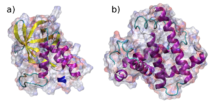
The structures of metmyoglobin and staphylococcal nuclease proteins are shown in Fig. 1. These are relatively small globular proteins consisting of 150 amino acids. Under certain experimental conditions (salt concentration and pH) the metmyoglobin and the staphylococcal nuclease experience two foldingunfolding transitions, which induce two peaks in the dependency of heat capacity on temperature (see further discussion). The peaks at lower temperature are due to the cold denaturation of the proteins. The peaks at higher temperatures arise due to the ordinary foldingunfolding transition. The availability of experimental data for the heat capacity profiles of the mentioned proteins, the presence of the cold denaturation and simple two-stage-like folding kinetics are the reasons for selecting these particular proteins as case studies for the verification of the developed theoretical model.
III.1 Heat capacity of staphylococcal nuclease
Staphylococcal or micrococcal nuclease (S7 Nuclease) is a relatively nonspecific enzyme that digests single-stranded and double-stranded nucleic acids, but is more active on single-stranded substrates SNase79 . This protein consists of 149 amino acids. It’s structure is shown in Fig. 1a.
To calculate the SASA of staphylococcal nuclease in the folded state the 3D structure of the protein was taken from the Protein Data Bank PDB (PDB ID 1EYD). Using CHARMM27 CHARMM forcefield and NAMD program NAMD we performed the structural optimization of the protein and calculated SASA with the solvent probe radius 1.4 Å.
The value of SASA of the side-chain radicals in the folded protein conformation is equal to 6858 Å2. In order to calculate SASA for an unfolded protein state, the value of all angles and were put equal to , corresponding to a fully stretched conformation. Then, the optimization of the structure with the fixed angles and was performed. The optimized geometry of the stretched molecule has a minor dependence on the value of dielectric susceptibility of the solvent, therefore the value of dielectric susceptibility was chosen to be equal to 20, in order to mimic the screening of charges by the solvent. SASA of the side-chain radicals in the stretched conformation of the protein is equal to 15813 Å2.
The change of the number of water molecules those interacting with the protein due to the unfolding process can be calculated as follows:
| (20) |
where Å2 and Å2 are the SASA of the protein in unfolded and in folded conformations, respectively and is the density of the water molecules. The volume of one mole of water is equal to 18 cm3, therefore Å-3
To account for the effects caused by the electrostatic interaction of water molecules with the charged groups of the protein it is necessary to evaluate the strength of the average electrostatic field in Eq. (15). The strength of the average field can be estimated as , where is the dipole moment of a water molecule, is Bolzmann constant and T=300 K is the room temperature. According to this estimate the energy of characteristic electrostatic interaction of water molecules is equal to the thermal energy per degree of freedom of a molecule.
The total number of water molecules that interact with the electrostatic field of the protein can be estimated from the known Debye screening length of a charge in electrolyte as follows:
| (21) |
where is the number of charged groups in the protein, is the density of water and is the Debye screening length. Debye screening length of the symmetric electrolyte can be calculated as follows Russel :
| (22) |
where is the permittivity of free space, is the dielectric constant, is the Avogadro number, is the elementary charge and is is the ionic strength of the electrolyte.
The experiments on denaturation of staphylococcal nuclease and metmyoglobin were performed in 100 mM ion buffer of sodium chloride and 10mM buffer of sodium acetate respectively Griko88 ; Privalov97 . The Debye screening length in water with 10 mM and 100 mM concentration of ions is 30 Å and 10 Å at room temperature respectively.
The described method allows to estimate the number of water molecules () interacting with electric filed created by the charged groups of a protein. It should be considered as qualitative estimate since we have assumed the average electric field as being constant within a sphere of the radius , but in fact it experiences some variations. Thus, at the distances 15 Å from the point charge the interaction energy of a H2O molecule with the electric field becomes equal to kT (for this estimate we have used the linear growing distance-dependent dielectric susceptibility as derived in Mallik02 for the atoms fully exposed to the solvent). However, we expect that the more accurate analysis accounting for the spatial variation of the electric field will not change significantly the results of the analysis reported here, because it is based on the physically correct picture of the effect and the realistic values of all the physical quantities. At physiological conditions staphylococcal nuclease has 8 charged residues Zhou02 . The value of for this protein varies within the interval from 1.29 to 31.27 for [10..30] Å. In our numerical analysis we have used the characteristic value of equal to 2.5.
Note that number of molecules interacting with the electrostatic field and the strength of the electrostatic field should be considered as the parameters of our model. In this work we do not perform accurate accounting for the spatial dependence of the electrostatic field. Instead, we introduce the parameters and that can be interpreted as effective values of the number of H2O molecules and the strength of the electrostatic field correspondingly. Let us stress that the number of water molecules and the strength of the field are not independent parameters of our model because by choosing the higher value of and smaller value of or vice versa one can derive the same heat capacity profile.
In this work we do not investigate the dependencies of the heat capacity profiles on the values of the parameters and . Below we focus on the investigation of the dependence of the protein heat capacity on the energy at the fixed value of and equal to 2.5 and 0.58 kcal/mol respectively.
An important parameter of the model is the energy difference between the two states of the protein normalized per one amino acid, introduced in Eq. (6). This parameter describes both the energy loss due to the separation of the hydrophobic groups of the protein which attract in the native state of the protein due to Van-der-Waals interaction and the energy gain due to the formation of Van-der-Waals interactions of hydrophobic groups of the protein with H2O molecules in the protein’s unfolded state. Also, the difference of the electrostatic energy of the system in the folded and unfolded states is accounted for in . The difference of the electrostatic energy may depend on various characteristics of the system, such as concentration of ions in the solvent and its pH, on the exact location of the charged sites in the native conformation of the protein and on the probability distribution of distances between charged amino acids in the unfolded state. Thus, exact calculation of is rather difficult. It is a separate task which we do not intend to address in this work. Instead, in the current study the energy difference between the two phases of the protein is considered as a parameter of the model. We treat as being dependent on external properties of the system, in particular on the pH value of the solution.
Another characteristic of the protein foldingunfolding transition is its cooperativity. In the model it is described by the parameter in Eq. (4). describes the number of amino acids in the flexible regions of the protein. The staphylococcal nuclease possesses a prominent two-stage folding kinetics, therefore only 5-10% of amino acids is in the protein’s flexible regions. Thus, the value of for this protein is small. It can be estimated as being equal to amino acids.
The values of for staphylococcal nuclease at different values of pH are given in Table 2.
| pH value | 7.0 | 5.0 | 4.5 | 3.88 | 3.23 |
|---|---|---|---|---|---|
| (kcal/mol) | 0.789 | 0.795 | 0.803 | 0.819 | 0.890 |
For the analysis of the variation of the thermodynamic properties of the system during the folding process one can omit all the contributions to the free energy of the system that do not alter significantly in the temperature range between -50∘C and 150∘. Therefore, from the expression for the total free energy of the system we can subtract all slowly varying contributions as follows:
| (23) |
From Eq. (23) follows that the subtraction of corresponds to the division of the total partition function by the partition function of the subsystem () with slowly varying thermodynamical properties. Therefore, in order to simplify the expressions, one can divide the partition function in Eq. (II.2) by the partition function of fully unfolded conformation of a protein (by ) and by the partition function of free water molecules (by ). Thus, Eq. (II.2) can be rewritten as follows:
| (25) |
With the use of Eq. (19) on can calculate the heat capacity of the system as follows:
| (26) |
where the factors and are responsible for the absolute value and the inclination of the heat capacity curve respectively. These factors account for the contribution of stiff harmonic vibrational modes in the system (factor ) and for the unharmonic correction to these vibrations (factors and ). The contribution of protein’s stiff vibrational modes and the heat capacity of the fully unfolded conformation of protein is also included into these factors. In our numerical analysis we have adjusted the values of , and in order to match experimental measurements. However, factors , and should not be considered as parameters of our model since their values are not related to the thermodynamic characteristics of the foldingunfolding transition and depend not entirely on the properties of the protein but also on the properties of the solution, protein and ion concentrations, etc.
In our calculations for staphylococcal nuclease we have used the values of JK-1g-1, JK-2g-1 and 323 in Eq. (26).
The dependence of heat capacity on temperature calculated for staphylococcal nuclease at different pH values are presented in Fig. 2 by solid lines. The results of experimental measurements form Ref. Griko88 are presented by symbols. From Fig. 2 it is seen that staphylococcal nuclease experience two foldingtransitions in the range of pH between 3.78 and 7.0. At the pH value 3.23 no peaks in the heat capacity is present. It means that the protein exists in the unfolded state over the whole range of experimentally accessible temperatures.
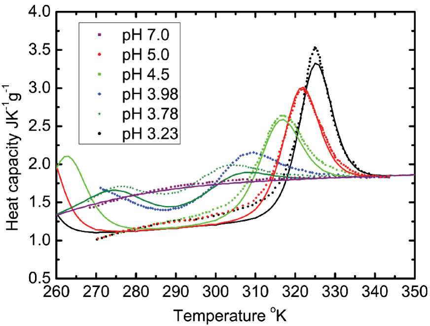
Comparison of the theoretical results with experimental data shows that our theoretical model reproduces experimental behavior better for the solvents with higher pH. The heat capacity peak arising at higher temperatures due to the standard foldingunfolding transition is reproduced very well for pH values being in the region 4.5-7.0. The deviations at low temperatures can be attributed to the inaccuracy of the statistical mechanics model of water in the vicinity of the freezing point.
The accuracy of the statistical mechanics model for low pH values around 3.88 is also quite reasonable. The deviation of theoretical curves from experimental ones likely arise due to the alteration of the solvent properties at high concentration of protons or due to the change of partial charge of amino acids at pH values being far from the physiological conditions.
Despite some difference between the predictions of the developed model and the experimental results arising at certain temperatures and values of pH the overall performance of the model can be considered as extremely good for such a complex process as structural folding transition of a large biological molecule.
III.2 Heat capacity of metmyoglobin
Metmyoglobin is an oxidized form of a protein myoglobin. This is a monomeric protein containing a single five-coordinate heme whose function is to reversibly form a dioxygen adduct Myo04 . Metmyolobin consists of 153 amino acids and it’s structure is shown in Fig. 1 on the right.
In order to calculate SASA of side chain radicals of metmyoglobin exactly the same procedure as for staphylococcal nuclease was performed (see discussion in the previous subsection). SASA in the folded and unfolded states of the protein has been calculated and is equal 6847 Å2 and 16926 Å2 respectively. Thus, there are H2O molecules interacting with protein’s hydrophobic surface in its unfolded state.
The electrostatic interaction of water molecules with metmyoglobin was accounted for in the same way as for staphylococcal nuclease. The parameter in Eq. (15) was chosen to be equal to 2.5. With this we derive that 10950 H2O molecules involve in the interaction with the electrostatic field of metmyoglobin in its folded state. The strength of the field was chosen the same as for staphylococcal nuclease.
The parameter for metmyoglobin in Eq. (4), describing the cooperativity of the foldingunfolding transition, differs significantly from that for staphylococcal nuclease. The transition in metmyoglobin is less cooperative than the transition in staphylococcal nuclease because metmyoglobin has intermediate partially folded states Schortle01 . Thus, while the rigid native-like core of the protein is formed, a significant fraction of amino acids in the flexible regions of the protein can exist in the unfolded state. We assume that 1/3 of metmyoglobin’s amino acids are in the flexible region, i.e. the parameter in Eq. (4) equal to 50.
| pH value | 4.10 | 3.70 | 3.84 | 3.5 |
|---|---|---|---|---|
| (kcal/mol) | 1.128 | 1.150 | 1.165 | 1.2 |
The values of in Eq. (6) differ from that for staphylococcal nuclease and are compiled in Table 3. In our calculations for metmyoglobin we have used the values of JK-1g-1, JK-2g-1 and =323 in Eq. (26).
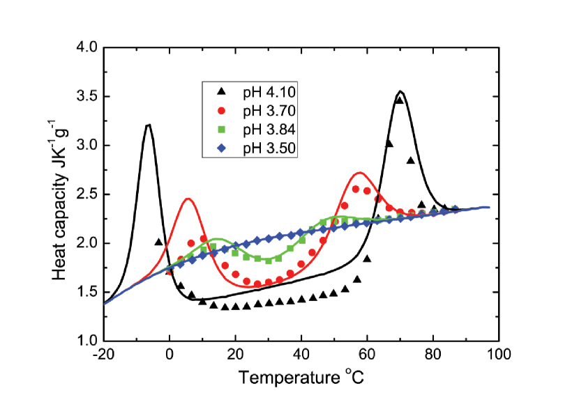
Solid lines in Fig. 3 show the dependence of the metmyoglobin’s heat capacity on temperature calculated using the developed theoretical model. The experimental data from Ref. Privalov97 are shown by symbols.
Metmyoglobin experiences two foldingunfolding transitions at the pH values exceeding 3.5 which can be called as cold and heat denaturations of the protein. The dependence of the heat capacity on temperature therefore has two characteristic peaks, as seen in Fig. 3. Figure 3 shows that at pH lower than 3.84 metmyoglobin exists only in the unfolded state.
The comparison of predictions of the developed theoretical model with the experimental data on heat capacity shows that the theoretical model is well applicable for metmyoglobin case as well. The good agreement of the theoretical and experimental heat capacity profiles over the whole range of temperatures and pH values shows that the model treats correctly the thermodynamics of the protein folding process.
Our theory includes a number of parameters, namely the energy difference between two phases , strength of the electrostatic field , number of interacting H2O molecules , the parameter describing the cooperativity of the phase transition , as well as other parameters introduced in Ref. Scheraga_water04 to treat the partition function of water. Three parameters, , and , are dependent on the properties of a particular protein and on the pH of the solvent. We have adjusted the values of these parameters in order to reproduce the experimental data. All other parameters of the model describing the structure of energy levels of water molecules, their vibrational and librational frequencies, etc. are considered as fixed, being universal for all proteins.
In spite of the model features of our approach, we want to stress that the complex behavior and the peculiarities in dependencies of the heat capacity on temperature are all very well reproduced by the developed model with only a few parameters. This was demonstrated for two proteins and we consider this result as a significant achievement. This fact supports our conclusion that the developed model can be used for the prediction of new features of phase transitions in various biomolecular systems. Indeed, from Figs. 2 and 3 one can extract a lot of useful information on the heat capacity profiles: the concave bending of the heat capacity profile for a completely unfolded protein, the temperature of the cold and heat denaturation, the absolute values of the heat capacity at the phase transition temperature, the broadening of heat capacity peaks. Another peculiarity which is well reproduced by our statistical mechanics model is the decrease of the heat capacity of the folded state of the protein in comparison with that for unfolded state and asymmetry of the heat capacity peaks.
IV Conclusions
We have developed a novel statistical mechanics model for the description of foldingunfolding processes in globular proteins obeying simple two-stage-like folding kinetics. The model is based on the construction of the partition function of the system as a sum over all statistically significant conformational states of a protein. The partition function of each state is a product of partition function of a protein in a given conformational state, partition function of water molecules in pure water and a partition function of H2O molecules interacting with the protein.
The introduced model includes a number of parameters responsible for certain physical properties of the system. The parameters were obtained from available experimental data and three of them (energy difference between two phases, cooperativity of the transition and the average strength of the protein’s electrostatic field) were considered as being variable depending on a particular protein and pH of the solvent.
We have compared the predictions of the developed model with the results of experimental measurements of the dependence of the heat capacity on temperature for staphylococcal nuclease and metmyoglobin. The experimental results were obtained at various pH of solvent. The suggested model is capable to reproduce well within a single framework a large number of peculiarities of the heat capacity profile, such as the temperatures of cold and heat denaturations, the corresponding maximum values of the heat capacities, the temperature range of the cold and heat denaturation transitions, the difference between heat capacities of the folded and unfolded states of the protein.
The good agreement of the results of calculations obtained using the developed formalism with the results of experimental measurements demonstrates that it can be used for the analysis of thermodynamical properties of many biomolecular systems. Further development of the model can be focused on its advance and application for the description of the influence of mutations on protein stability, analysis of assembly and stability of protein complexes, protein crystallization process, etc.
V Acknowledgments
We acknowledge support of this work by the NoE EXCELL. We are grateful to Dr. Ilia Solov’yov for the careful reading of the manuscript and helpful advice.
References
- (1) V. Muñoz, Annu. Rev. Biophys. Biomol. Struct. 36, 395 (2007).
- (2) K. A. Dill, S. B. Ozkan, M. S. Shell, and T. R. Weikl, Annu. Rev. Biophys. 37, 289 (2008).
- (3) J. N. Onuchic and P. G. Wolynes, Curr. Op. Struct. Biol. 14, 70 (2004).
- (4) E. Shakhnovich, Chem. Rev. 106, 1559 (2006).
- (5) N. V. Prabhu and K. A. Sharp, Chem. Rev 106, 1616 (2006).
- (6) A. Yakubovich, I. Solov’yov, A. Solov’yov, and W. Greiner, Eur. Phys. J. D 46, 215 (2007), arXiv:0704.3079v1 [physics.bio-ph], 23 Apr 2007.
- (7) A. Yakubovich, I. Solov’yov, A. Solov’yov, and W. Greiner, Europhys. News 38, 10 (2007).
- (8) I. Solov’yov, A. Yakubovich, A. Solov’yov, and W. Greiner, Eur. Phys. J. D 46, 227 (2008), arXiv:0704.3085v1 [physics.bio-ph], 23 Apr 2007.
- (9) A. Yakubovich, I. Solov’yov, A. Solov’yov, and W. Greiner, Eur. Phys. J. D 40, 363 (2006).
- (10) A. Yakubovich, I. Solov’yov, A. Solov’yov, and W. Greiner, Eur. Phys. J. D 39, 23 (2006).
- (11) A. Yakubovich, I. Solov’yov, A. Solov’yov, and W.Greiner, Khimicheskaya Fizika (Chemical Physics) (in Russian) 25, 11 (2006).
- (12) A. Yakubovich, I. Solov’yov, A. Solov’yov, and W. Greiner, Eur. Phys. J. D DOI: 10.1140/epjd/e2008-00126-y (2008).
- (13) I. Solov’yov, A. Yakubovich, A. Solov’yov, and W. Greiner, J. Exp. Theor. Phys. 103, 463 (2006).
- (14) I. Solov’yov, A. Yakubovich, A. Solov’yov, and W. Greiner, Phys. Rev. E 73, 021916 (2006).
- (15) I. Solov’yov, A. Yakubovich, A. Solov’yov, and W. Greiner, J. Exp. Theor. Phys. 102, 314 (2006).
- (16) A. Yakubovich, A. Solov’yov, and W. Greiner, Int. J. Quant. Chem. 110, 257 (2010).
- (17) A. Yakubovich, A. Solov’yov, and W. Greiner, AIP Conf. Proc. 1197, 186 (2009).
- (18) B. Noetling and D. A. Agard, Proteins 73, 754 (2008).
- (19) S. Kumar, C.-J. Tsai, and R. Nussinov, Biochemistry 41, 5359 (2002).
- (20) J. H. Griffith and H. Scheraga, J. Mol. Struc. 682, 97 (2004).
- (21) A. Bakk, J. S. Hye, and A. Hansen, BJ 82, 713719 (2002).
- (22) Y. Griko, P. Privalov, J. Aturtevant, and S. Venyaminov, Proc. Natl. Acad. Sci. USA 85, 3343 (1988).
- (23) P. Privalov, J. Chem. Thermodyn. 29, 447 (1997).
- (24) S. He and H. A. Scheraga, J. Chem. Phys. 108, 271 (1998).
- (25) S. He and H. A. Scheraga, J. Chem. Phys. 108, 287 (1998).
- (26) W. Scott and W. van Gunsteren, in Methods and Techniques in Computational Chemistry: METECC-95, edited by E. Clementi and G. Corongiu (STEF, Cagliari, Italy, 1995) pp. 397–434.
- (27) W. Cornell, P. Cieplak, C. Bayly, and et al, J. Am. Chem. Soc. 117, 5179 (1995).
- (28) A. MacKerell, D. Bashford, R. Bellott, and et al, J. Phys. Chem. B 102, 3586 (1998).
- (29) S. Krimm and J. Bandekar, Biopolymers 19, 1 (1980).
- (30) M. Cubrovic, O. Obolensky, and A. Solov’yov, Eur. Phys. J. D 51, 41 (2009).
- (31) A. Finkelstein and O. Ptitsyn, Protein Physics. A Course of Lectures (Elsevier Books, Oxford, 2002).
- (32) G. Makhatadze and P. Privalov, J. Mol. Biol. 232, 639 (1993).
- (33) J. Chen, Z. Lu, J. Sakon, and W. Stites, J.Mol.Biol. 303, 125 (2000).
- (34) S. Evans and G. Brayer, J.Mol.Biol. 213, 885 (1990).
- (35) W. Humphrey, A. Dalke, and K. Schulten, J. Molec. Graphics 14, 33 (1996).
- (36) F. A. Cotton, J. Edward E. Hazen, and M. J. Legg, Proc. Natl. Acad. Sci. USA 76, 2551 (1979).
- (37) http://www.rcsb.org/(2009).
- (38) J. C. Phillips, R. Braun, W. Wang, and et al, J. Comp. Chem. 26, 1781 (2005).
- (39) W. Russel, D. Saville, and W. Schowalter, Colloidal Dispersions (Cambridge University Press, 1989).
- (40) B. Mallik and T. L. A. Masunov, J. Comp. Chem. 23, 1090 (2002).
- (41) H.-X. Zhou, BJ 83, 2981 – 2986 (2002).
- (42) J. P. Collman, R. Boulatov, C. J. Sunderland, and L. Fu, Chem. Rev. 104, 561 (2004).
- (43) D. Shortle and M. S. Ackerman, Science 293, 487 (2001).