Combining high pressure and coherent diffraction: a first feasibility test
Abstract
We present a first experiment combining high pression and coherent X-ray diffraction. By using a dedicated diamond anvil cell, we show that the degree of coherence of the X-ray beam is preserved when the X-ray beam passes through the diamond cell. This observation opens the possibility of studying the dynamics of slow fluctuations under high pressure.
I Introduction
Coherent diffraction techniques provide a new insight into a wide variety of phenomenon. In particular, disordered samples which are coherently illuminated give rise to speckle pattern and speckle fluctuations analysis gives access to the dynamic of fluctuations of matter.
In situ experiments using various external constraints as temperature, external current or magnetic field can be easily combined with coherent x-rays as far as the sample environment does not destroy the coherence properties of the beam. Up to now this has not been demonstrated for diamond anvil cell. In classical anvil cell, the x-ray beam goes through the diamonds or the gasket which may induce an irreparable loss of coherence.
We show in this paper that high pression and coherent diffraction can both be combined, going through the diamond anvils. We exclude the other geometry, through a Be gasket for example, because such materials with non perfect surfaces strongly affect the degree of coherence of the beam. This is not the case of the diamond anvils which are nearly perfect single crystals with a well polished surface. In that case, we show that the degree of coherence of the beam is almost not perturbated when the beam passes through the diamond cell. This setup opens the possibility to study time fluctuations under high pressure.
II Propagation properties of the transverse coherence length
Thanks to third generation synchrotron radiation sources, it is possible to obtain X-ray beams with an excellent degree of coherence. This experimental fact may be surprising since synchrotron sources are only weakly coherent. In fact, this is possible thanks to the propagation properties of the transverse coherence length. The propagation properties of a partially coherent beam can be obtained from the mutual coherence function
which characterizes the spatial coherence properties of the amplitude of the field at a pair of points , located in the plane perpendicular to the direction of propagation. If the beam profile at is assumed to be Gaussian, in the Schell approximationborn (1), an analytical expression of the transverse coherence length can be obtained:
This last equation gives the transverse coherence length of the beam at any distance from the source as a function of the beam size and of the coherence properties of the source . For example, in the case of a totally incoherent source , the transverse coherence length will not be zero:
| (1) |
As an example, if the source is totally incoherent and located at 40m from the sample position, we should obtain transverse coherence lengths up to (with and ). Unfortunatly, optical aberrations, mainly due to focussing mirrors, reduce considerably this value. In practice, secondary slits are located upstream from the sample positon with 100-250 apertures in order to clean the wavefront. We then obtained around at the sample position. We just need to reduce the beam size close to to obtain a degree of coherence which tends towards 1.
III Diffraction pattern of a squared slit from a pratially coherent X-ray beam
The mutual coherence function can be used to reproduce the diffraction pattern of a squared slit as a function of the coherence properties of the x-ray beam. In the case of a totally coherent beam, we obtain the well-known cross-like intensity profile:
| (2) |
where is the slit aperture. As decreases, the contrast of fringes is getting more and more reduced as displayed in figure 1a). Provided that the slit blades are carefully polishedlebolloch (2), the measurement of the contrast of fringes gives a good estimation of the degree of coherence. In the following, we have measured how a coherent beam propagates through the anvil diamond cell from the contrast of fringes of a rectangular slits diffraction pattern.
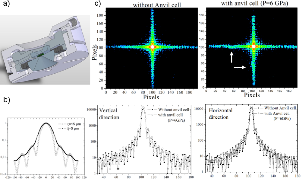
III.1 Combining pressure and coherent diffraction
To combine high pressure and coherent diffraction will provide an unique way to measure fluctuations under high pressure. From an experimental point of view, the combination of both techniques is not obvious. A good compromise has to be found between the degree of coherence (which decreases with increasing photons energy), the absorption of the anvils, the opening of the cell and the maximum pressure expected. The geometry of the diamond anvil cell is displayed in figure 1b). We use 1.2 mm thick diamonds and 10 keV x-rays. Compared to 8 keV x-rays usually used in coherent diffraction experiments, the degree of coherence is reduced by (see Eq.1) but the transmission is increased by a factor 3. The cell is designed to have an opening of with such diamonds. Diffraction patterns of rectangular slit has been recorded with and without the presence of the anvil cell at the sample position (see figure 1c). The secondary slits were closed at and the slit located at the sample position at . The 2D images have been recorded with a CCD camera ( pixels size) located at 2m from the sample position. The degree of coherence without diamond cell has been estimated from the contrast of fringes around .
The main result of this paper is to establish that when using such anvil cell, with a residual pressure of 0.2GPa, similar contrast of fringes is observed. The coherence properties of the beam are not altered by our anvil cell. This opens the possibility to do coherent diffraction experiment under high pressure. In fact, the contrast of fringes is slightly stronger with the anvil cell (see figure 1c). This is probably due to the fact that the anvil cell absorbs X-rays scattered by air or kapton foils upstream. Note also that the diffraction pattern is locally perturbed at two positions along the cross-like profile (see arrows in figure 1c).
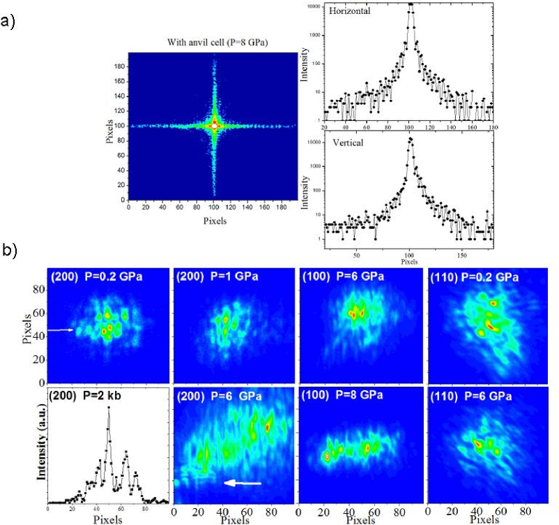
A similar experiment has been performed at higher pressure, up to 8GPa. Unfortunatly, the figure 2a has not been exactly obtained in the same configuration. The secondary slits were opened at and the last slits at . The contrast of fringes is thus smaller. Nevertheless, it seems that for larger pressure, the contrast decreases. We can not exclude that the degree of coherence decreases for higher pressure due to larger deformations of the diamond cell.
As an example of coherent diffraction at wide angles, we choose the system which has been recently study by this method close to its displacive transitionravy (3). We have measured different fundamental Bragg peaks, in the scattering plane (the (100) and (200) reflections) and out-of-plane (the (110) reflection), at several pressures (see figure 2b). Reflections up to the (200) are reachable thanks to the large angular aperture of our diamond cell. We obtained speckles with a good contrast up to 8GPa. The micrometer-size sample was not a perfect monocrystal. Speckles are probably due to interferences between misoriented domains. In addition, the crystal broke up into several pieces under higher pressure.
References
- (1) M. Born and E. Wolf, Principles of optics, Pergamon Press, Oxford; L. Mandel and E. Wolf, Optical coherence and quantum optics, Cambridge University Press (1995).
- (2) D. Le Bolloc’h, et al., J. Synchrotron Rad. 9, 258 (2002).
- (3) S. Ravy,D. Le Bolloc’h, R. Currat,A. Fluerasu,C. Mocuta and B. Dkhil, Phys. Rev. Lett. 98 105501 (2007).