Optical control of DNA-base radio-sensitivity
Abstract
Purpose: Manipulation of the radio-sensitivity of the nucleotide-base driven
by the spin blockade mechanism of diffusive free radicals against ionizing
radiation.
Materials and methods: We theoretically propose a mechanism which uses the
simultaneous application of circularly polarized light and an external
magnetic field to control the polarization of the free radicals and create
S=1 electron-hole spin excitations (excitons) on nucleotide-base. We deploy
an ab-initio molecular dynamics model to calculate the characteristic
parameters of the light needed for optical transitions.
Results: As a specific example, we present the numerical results calculated
for a Guanine, in the presence of an OH free radical. To increase the
radio-resistivity of this system, a blue light source for
the optical pumping and induction of excitons on guanine
can be used.
Conclusions: The effect of spin-injection on the formation of a free energy
barrier in diffusion controlled chemical reaction pathways leads to the
control of radiation-induced base damage. The proposed method allows us to
manipulate and partially suppress the damage induced by ionizing radiation.
I Introduction
Ionizing radiation is both hazardous and beneficial to living organisms, and is extensively used for cancer treatment in radiation therapy Khan:book . A major problem in the application of ionizing radiation to cancer treatment is the protection of normal cells and tissues against unavoidable exposure to radiation during radiation treatment. It is now well understood that the ionization or excitation of the DNA molecules, either directly or indirectly, can lead to DNA single or double strand breaks. As a result, misrepaired DNA molecules can lead to specific genetic aberrations and/or mutations which could cause carcinogenesis in normal cells or lead to fatal damage in normal or cancer cells Lyudmila2008:Science ; EricHall:book . It has been shown that low linear-energy-transfer (LET) ionizing radiation creates approximately 1,000 single strand breaks (SSBs) and 40 double strand breaks (DSBs) per gray (1Gy=1J/Kg) in typical mammalian cells Ward1988:RR ; Goodhead1994:IJRB ; Nikjoo1997:IJRB ; Semenenko2004:RR . The level of DNA molecular base damage is around 2,500 to 25,000 per Gy in a cell, which is about 2.5 to 25 times the yield of sugar-phosphate induced damage in the DNA backbone Ward1988:RR ; Semenenko2004:RR . In indirect mechanisms, the water molecules surrounding the DNA molecule which compose 80% of a cell, may be excited by ionizing radiation in form of free radicals, e.g., a charged neutral hydroxyl (OH). The motion of OH-radicals which are randomly produced throughout the cell is governed by diffusion processes. Massive DNA damage can result from a large number of DNA dehydrogenations caused by free radicals. For example, a free radical can diffuse to reach a DNA molecule and remove a hydrogen ion from it to form a water molecule. Detailed studies at the molecular level is necessary to bring the radiation-induced DNA damage under control.
II Method
In this work, we apply a quantum physical description of molecular interactions to propose a mechanism that could allow the manipulation of DNA radio-sensitivity. In particular the Pauli exclusion principle LandauLifshitzQM:book which prevents two electrons with parallel spin form occupying a single spatial orbital, plays a major role and is used to magnetically manipulate the diffusion of hydroxyl radicals and the OH-DNA relative motion. It has been shown in studies in semiconductor physics and quantum optics that the Pauli exclusion principle can be used to rectify electrical currents passing through weakly coupled quantum dots Ono2002:Science and to induce ferromagnetic ordering by photo-generated carriers in magnetic semiconductor hetero-structures Oiwa2002:PRL .
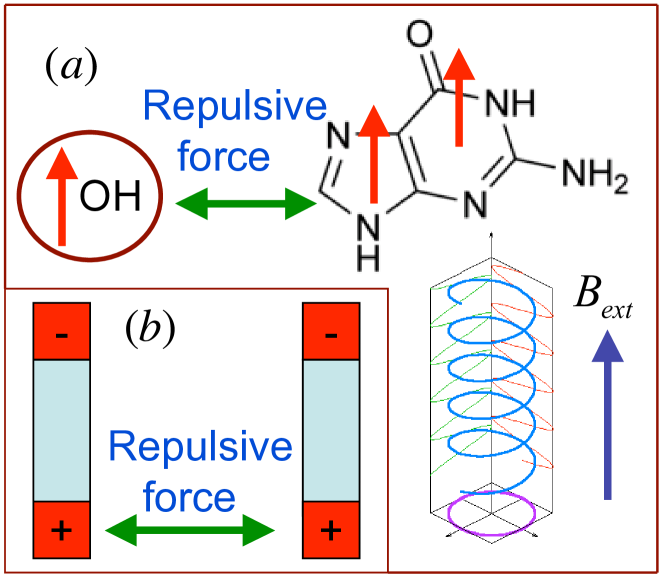
A free radical carries an odd number of electrons with an unpaired spin in the outermost open shell. Due to the reduction of the exchange interaction, the pairing of opposite spin electrons in the open orbital of the free radical with an electron in a DNA molecule makes free radicals highly reactive. In the process of dehydrogenation of a DNA molecule by free radicals an unpaired hole (a half-empty orbital) is transferred to the DNA. In the absence of spin-orbit coupling and hyperfine interaction the spin of transfered electron is conserved. The electronic ground state of DNA-molecule is spin-singlet (in the absence of an external magnetic field). The OH-radical which contains nine electrons is a doubly degenerate ground state with , where we have conveniently taken the quantization axis along the -axis. The degeneracy of the ground state can be lifted by applying a weak magnetic field which couples to the electron spin through the Zeeman interaction LandauLifshitzQM:book , . Here is the Zeeman energy, is the electron -factor (), is the Bohr magneton ( eV/Tesla), and is the strength of external magnetic field.
In a random interaction of radiation with a biological system the initial direction of OH-radical magnetic moment immediately after its generation is also random. However, by applying a weak external magnetic field () (which defines the quantization axis) and using a circularly polarized light field parallel to the direction of the light propagation, as shown in Fig.1a, a molecular transition corresponding to can be induced by means of optical pumping Walker1997:RMP of the OH-radicals Meerakker2005:PRL . Here denotes the total angular momentum of diatomic OH-radical Herzberg:Bppk . Alternatively, tchniques such as electron spin resonance (ESR) can be used to achieve strong polarization of free radicals, as recent advances in ESR have demonstrated the capability of detecting the transfer of electron spin polarization between radicals Jenks1999:JACS ; Brocklehurst1979:FT . In this case microwaves can be used for the optical transitions. In a similar fashion, by applying a second circularly polarized light field one may excite an electron-hole pair (exciton) in the DNA molecule. Because the circularly polarized light carries angular momentum , the exciton has a particular spin polarization. Here the spin of exciton is with polarization along the light propagation direction (because of angular momentum selection rules). Fig. 1 schematically shows the generation of the optically pumped exciton by circularly polarized light. The injection of photo-electrons with the spin out of equilibrium may lead to a dramatic effect in the collective dynamical behavior of DNA-molecules and the interaction with OH-radicals. For example the OH-DNA repulsive magnetic force provides a potential barrier which blocks the diffusion pathway (see Fig. 1b) of OH-radicals toward the DNA-molecules. This is expected to hinder the DNA dehydrogenation and consequently increase the cell radio-resistivity. To verify this hypothesis, an ab-initio molecular dynamical model, which is the mathematical formulation that governs the appropriate dynamics of the molecular system Marx_Hutter is deployed. We have used the Car-Parrinello molecular dynamics (CPMD) Car1985:PRL ; CPMD model, in which the potential energy of the system can be calculated on-the-fly, as needed for the conformations of the dynamical trajectory to simulate the chemical reaction pathways. Because the absorption of a circularly polarized photon alters the local electronic state of a DNA-molecule, we confine our simulation to a particular segment, e.g., only a part of the DNA where the injected exciton is localized and the optical transition takes place. To illustrate this, let us consider a system of interest consisting of a DNA nucleotide base, (e.g., guanine) in the presence of the OH-radical. We assume that a photon with circular polarization interacting with guanine can induce an optical transition in the form of an exciton. Here we investigate the effect of an exciton produced in this way on the guanine-dehydrogenation pathway, assuming that another photon generated through interactions with ionizing radiation (such as radiotherapy x-rays or cosmic rays) creates a free radical in the vicinity of guanine. Because the local density of free radicals and excitons are large and are comparable, the events described in our calculation can be observed with reasonable probability. We adopt computational parameters and variables needed for the CPMD calculation of the dynamical trajectory of the gas phase nucleotide bases in the presence of OH-radicals following Refs. Mundy2002:JPC , where the consistency of CPMD results for guanine with other quantum chemistry approaches has been investigated.
III Results
We identify the dehydrogenation of the nucleotide bases as a function of their spin multiplicity. The ground and excited states of the nucleotide correspond to spin singlet (), and spin triplet () states. The latter can be realized through the application of circularly polarized light as discussed above (see Fig. 1). Our CPMD is implemented in a plane-wave basis within local spin density approximation (LSDA) with an energy cutoff of 70 Rydberg (Ry), and with Becke Becke1988:PRA exchange and Lee-Yang-Parr (BLYP) gradient-corrected functional Lee1988:PRB . Norm conserving ultrasoft Vanderbilt pseudo-potentials were used for oxygen, hydrogen, nitrogen and carbon. The CPMD micro-canonical dynamics (constant energy ensemble) were performed after wave-function optimization following dynamical equilibration at T=300K and re-quenching of the wave-function. An isolated cubic cell of length 13.229 with Poisson solver of Martyna and Tuckerman Martyna1999:JCP was used. Our CPMD studies consist of two classes of spin-restricted calculations, as the total spin along the quantum axis is subjected to the constraints , and , corresponding to doublet and quartet spin configurations. In both calculations the initial distance between OH-radical and nucleotide is considered to be about 1.5 . We selectively choose an initial coordinate for OH-radical in the neighborhood of the nucleotide where the Hydrogen transfer shows a reactive path in normal state of DNA (the doublet spin configuration in the absence of circularly polarized light and magnetic field).
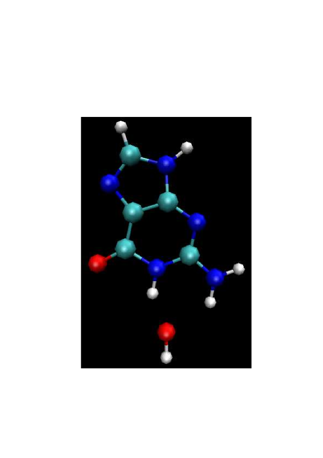
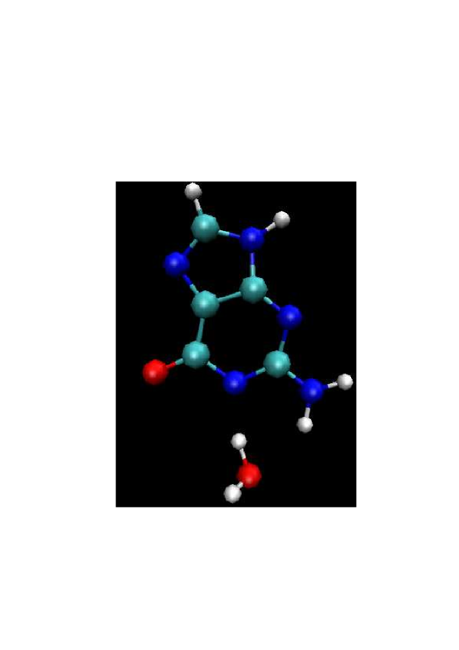

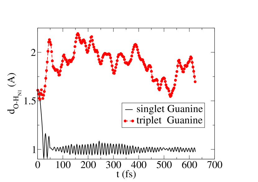
The initial and final states of the molecules are shown in Figs. 2-4. The final configurations of the molecules have been obtained after 0.6 ps where the rearrangement of the atomic coordinates have been deduced from a dynamical trajectory calculated by CPMD. According to our results, a rapid dehydrogenation of the nucleotides takes place for a system with (total spin-doublet) as shown in Fig. 3. This process leads to the formation of a water molecule. In contrast, as shown in Fig. 4, in the quartet spin configuration the repulsive exchange interaction, analogous to Heisenberg anti-ferromagnetic coupling which originates from the Pauli exclusion principle, blocks the exchange of hydrogen and hence the chemical reaction. In Fig. 5 the evolution of the N1 Hydrogen in the guanine and free radical oxygen distance is shown. As it is seen the abstraction of Hydrogen occurs around fs in the spin singlet state of guanine, and the injection of exciton in guanine blocks the hydrogen abstraction. Fig. 6 shows the Kohn-Sham energies (equivalent to potential energy in classical molecular dynamics) of the spin singlet and spin triplet of the Guanine in the presence of the OH free radical as a function of time, calculated by the CPMD at T=300K corresponding to a canonical dynamics (constant temperature ensemble). A drop in Kohn-Sham energy in spin singlet multiplicity is indication of dehydrogenation of HN1 in the guanine by OH free radical. To systematically check the convergence of the results, we increased the size of the molecule by adding sugar-phosphate rings to guanine and found that this has no influence on the spin-blocking effect. To estimate the energy needed for the polarization of the nucleotide in the absence of OH-radicals, we calculated the energy of the ground and excited states of the gas-phase nucleotide in spin singlet and triplet multiplicities. For guanine we calculated the spin singlet-spin triplet energy gap eV. This provides an estimate for the frequency of the circularly polarized light, which is within the range of the visible spectrum of the electromagnetic waves, nm (light blue). To calculate the stored magnetic energy due to the optical injection of spin, we calculated the energy of the gas-phase nucleotide in the presence of one OH-free radical with spin doublet and quartet multiplicities. For the molecules shown in Fig. 2, we find the energy gap eV. Here the excessive magnetic energy which originated from spin-spin repulsive interactions (which resemble the anti-ferromagnetic exchange interaction in the Heisenberg model) can be deduced to be eV. This energy can be interpreted as the excessive energy barrier due to the alignment of the spins in the DNA molecule and OH, and is the source of the magnetic repulsive force which makes the diffusion of OH toward DNA-molecules less likely. This is in agreement with the results obtained from CPMD, shown in Figs. 2-4. In addition, by switching the polarization of one of the light sources to the opposite direction, the relative direction of the DNA-OH polarization switches to antiparrallel, and hence the magnetic repulsive force changes to an attractive force that lowers the OH diffusion barrier and decreases the radio-sensitivity of the DNA-molecule.

After photon absorption, the nucleotide is spin polarized along the direction determined by the polarization state and the propagation direction of the circularly polarized light. The polarized state of the nucleotide then decays quantum mechanically to its unpolarized ground state either by spontaneous photon emission (electron-hole recombination) or through photon-electron spin decoherence. There are radiative and non-radiative channels that contribute to this process. Since spin-orbit coupling governs one of the decay mechanisms in non-radiative channel, we use Fermi’s golden rule to estimate the life-time of the triplet state. It then follows that LandauLifshitzQM:book . Here, is the transition rate from the spin-triplet () to the spin-singlet () state, is the volume, is the emitted photon wave-number, is the spin-orbit Hamiltonian, is the electron mass, is the speed of light, , are the spin and momentum of the th electron, is the Kohn-Sham effective potential, and . This calculation shows that ps. However, the electronic relaxed excitonic states with emperical lifetime of several 100 ps has been reported recently Buchvarov2007:PNAS . The spin-triplet life time of nucleotide turns out to be significantly larger than the dehydrogenation time scale. It is therefore possible to increase the radio-resistivity of the DNA molecule within this time scale through optically pumped spin polarization. It is important to compare with other time-scales in the process. The initial ionization takes place in about 1 fs ( second). The primary free radicals produced by ejection of electrons have a life time of nearly 100 ps, and the reported OH radical life-time is about 1 ns ( second) EricHall:book . The electron spin-lattice relaxation time of the OH radical has been estimated to be approximately between 0.1 and 0.5 ns in water at room temperature Brocklehurst1979:FT .
In order to estimate the technical requirements for the above described approach one could assume that an aqueous solution of DNA will be irradiated with a dose of 1Gy (1J/Kg). It is known Hiroshi2005:JRR that 100eV of absorbed photon/electron energy produce about 6 OH radicals. Therefore 1Gy of radiation produce about OH radicals in 0.1 cm3 of water. If the number of injected excitons can be exceeded up to at least ten times, by applying a laser pump with moderate intensity it is possible to increase significantly the resistance of DNA-molecules against irradiation. For example at a dose rate of 1.4 Gy/min a laser pump power of watt would be required, which is well within the technically achievable limits.
IV Conclusion
In conclusion, we have theoretically explored a mechanism which involves the injection of spin polarized excitons in DNA molecules to control and manipulate the radio-sensitivity of cells by using a circularly polarized light field and external magnetic field. The mechanism proposed here is based on the selection rules applicable to optical transitions between energy levels of the DNA-molecules and optical pumping of the OH-radicals, and we have employed a microscopic ab-initio molecular dynamics model to computationally study the dehydrogenation mechanism at the molecular level. The results of this study may be used as a guideline to develop new techniques for radiation therapy and radiation protection purposes.
The author thanks Homayoun Hamidian, Reinhard Kodym, Lech Papiez, and Tim Solberg for the comments and useful discussions.
References
- (1) Faiz M. Khan, The Physics of Radiation Therapy, (Williams & Wilkins, Baltimore, 1994).
- (2) L. G. Burdelya, V. I. Krivokrysenko, T. C. Tallant, E. Strom, A. S. Gleiberman, D. Gupta, O. V. Kurnasov, F. L. Fort, A. L. Osterman, J. A. DiDonato, E. Feinstein, and A. V. Gudkov, Science 320 226-230 (2008).
- (3) Eric J. Hall, Radiobiology for the Radiologist, (Lippincott Williams & Wilkins, Baltimore, Fifth Edition, 2000).
- (4) J.F. Ward, Prog. Nucleic Acid Res. Mol. Biol., 35 95 (1988); Radiat. Res. 142, 362 (1995); Errata, Radiat. Res. 143, 355 (1995).
- (5) D. T. Goodhead, Int. J. Radiat. Biol. 65, 7 (1994).
- (6) H. Nikjoo et al., Int. J. Radiat. Bio. 71, 467 (1997).
- (7) V. A. Semenenko, and R. D. Stewart, Radiat. Res. 161, 451 (2004).
- (8) L. D. Landau, and E. M. Lifshitz, Quantum Mechanics: Non-Relativistic Theory (Pergamon, Oxford, 2003).
- (9) D. M. Bartels, A. D. Trifunac, R. G. Lawler, Chem. Phys. Lett. 152, 109 (1988); William S. Jenks and Nicholas J. Turro, J. Am. Chem. Soc. 112 9009 (1990).
- (10) N. C. Verma and R. W. Fessenden, J. Chem. Phys. 65, 2139 (1976); B. Brocklehurst, J. Chem. Soc. Faraday Trans. II 75, 123 (1979); B. Bhattacharjee, and Ranjan Das, Molecular Physics, 105, 1053 (2007).
- (11) K. Ono, K., D. G. Austing, Y. Tokura, S. Tarucha, Science 297 1313 (2002).
- (12) S. Koshihara, A. Oiwa, M. Hirasawa, S. Katsumoto, Y. Iye, C. Urano, H. Takagi, and H. Munekata, Phys. Rev. Lett. 78, 4617 (1997); I. Žutić, J. Fabian, and S. Das Sarma, Rev. Mod. Phys. 76, 323 (2004).
- (13) Thad G. Walker and William Happer, Rev. Mod. Phys. 69, 629 (1997); William Happer, Rev. Mod. Phys. bf 44, 169 (1972).
- (14) G.H. Dieke, and H. M. Crosswhite, J. Quant. Spectrosc. Radiat. Transf. 2, 97 (1962); Sebastiaan Y. van de Meerakker, Nicolas Vanhaecke, Mark P. van der Loo, Gerrit C. Groenenboom, and Gerard Meijer, Phys. Rev. Lett. 95, 013003 (2005).
- (15) G. Herzberg, The Spectra and Structures of Simple Free Radicals (Dover Publications Inc. New York, 1971).
- (16) D. Marx, D., and J. Hutter, Ab initio molecular dynamics: Theory and Implementation, Modern Methods and Algorithms of Quantum Chemistry, J. Grotendorst (Ed.), (John von Neumann Institute for Computing, Jülich, NIC Series, Vol. 1, page 301-449, 2000).
- (17) C. J. Mundy, M. E. Colvin, A. A. Quong, J. Phys. Chem. A. 106 10063 (2002); Y. Wu, C. J. Mundy, M. E. Colvin, R. Car, J. Phys. Chem. A. 108 2922 (2004).
- (18) R. Car, and M. Parrinello, Phys. Rev. Lett. 55, 2471 (1985).
- (19) J. Hutter, P. Ballone, M. Bernasconi, P. Focher, E. Fois, S. Goedecker, M. Parrinello, M. E. Tuckerman, CPMD code, version 3.13, MPI fuer Festkoerperforschung, Stuttgart IBM Zurich Research Laboratory, 1990-2008.
- (20) A. D. Becke, Phys. Rev. A38, 3098 (1988).
- (21) C. Lee, W. Yang, and R. G. Parr, Phys. Rev. B37, 785 (1988).
- (22) G. J. Martyna, M. E. Tuckerman, J. Chem. Phys. 110, 2810 (1999).
- (23) I. Buchvarov, Q. Wang, M. Raytchev, A. Trifonov, T. Fiebig, Proc. Natl. Acad. Sci. 104, 4794 (2007); Q. Wang, and T. Fiebig in Charge Migration in DNA, Ed. T. Chakraborty (Springer-Verlag, Berlin, 2007).
- (24) Yamaguchi Hiroshi et al., J. Radiat. Res. 46, 333 (2005).