Dynamics in Soft-Matter and Biology Studied by Coherent Scattering Probes
Abstract
Neutrons and x-rays are coherent probes, and their coherent properties are used in scattering experiments. Only coherent scattering probes can elucidate collective molecular motions. While phonons in crystals were studied for half a century now, the study of collective molecular motions in soft-matter and biology is a rather new but upcoming field. Collective dynamics often determine material properties and interactions, and are crucial to establish dynamics-function relations. We review properties of neutrons and x-rays and derive the origin of coherent and incoherent scattering. Taking molecular motions in membranes and proteins as example, the difference between coherent and incoherent dynamics is discussed, and how local and collective motions can be accessed in x-ray and neutron scattering experiments. Matching of coherent properties of the scattering probe may become important in soft-matter and biology because of (1) the missing long ranged order and (2) the large length scales involved. It is likely to be important in systems, where fluctuating nanoscale domains strongly determine material properties. Inelastic scattering can provide very local structural information in disordered systems. Inelastic neutron scattering experiments point to a coexistence of short-lived nanoscale gel and fluid domains in phospholipid bilayers in the range of the gel-fluid phase transition, which may be responsible for critical behavior and determine elastic properties.
I Introduction
Neutron and x-ray scattering techniques were very successfully applied to investigate structure and dynamics in crystalline systems. The challenge we face is how to apply these powerful techniques to soft-matter and biology, i.e., systems with a high degree of static and dynamic disorder. Even though for instance biological membranes were studied for decades, very few biologically relevant processes were revealed on a molecular level. The reason is the combination of very small nanometer length scales and very fast dynamics of pico- and nanoseconds, which poses particular experimental challenges. Neutron and x-ray scattering is an ideal microscope to study structure and dynamics in these systems, because they give access to the relevant length and time scales. Neutrons and x-rays are coherent probes, and their coherent properties are used in scattering experiments.
Often, the structure of soft-matter and biomolecular systems is relatively well known and the corresponding experimental techniques, such as x-ray crystallography, Nuclear Magnetic Resonance (NMR), and also Atomic Force Microscopy (AFM), developed standard techniques in many disciplines. Dynamical properties are often less well understood, but are important for many fundamental properties such as elasticity and interaction forces. They may determine or strongly affect certain functional aspects, such as diffusion and transport, and be relevant for protein function. In particular collective molecular motions may play a key role to establish dynamics-function relations in complex soft-matter and biological systems. In this article, we review properties of neutrons and x-rays and the origin of coherent and incoherent scattering. The difference between coherent and incoherent dynamics is discussed, and how local and collective motions can be accessed in x-ray and neutron scattering experiments. Matching of coherent properties of probe and sample may become important in soft-matter and biology because of (1) the missing long ranged order and (2) the large length scales involved. Short-lived nanoscale domains in lipid membranes are believed to strongly determine material properties but their existence is very difficult to prove. We discuss how inelastic scattering experiments can provide very local structural information.
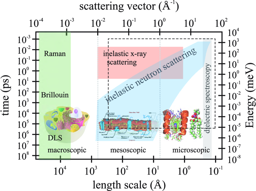
Biomolecular and soft-matter materials can be considered as multi-scale materials because relevant dynamics occur on a large range of length and time scales. Experimentally, to address the multi-scale character, different techniques and probes must be applied. Figure 1 depicts the length and time scales accessible by inelastic x-ray, inelastic neutron, dynamical light scattering (DLS), Brillouin and Raman scattering, and dielectric spectroscopy. By combining different techniques, macroscopic down to microscopic dynamics can be investigated. The motions cover slow conformational changes in the millisecond to microsecond time range. Nanosecond motions include side chain rotations and backbone torsional reorientations. Rotations of small side-chains and local vibrational modes motions occur in the fast picosecond time range. The relevant length scale for dielectric spectroscopy is in the order of an elementary molecular electric dipole, which can be estimated by the bond length of a C–O bond (about 140 picometer), and frequencies from kiloHertz to Gigahertz can be measured. Because of the large wavelength of the probe ( 510 nm, 632 nm), light scattering techniques are limited to small momentum transfers of about Å-1 to Å-1, corresponding to a length scale of about 100 nanometers. Inelastic neutron and x-ray scattering access length scales from smaller than Angstrom to more than 100 nm and time scales from picoseconds to almost one microsecond. While inelastic x-ray scattering is the perfect tool to measure fast dynamics at larger distances (small q-values), inelastic neutron scattering can more easily access slow dynamics at smaller length scales, as discussed in more detail in Section II. Molecular Dynamics (MD) simulations are an invaluable tool to develop models for molecular structure and dynamics in membranes and proteins. Because of increasing computer power and optimized algorithms, large system sizes and long simulation times, and also more and more complex systems can be addressed Smith (1991); Hayward and Smith (2002); Tarek et al. (2001); Tarek and Tobias (2002); Wood et al. (2007); Meinhold et al. (2007). The dashed rectangle in Figure 1 marks the dynamical range accessed by computer simulations. The elementary time scale in simulations is in the order of femtoseconds.
Motions in proteins and biological membranes occur on various length and time scales Frauenfelder et al. (1991); Fenimore et al. (2004). The functional behavior of membrane proteins is likely to depend on the lipid bilayer composition and physical properties, such as hydrophobic thickness and elastic moduli. Dynamics in complex membranes involve interactions between the different constituents, such as lipids, cholesterol, peptides and proteins. How the variety of inter- and intra protein motions, occuring over different time and length scales, interact to result in a functioning biological system remains an open field for those working at the interface of physics and biology. Collective molecular motions in membranes and proteins are attracting increasing attention Meinhold et al. (2007); Liu et al. (2008); Rheinstädter et al. (2009). The reason is that they may be responsible for certain functionalities, such as transport of small molecules, pore opening and membrane fusion processes. New developments and improvements in scattering instrumentation, sample preparation and environments and, eventually, the more and more powerful sources make it possible to access the usually very weak coherently scattered signals. Collective motions of functional groups may drive protein function. In a cellular context they may contribute to the understanding of macromolecular function because they can lead to an effective coupling and communication between the different constituents. In contrast to other spectroscopic techniques, such as dielectric spectroscopy, inelastic x-ray and neutron scattering give a wave vector resolved access to molecular dynamics. Excitation frequencies and relaxation rates are measured at different internal length scales of the system. A typical dynamical scattering experiment measures (q,) pairs, it delivers a frequency together with a corresponding length scale and possibly also corresponding direction, such as parallel or perpendicular to the protein axis. This additional information is very important to assign the measured frequencies to certain molecules or molecular components.
II Local and Collective Molecular Motions
Atomic and molecular motions can be classified as local, self-correlated, and collective, pair-correlated dynamics. Local dynamics is the motion of molecules or functional groups in local energy potentials. The force and time constants involved are determined by the local friction and restoring forces. This type of dynamics is called incoherent because the particles move independently in their local environments and examples are vibration, rotation, libration (hindered rotation) and diffusion of individual lipid molecules.
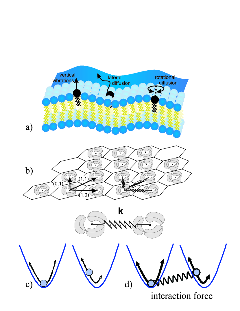
Coupled, collective molecular motions need an interaction between neighboring particles or functional groups (pictured as a spring). Coupled particles basically behave like coupled pendulums. This type of dynamics is called coherent. Collective molecular motions determine for instance elasticity of membranes, interactions between membrane embedded proteins, and may impact on transport processes. In biology, any dynamics will most likely show a mixed behavior of particles moving in local potentials but with a more or less pronounced coherent (interacting) character. Figure 2 exemplary depicts some of the local and collective modes in a lipid bilayer. Rotational and lateral diffusion, vibrations and rotations of the single lipid molecules can be investigated by, e.g., incoherent inelastic neutron scattering, nuclear magnetic resonance or dielectric spectroscopy (Figure 2 (a) and (c)). Only coherent probes, such as coherent inelastic neutron scattering or inelastic x-ray scattering, can elucidate collective molecular motions, as will be discussed below. Collective molecular motions can for instance be depicted by a system of coupled membrane embedded proteins in Figure 2 (b). The corresponding interaction forces (Figure 2 (d)) can be determined in scattering experiments.
Coherent and Incoherent Scattering
The peculiarity of neutrons is that they can scatter incoherently or coherently, and therefore give access to local or collective dynamics. If a single atom of an element is hit by a photon or neutron represented by a planar wave, , the scattering can be analyzed in terms of an emerging spherical S-wave , . The amplitude of the scattered spherical wave, b, is defined as the scattering length of the atom. The scattering length of an element is different for x-rays and neutrons. Because x-rays are electromagnetic waves and scattered by the electrons in the electron shell, the scattering length depends on the number of electrons Z, and is proportional to the element number, bZ Als-Nielsen and McMorrow (2001).
The case is different for the interaction of neutrons with matter. Neutrons are microscopic particles. They carry a mass of m=1.675 10-27 kg and move with velocities between about 500 m/s (cold neutrons) to 2,200 m/s (thermal neutrons). Neutrons carry no charge, but a magnetic moment, a spin Squires (1978). In contrast to x-rays, neutrons are scattered by the atomic nucleus. The scattering length b depends on the spin of the nucleus-neutron system. If the spin of the nucleus is I, then every nucleus with non zero spin has two values of b, namely for I and I, depending on the orientation of neutron and nucleus spin. The scattering length is not simply a monotonic function of the atomic number, as it is for x-rays, but depends on the spin configurations of the nuclei. In addition, different isotopes of the same element have different scattering lengths. Isotopes have nuclei with the same number of protons (the same atomic number) but different numbers of neutrons and also different nuclear spins I. So while for x-rays, all atoms of the same element look the same, they may look different for neutrons because of (1) different orientations of the nuclear spin, and (2) different isotopes (nuclides). The most pronounced difference between the x-ray and the neutron probe is therefore that x-ray scattering is always coherent, while neutron scattering may contain contributions from coherent and incoherent scattering, as will be discussed in the following.
The coherent and incoherent neutron cross sections, bcoh and binc, of an element can be illustrated by two extreme cases. If all the nuclei in a sample have different b’s, there is no interference between the waves scattered by the different atoms. The incoherent scattering therefore depends only on correlation between the positions of the same nucleus at different times. Incoherent scattering therefore probes the local atomic or molecular environment. If all scattering lengths are the same, i.e., all nuclei are identical for the neutron probe, the coherent scattering still depends on the correlation between the positions of the same nucleus at different times, but also on the correlation between the positions of different nuclei at different times. It therefore gives interference effects and allows to measure interaction forces. In general, every element has coherent and incoherent scattering lengths. Table 1 lists the scattering lengths for selected elements, which are common in biological material.
| Nuclide | bcoh (fm) | binc (fm) | Nuclide | bcoh (fm) | binc (fm) |
|---|---|---|---|---|---|
| 1H | -3.7406 | 25.274 | 12C | 6.6511 | 0 |
| 2H | 6.671 | 4.04 | 13C | 6.19 | -0.52 |
| 3H | 4.792 | -1.04 | 16O | 5.803 | 0 |
| 14N | 9.37 | 2.0 | 17O | 5.78 | 0.18 |
| 15N | 6.44 | -0.02 | 18O | 5.84 | 0 |
bcoh is defined as the average scattering length, . The terms and can be used to define the coherent and incoherent scattering cross sections, and by and , which are more commonly used. The unit of the cross section is 1 barn (1 b), equivalent to an area of 10-28 m2. The coherent scattering is then the scattering of a system where all scattering lengths are equal to . The incoherent scattering stems from the random distribution of deviations of the scattering length from the mean value . Table 2 lists values for the scattering cross sections for selected elements. Most noticeable are the very large incoherent cross section for hydrogen, 1H, and the relatively large coherent cross section for deuterium, 2H. This is important for selective deuteration and labeling, as will be discussed in one of the next paragraphs.
| Nuclide | (b) | (b) | Nuclide | (b) | (b) |
|---|---|---|---|---|---|
| 1H | 1.7583 | 80.27 | 12C | 5.559 | 0 |
| 2H | 5.592 | 2.05 | 13C | 4.81 | 0.034 |
| 3H | 2.89 | 0.14 | 16O | 4.232 | 0 |
| 14N | 11.03 | 0.5 | 17O | 4.2 | 0.004 |
| 15N | 5.21 | 0.00005 | 18O | 4.29 | 0 |
Scattering Functions
To more accurately define coherent and incoherent scattering, the corresponding scattering functions shall be introduced briefly. The differential cross section of the quasi-elastic scattering of neutrons in a solid angle with an energy transfer can be expressed as Lovesey (1984)
| (1) |
where are atom-type indices, while [] and [] are, respectively, the incoherent [coherent] scattering length and dynamic structure factor. is directly measured in inelastic x-ray and neutron scattering experiments.
is obtained from the Fourier transform of the self intermediate scattering function
| (2) |
where the summation goes over all atoms of type in the system. The coherent part, , involves correlations between different particles and can be obtained from the Fourier transform of the intermediate scattering function Squires (1978)
| (3) |
The coherent scattering function describes the time evolution of positional correlations between different molecules or atoms in a system. In the incoherent scattering function, , positional correlations of the same particle at different times are considered, only. While describes pair-correlated dynamics, the incoherent scattering, , describes self-correlated motions. Interactions between proteins in a membrane are an example for coherent dynamics and the corresponding (coherent) scattering experiments measure the interaction force between the proteins, as depicted in Figure 2 (d). Diffusion of a single lipid molecule for instance is observed as incoherent scattering. An (incoherent) scattering experiment probes the local energy potential, as sketched in Figure 2 (c). But how can neutron scattering experiments be tuned to measure coherent respective incoherent dynamics and what is the difference between x-ray and neutron spectra?
Selective Deuteration and Labeling Techniques
The total scattering of a sample in neutron scattering experiments will contain coherent and incoherent scattering contributions. The fraction of coherent and incoherent scattering depends on the atomic composition and the respective scattering lengths. Substitution of certain elements in a compound by their isotopes may increase contributions of certain molecules or functional groups to the coherent or incoherent scattering contribution.
The incoherent cross section of hydrogen atoms is about 40 times larger than that of deuterium (see Table 2), and of all other atoms present in biological macromolecules. Consequently, hydrogen atoms dominate the incoherent scattering signal of biological samples. The hydrogen atoms reflect the movements of larger groups to which they are attached, such as amino acid side chains. Deuteration, i.e., the substitution of protons by deuterium (2H), is often used to increase or suppress the incoherent scattering contribution of certain functional groups to the total scattering.
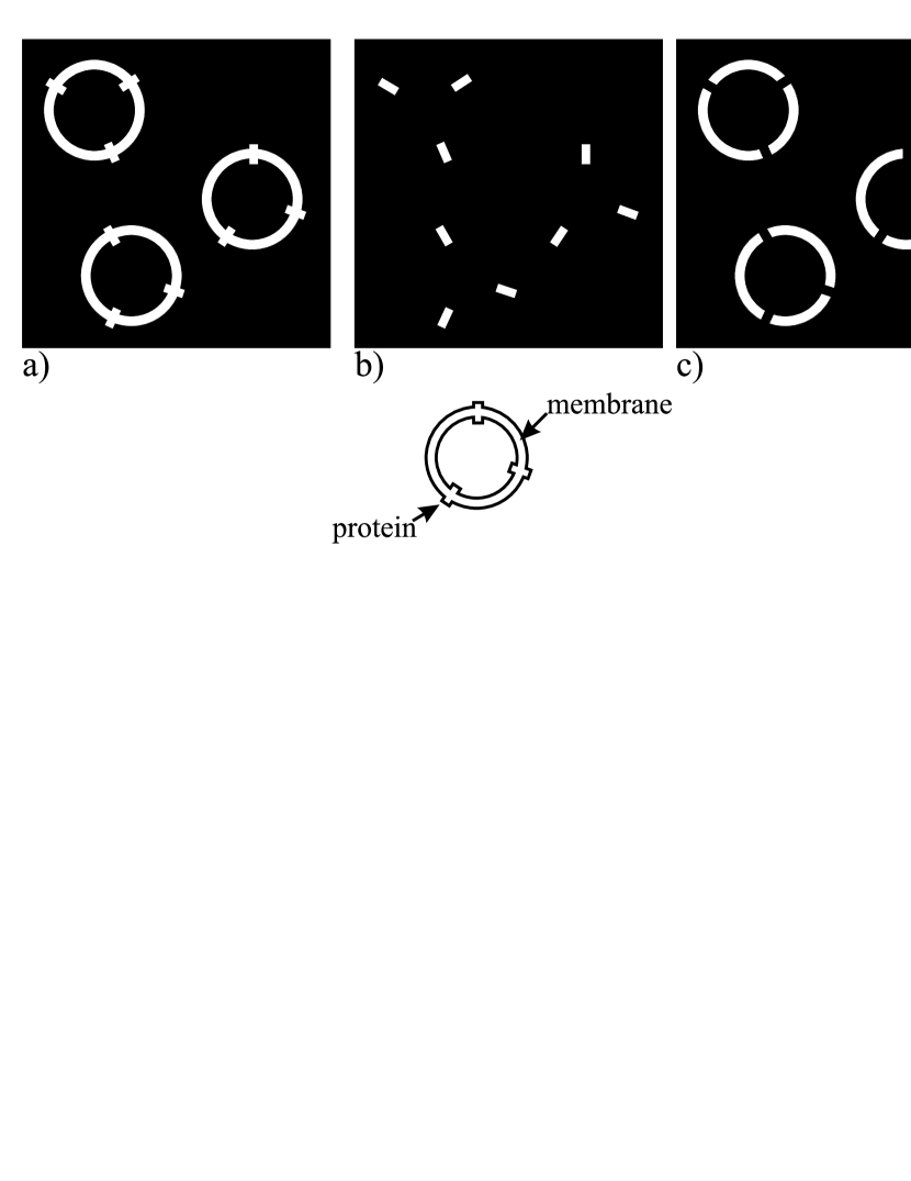
While in protonated samples the incoherent scattering is usually dominant and the time self-correlation function of individual scatterers is accessible, (partial) deuteration emphasizes the coherent scattering and gives access to collective motions by probing the pair correlation function. In a membrane sample with protonated proteins, the experiments would be sensitive to the diffusive motions of the proteins. Deuteration of the proteins increases the coherent scattering and allows measuring the interaction forces between the embedded proteins. The effect of labeling is sketched in Figure 3. In Figure 3 (a), self-correlated, diffusive motions of membranes and embedded proteins can be accessed when the membrane-protein system is labeled, and deuterated solvent is used. Diffusive dynamics of the proteins is highlighted when solvent and membrane are deuterated, and hydrogenated proteins are used. The effect of protein insertion on membrane dynamics can be studied in Figure 3 (c), with deuterated solvent and proteins. Note that at the same time, the interfaces between protonated and deuterated areas scatter coherently. The preparation in Figure 3 (b) can therefore be used to study possible protein-protein interactions.
Excitation Spectra: The Finger Print of Dynamics
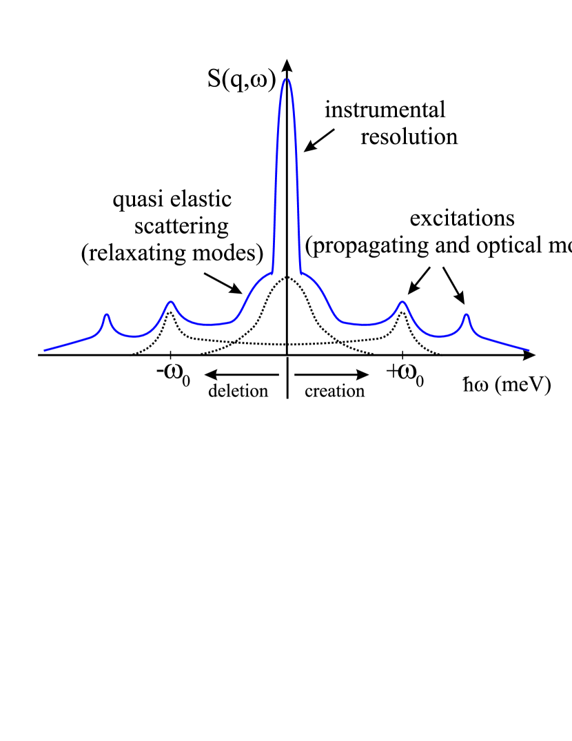
While Bragg scattering, i.e., the occurrence of diffraction peaks, stems from purely coherent scattering, inelastic spectra may contain contributions from coherent and incoherent scattering. A typical excitation spectrum , measured by inelastic x-ray or neutron scattering at a particular q-vector, is shown in Figure 4. The total signal typically consists of a sharp peak at energy transfer zero, which marks the instrumental resolution. The resolution is often well described by a Gaussian peak shape. Quasielastic scattering is observed as a broadening of the instrumental resolution. In addition, inelastic peaks may be observed. Excitations always occur as pairs of inelastic peaks at frequencies of and , caused by creation and deletion of dynamical modes. Starting from the simple models for local and collective modes in Figure 2 (c) and (d), assumptions for peak shapes of the corresponding modes can be developed. Typical local modes, such as diffusion and libration (hindered rotation), are examples for relaxators, i.e., overdamped oscillations. The characteristic frequency of a relaxator is , so that the response is centered on the elastic line. The width of the response is determined by the relaxation time as . In the time domain a relaxation is described by an exponential decay , the Fourier transform of which is a Lorentzian function. So local, incoherent dynamics give rise to a quasielastic broadening in the energy domain and are described by Lorentzian line shapes, centered at energy transfer zero. Coherent dynamics, probe the elastic springs between molecules or atoms. They can be considered as particles bound by an isotropic harmonic force and damped through hybridization with a phonon bath. Those propagating (acoustical) or oscillating (optical) modes have well defined eigenfrequencies and lead to inelastic excitations at energy values . They are described by a damped harmonic oscillator model (dho). The width of the inelastic peak is related to the life time of the excitation. A strict derivation of the peak profiles can be found in the books by Squires Squires (1978) and Lovesey Lovesey (1984).
The excitation spectrum looks identical for the x-ray and neutron probes. The origin of quasielastic and inelastic scattering is purely coherent for x-rays. In the case of liquids for instance, the spectrum can be evaluated by a generalized effective eigenmode theory Liao et al. (2000) in terms of a heat mode (a collective diffusion process) and sound modes. For neutrons, the quasielastic part of the spectrum can have contributions from coherent and incoherent scattering, but is most likely dominated by the incoherent scattering of hydrogen atoms. The corresponding dynamical modes are relaxating local modes, such as diffusion of molecules or functional groups. The relaxation times can be determined from the width of a Lorentzian peak shape. Excitations stem from coherent scattering and involve dynamics between different particles from interference effects. So by preparing a hydrogenated or deuterated sample and tuning the spectrometer to quasi- or inelastic scattering, a neutron scattering experiment can be made sensitive to local or collective dynamics of certain components to probe local environments or interactions.
Scattering Probes and Dispersion Relation
Collective motions can be modeled by interacting particles, as sketched in Figures 2 (b) and (d). The dynamics must be described by a set of coupled pendulums (particles bound by an isotropic harmonic force). In inelastic scattering experiments, excitation energies are probed at different internal length scales and different directions. No trivial q-dependence of the energies can usually be expected. The energies will depend on the interaction constant, k, but also on the geometrical arrangement of the objects. It will depend on the length scale, the q-value, but critically also on the coupling path, i.e., the direction, which is probed, as depicted in Figure 2 (b). A typical experiment to measure collective dynamics records excitation frequencies at different q-values over a coherent q-range. The corresponding curve is called a dispersion relation. It describes the complex dynamics of a (oriented) system of coupled oscillators. Technically, it describes the way waves with a certain wavelength, and certain coherence length, propagate through the system. Note that only coherent probes are capable to elucidate coherent dynamics.
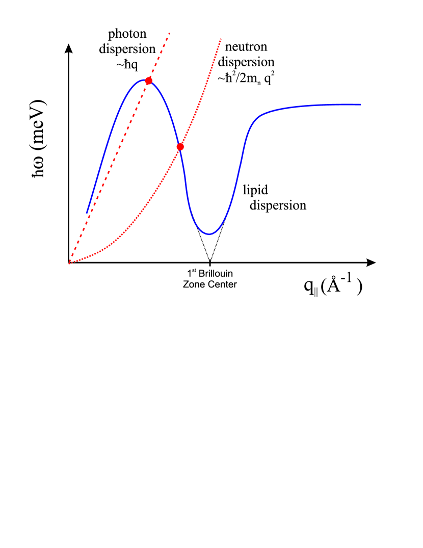
Figure 5 exemplary depicts the dispersion relation of the collective short wavelength density fluctuations in a phospholipid bilayer, as adapted from Rheinstädter et al. (2004). is the lateral momentum transfer, i.e., the projection of the scattering vector into the plane of the membrane. The particular shape of the corresponding dispersion relation can qualitatively be explained. The basic scenario is the following: at small , longitudinal sound waves in the plane of the bilayer are probed and give rise to a linear increase of , saturating at some maximum value, before a pronounced minimum is observed at 1.4 Å-1, the first maximum in the static structure factor (the inter acyl chain correlation peak). can be interpreted as the quasi- Brillouin zone of a twodimensional liquid. Collective modes with a wavelength of the average nearest neighbour distance are energetically favorable. The static and dynamic disorder in the lipid bilayers finally leads to a minimum at finite energy values (softmode). The lipid dispersion relation is similar to those in ideal liquids, as, e.g. liquid argon de Schepper et al. (1983); van Well and de Graaf (1985a), liquid neon van Well and de Graaf (1985b) or liquid helium Glyde (1994). The interior of a lipid bilayer, the C-atoms or C H groups of the lipid acyl chains, behave like a quasi liquid. In contrast to real liquids, the chain atoms of the lipid molecules are chemically bound to each other, leading to smaller mobility and diffusion and, as a consequence, more pronounced excitations. Higher Brillouin zone centers are usually not well developed in soft-matter and biology because of the high degree of disorder. Scattering experiments are therefore usually limited to the 0th Brillouin zone, only. This is true in particular for inelastic scattering experiments and probably one of the most fundamental differences to scattering experiments in crystalline systems. One of the consequences is that momentum transfer in soft-matter and biology occurs mainly in the forward direction. The transverse component is usually very small, in contrast to crystals, where longitudinal and transverse branches are found.
The dispersion relations of the neutron and x-ray scattering probes are also plotted in Figure 5. The dispersion of the photon probe is proportional to (with photon energies of keV), i.e., a very steep straight line as compared to typical thermal excitation energies in biological materials in the meV range. Because of the distinct particle character of neutrons, the corresponding dispersion relation is proportional to (with neutron mass ). Neutrons carry kinetic energies comparable to thermal energy (“thermal neutrons”). For soft-matter and biological studies, which usually involve large distances and low excitation energies, neutrons are often slowed down by a cold moderator to produce “cold (slow) neutrons” to further enhance the energy and -resolution. Momentum and energy are conserved during the scattering process. As a graphical interpretation of the conservation laws, the dispersion curves of probe and lipids must intersect, as shown in Figure 5. The range of energy and momentum transfers accessible by the respective neutron or x-ray instrument depends on mechanical restrictions, such as angular range, but also on the properties of the scattering probe. X-rays can easily access faster dynamics at small -values (Brillouin scattering), but offer lower energy resolution because of the high incident photon energies. Thermal and cold neutrons offer a high energy resolution because incident energies are comparable to the energies of dynamical modes. The parabolic shape of the neutron dispersion prevents inelastic experiments at small -values and at the same time high energy transfers. This is called the kinematic restriction.
Coherence
While in most natural systems, waves with only partial coherence are more common, x-rays and neutrons both are “coherent” probes with coherence lengths significantly longer than common lattice spacings, and even protein-protein distances in biological membranes. The coherence of waves in periodic systems (lattices) is crucial to their dynamics, as interference effects, such as Bragg reflections, largely determine their propagation. Also in less well ordered systems, the coherence length of the probe, , may play an important role for the investigation of small structures, comparable to the size of . The coherence properties of x-rays and neutrons depend on their wave character.
A collimated beam of neutrons or x-rays prepared by Bragg reflection from a single crystal, or by passage through a pair of phased Fermi choppers, is never precisely monochromatic. From the wave properties, the longitudinal coherence length, , can be estimated to be Rauch (1993). A longitudinal coherence length of 100 Å was reported for thermal neutrons with wavelength of =1.8 ÅRauch et al. (1996). For cold neutrons with wavelengths of about 5 Å and a typical monochromaticity by single crystal reflection of about 5%, the longitudinal coherence length can be estimated to be about 500 Å. Note that the reason for the typically rather low monochromaticity of neutron beams is not to compromise the relatively low intensity of the neutron sources, as compared to x-ray synchrotron sources. Longitudinal coherence for x-rays reflected from a Si(111) monochromator with a wavelength resolution of and a wavelength of =1 Å is in the order of =10,000 Å. The coherence properties of the scattering probe may play an important role for the investigation of small structures, such as nanoscale domains, comparable to or smaller than the size of . Structures smaller than may lead to spatially averaged values for, e.g., peak positions and widths. As will be shown below, inelastic data may still show separate excitations for the different nanodomains and offer a very high spatial resolution, as inelastically scattered waves with different wavelength no longer interfere constructively or destructively.
III Matching of coherent properties
Clusters, rafts, nanodomains, and patches have become a central issue in cell membrane studies. The heterogeneous organization of membrane constituents is believed to be essential for cellular functions such as signalling, trafficking and adhesion Lenne and Nicolas (2009); Apajalahti et al. (2009). The experimental observation of those often very short-lived nanoscale structures is extremely difficult. Experimental techniques must be capable to simultaneously access the small length and the fast time scales. In particular inelastic scattering may be capable to provide the spatial resolution needed to investigate small nanometer structures, as will be discussed in the following. Inelastic neutron scattering experiments point to a possible coexistence of short-lived nanoscale gel and fluid domains in lipid bilayers, which may be responsible for critical behavior and determine material properties.
The transition from the rigid gel into the fluid phase in phospholipid bilayers was reported to show a critical behavior (“critical swelling”) Chen et al. (1997). The lamellar spacing of the membrane stacks, i.e., the distance between neighboring stacked bilayers (normal to the plane of the membrane), but also the nearest neighbor distance of lipid molecules in the plane of the membranes and the corresponding coherence length mimics the critical behavior known from for instance second order magnetic order-disorder transitions. Inelastic neutron experiments in 1,2-dimyristoyl-sn-glycero-3-phosphocholine (DMPC) showed evidence for a coexistence of very small nanometer gel and fluid domains in the range of the main phase transition Rheinstädter et al. (2004, 2006). Coexisting gel and fluid excitations were found. The long wavelength dispersion relation showed showed a soft mode at a length scale comparable to the expected domain size. It was speculated that these domains are responsible for the extreme softness, the very low value of the bending modulus of phospholipid bilayers in the range of the phase transition, the well known “critical softening” Chu et al. (2005). Bending of the membrane would occur mainly in the interface between two nanodomains, which costs less energy than bending a gel or fluid domain. A corresponding soft mode was found in the long wavelength spectrum at a length scale of about 45 nm Rheinstädter et al. (2006). A structural feature was also found in AFM experiments in thick membrane stacks Schäfer et al. (2008). The existence of gel and fluid nanodomains is very difficult to prove experimentally. The reason is the combination of small nanometer length scales and the high dynamics in the fluid phase of the membranes with a rapidly changing domain pattern.
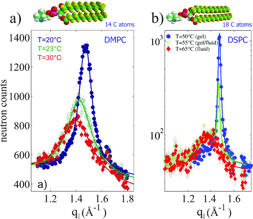
To clarify this point, we investigated the packing and correlations of the lipid acyl chains in phospholipid bilayers with different chain length using neutron diffraction. DMPC, with 14 C-atoms in the acyl chains, and 1,2-distearoyl-sn-glycero-3-phosphocholine (DSPC), with 18 C-atoms, were chosen for this study. The experiment was carried out at the cold triple-axis spectrometer V2 at the Hahn-Meitner-Institute, Berlin, Germany. Partially, chain deuterated lipids (DMPC-d54 and DSPC-d70), hydrated by heavy water (D2O), were used to enhance the intensity of the the lipid acyl chain correlation peak. The samples were kept in a closed temperature and humidity controlled aluminum chamber. Hydration of the lipid membranes was achieved by separately adjusting two heating baths, connected to the sample chamber and to a heavy water reservoir, hydrating the sample from the vapor phase. Temperature and humidity sensors were installed close to the sample. -spacings of 53 Å in DMPC-d54 were achieved. The corresponding relative humidity (RH) can be determined from –RH curves (for instance from Fig. 3 in Chu et al. (2005)), to be 99.5%. No –RH curves are published for DSPC but we argue that a similar level of hydration was achieved for the DSPC sample, too. Note that even that reasonable hydration levels were achieved, the membranes can not be considered as fully hydrated, with water being in excess, what will be important for the discussion of the nanodomains below. The main transition in DMPC occurs at 23.4∘C Jenskea et al. (2008). The transition temperature, , is slightly lowered in the deuterated compound (DMPC-d54) to about 21∘C Aussenac et al. (2003). Position and intensity of the inter acyl chain correlation peak was used in neutron diffraction experiments to define the temperature of the main transition and for DMPC-d54 was determined to 21.5∘C Rheinstädter et al. (2004). The main transition in DSPC was reported to occur at about 54∘C Heimburg (2007). No lowering of the transition temperature was found in the deuterated compound DSPC-d70 by following the chain correlation peak by neutron diffraction, and was determined to 54∘C.
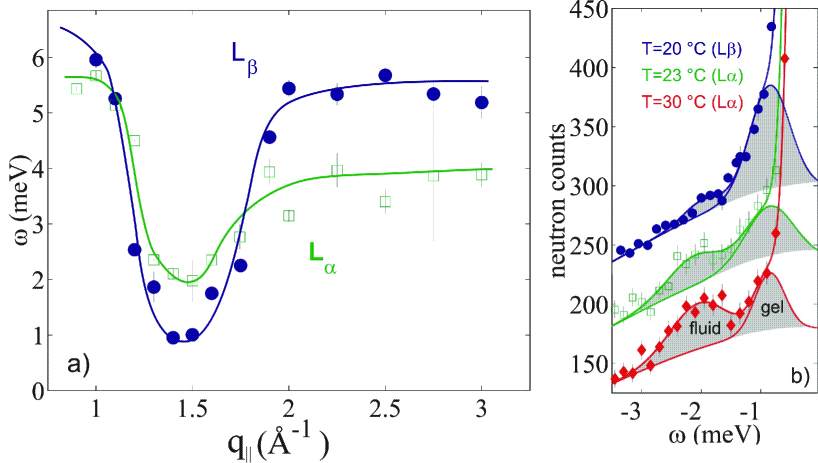
The static structure factor for DMPC and DSPC is shown in Figure 6 (a) and (b). Data were taken in the respective gel and fluid phases, and at an intermediate temperature slightly above the main transition temperature. DMPC showed the well known critical behavior, with a continuous shift of the peak position and continuous increase of the peak width when heating from the rigid low temperature gel into the fluid phase. For DSPC, there is a coexistence of gel- and fluid domains indicated by the existence of two well separated peaks in the range of the phase transition. The weight of the two peaks changes pointing to an increasing number of fluid domains when heating from the gel into the fluid phase. Figure 7 shows the collective short wavelength dynamics in DMPC-d54. Figure 7 (a) depicts the corresponding dispersion relations in the gel and fluid phase. Temperature dependent inelastic scans are shown in Figure 7 (b). The most striking observation is a coexistence of gel and fluid excitations (with changing population) in inelastic experiments, while elastic scattering showed a gradual change of the correlation peak position.
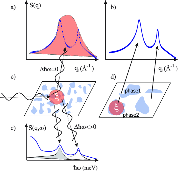
Taking into account a coherence length of the cold neutrons used for this study of about =500 Å, the results point to a coexistence of gel- and fluid domains in the range of the phase transition, but with distinctly different sizes for the two lipids. The DMPC domains must be distinctly smaller than to average over the different nearest neighbor distances. The mismatch of the two phases (and corresponding energies) seems to be larger for longer acyl chains and the system tries to avoid domain boundaries by larger domain sizes () in DSPC. The observations are summarized in Figure 8: The coexisting gel and fluid nanodomains in DMPC most likely have sizes smaller than the neutron coherence length (Figure 8 (c)). The elastic scan does not show two separated gel and fluid peaks, but averages over gel and fluid areas, as shown in Figure 8 (a). As more and more fluid domains are created, the peak gradually shifts from the gel to the fluid position. While constructive interference of elastically scattered neutrons leads to the correlation peaks in , inelastically scattered neutrons have slightly different final energies and thus show two separate excitations, as depicted in Figure 8 (e). In DSPC the coexisting nanodomains can be speculated to be larger than so that the elastic data show two distinct peaks. The weight of the gel peak decreases indicating the creation of more and more fluid domains when heating though the main transition (Figures 8 (b) and (d)). If it comes to soft-matter and biological materials, (highly coherent) high resolution radiation may average over small scale structures, such as nanodomains. Inelastically scattered waves do no longer interfere constructively or destructively so that the scattering volume is drastically reduced, and inelastic signals cna provide very local information.
The conclusion about existence and possible impact of nanodomains observed in phospholipid model membranes on real biological membranes critically depends on the hydration level achieved in the scattering experiments. In these experiments, the bilayers are often hydrated from the vapor phase because the additional scattering and absorption of bulk water around the membranes increases background and decreases sample signals considerably. Even that very reasonable hydration levels can be achieved (note that even full hydration was reported Katsaras and Watson (2000); Katsaras (1998)), the lipid/water phase diagram and corresponding phases critically depend on the level of hydration. Following the Gibb’s Phase Rule in condensed systems (without a coexisting gas phase) the number of independent intensive properties (F), such as temperature, depends on the number of components (C), and the number of coexisting phases (P): F=C-P+1. The observation of coexisting domains over a certain temperature interval implies F=1. The system is composed of lipids and water (C=2). The number of possible phases then follows to: P=C+1-F=2+1-1=2. The observation of two lipid phases would therefore exclude the existence of a coexisting bulk water phase. Conversely, a membrane completely immersed in a solvent may not show coexisting gel and fluid areas. However, the observation of small domains in AFM experiments under excess water Schäfer et al. (2008) possibly points to their existence in membranes in liquid environment. But scattering experiments of phospholipid bilayers immersed in water will be conducted in the future.
IV Conclusion
The coherent properties of photons and neutrons are used to study structure and dynamics in elastic and inelastic x-ray and neutron scattering experiments. A good monochromaticity is a prerequisite for atomic resolution in crystal and protein structure determinations. High resolution radiation is also highly coherent with large coherence lengths, possibly of several thousands of Angstroms. The large coherence length may average over small structures, such as nanoscale domains. While elastic scattering experiments show average values for peak positions for instance, inelastic experiments can still provide very local information. The well known critical behavior in phospholipid bilayers may stem from coexisting gel and fluid nanodomains in the range of the gel-fluid transition. Nanodomains are not only important to understand fundamental properties of model membranes, but also to better understand complex biological membranes and for instance the formation and function of lipid rafts Eggeling et al. (2009); Oelke et al. (2008); Risselada and Marrink (2008). Additional examples are the recently emerging nanoferroelectrics Scott (2007), and nanoscale magnetic domains in thin magnetic films Shpyrko et al. (2007). The development of neutron instrumentation, which allows to control the neutron coherence length, can be envisioned for the future. Such an instrument would not only be important for the investigation of biological materials, but in all systems, where fluctuating nanodomains determine material properties.
Acknowledgements.
It is my pleasure to thank Klaus Habicht, Helmholtz-Zentrum-Berlin, and Beate Brüning, who were involved in the DSPC data collection.References
- Smith (1991) J. C. Smith, Q. Rev. Biophys. 24, 227 (1991).
- Hayward and Smith (2002) J. A. Hayward and J. C. Smith, Biophysical Journal 82, 1216 (2002).
- Tarek et al. (2001) M. Tarek, D. Tobias, S.-H. Chen, and M. Klein, Phys. Rev. Lett. 87, 238101 (4 pages) (2001).
- Tarek and Tobias (2002) M. Tarek and D. Tobias, Phys. Rev. Lett. 88, 138101 (4 pages) (2002).
- Wood et al. (2007) K. Wood, M. Plazanet, F. Gabel, B. Kessler, D. Oesterhelt, D. J. Tobias, G. Zaccai, and M. Weik, Proc. Natl. Acad. Sci. U.S.A. 104, 18049 18054 (2007).
- Meinhold et al. (2007) L. Meinhold, J. C. Smith, A. Kitao, and A. H. Zewail, Proc. Natl. Acad. Sci. U.S.A. 104, 17261 (2007).
- Frauenfelder et al. (1991) H. Frauenfelder, S. Sligar, and P. Wolynes, Science 254, 1598 1603 (1991).
- Fenimore et al. (2004) P. Fenimore, H. Frauenfelder, B. McMahon, and R. Young, Proc. Natl. Acad. Sci. U.S.A. 101, 14408 14413 (2004).
- Liu et al. (2008) D. Liu, X.-Q. Chu, M. Lagi, Y. Zhang, E. Fratini, P. Baglioni, A. Alatas, A. Said, E. Alp, and S.-H. Chen, Phys. Rev. Lett. 101, 135501 (4 pages) (2008).
- Rheinstädter et al. (2009) M. C. Rheinstädter, K. Schmalzl, K. Wood, and D. Strauch, accepted for publication in Phys. Rev. Lett. (2009), http://arxiv.org/abs/0803.0959.
- Als-Nielsen and McMorrow (2001) J. Als-Nielsen and D. McMorrow, Elements of Modern X-Ray Physics (John Wiley & Sons, Ltd, New York, 2001), ISBN 0-471-498572.
- Squires (1978) G. Squires, Introduction to the theory of thermal neutron scattering (Dover Publications, Inc., Mineola, New York, 1978), ISBN 0-486-69447-X.
- Lovesey (1984) S. Lovesey, Theory of Neutron Scattering from Condensed Matter (Clarendon Press, Oxford, 1984).
- Liao et al. (2000) C. Liao, S. Chen, and F. Sette, Phys. Rev. E 61, 1518 (2000).
- Rheinstädter et al. (2004) M. C. Rheinstädter, C. Ollinger, G. Fragneto, F. Demmel, and T. Salditt, Phys. Rev. Lett. 93, 108107 (4 pages) (2004).
- de Schepper et al. (1983) I. de Schepper, P. Verkerk, A. van Well, and L. de Graaf, Phys. Rev. Lett. 50, 974 (1983).
- van Well and de Graaf (1985a) A. van Well and L. de Graaf, Phys. Rev. A 32, 2396 (1985a).
- van Well and de Graaf (1985b) A. van Well and L. de Graaf, Phys. Rev. A 32, 2384 (1985b).
- Glyde (1994) H. Glyde, Excitations in Liquid and Solid Helium, vol. 9 of Oxford Series on Neutron Scattering in Condensed Matter (Clarendon Press, Oxford, 1994).
- Rauch (1993) H. Rauch, Foundation of Physics 23, 7 (1993).
- Rauch et al. (1996) H. Rauch, H. Wölwitsch, H. Kaiser, R. Clothier, and S. A. Werner, Phys. Rev. A 53, 902 (1996).
- Lenne and Nicolas (2009) P.-F. Lenne and A. Nicolas, Soft Matter 5, 2841 2848 (2009).
- Apajalahti et al. (2009) T. Apajalahti, P. Niemel , P. N. Govindan, M. S. Miettinen, E. Salonen, S.-J. Marrink, and I. Vattulainen, Faraday Discuss. p. 2010 (2009).
- Chen et al. (1997) F. Chen, W. Hung, and H. Huang, Phys. Rev. Lett. 79, 4026 (1997).
- Rheinstädter et al. (2006) M. C. Rheinstädter, W. Häussler, and T. Salditt, Phys. Rev. Lett. 97, 048103 (4 pages) (2006).
- Chu et al. (2005) N. Chu, N. Kuc̆erka, Y. Liu, S. Tristram-Nagle, and J. F. Nagle, Phys. Rev. E 71, 041904 (8 pages) (2005).
- Schäfer et al. (2008) A. Schäfer, T. Salditt, and M. C. Rheinstädter, Phys. Rev. E 77, 021905 (8 pages) (2008).
- Jenskea et al. (2008) R. Jenskea, F. Lindström, G. Gröbner, and W. Vettera, Chemistry and Physics of Lipids 154, 26 32 (2008).
- Aussenac et al. (2003) F. Aussenac, M. Laguerre, J.-M. Schmitter, and E. J. Dufourc, Langmuir 19, 10468 (2003).
- Heimburg (2007) T. Heimburg, Thermal biophysics of membranes (Wiley-VCH, Weinheim, 2007), ISBN 3527404716.
- Katsaras and Watson (2000) J. Katsaras and M. Watson, Rev. Sci. Instrum. 71, 1737 (2000).
- Katsaras (1998) J. Katsaras, Biophysical Journal 75, 2157 (1998).
- Eggeling et al. (2009) C. Eggeling, C. Ringemann, R. Medda, G. Schwarzmann, K. Sandhoff, S. Polyakova, V. N. Belov, B. Hein, C. von Middendorf, A. Schönle, et al., Nature 457, 1159 (2009).
- Oelke et al. (2008) J. Oelke, A. Pasc, A. Wixforth, O. Konovalov, and M. Tanaka, Appl. Phys. Lett. 93, 213901 (2008).
- Risselada and Marrink (2008) H. J. Risselada and S. J. Marrink, PNAS 105, 17367 17372 (2008).
- Scott (2007) J. F. Scott, Science 315, 954 (2007).
- Shpyrko et al. (2007) O. G. Shpyrko, E. D. Isaacs, J. M. Logan, Y. Feng3, G. Aeppli, R. Jaramillo, H. C. Kim, T. F. Rosenbaum, P. Zschack, M. Sprung, et al., Nature 447, 68 (2007).