Energy transfer in nonlinear network models of proteins
Abstract
We investigate how nonlinearity and topological disorder affect the energy relaxation of local kicks in coarse-grained network models of proteins. We find that nonlinearity promotes long-range, coherent transfer of substantial energy to specific, functional sites, while depressing transfer to generic locations. Remarkably, transfer can be mediated by the self-localization of discrete breathers at distant locations from the kick, acting as efficient energy-accumulating centers.
pacs:
87.14.E-, 87.15.A-, 63.20.PwIt is now well established that the functional dynamics of proteins
is deeply rooted in the peculiar topological arrangement of their
native folds, as revealed by many experimental and computational
studies Best et al. (2007); Torchia and Ishima (2003).
In particular, the success of coarse-grained elastic network models (ENMs)
in describing atomic fluctuations at room temperature
have helped elucidate, at the harmonic level, the subtle interplay between structure and dynamics
on one side and biological function on the
other Tirion (1996); Bahar et al. (1997); Hinsen (1998); Tama and Sanejouand (2001); Micheletti et al. (2002); Delarue and Sanejouand (2002); Krebs et al. (2002); Kondrashov et al. (2007).
However, protein dynamics is strongly
anharmonic Xie et al. (2000); Edler et al. (2004), a property which has
to be taken into account in order to rationalize crucial biological processes such as
energy storage and transfer upon ligand binding, chemical reaction, etc Sagnella et al. (2000); Leitner (2008). Yet, even though many theoretical studies
suggest that nonlinear excitations may play an active role in
protein functioning Kopidakis et al. (2001); Archilla et al. (2002),
the rich phenomenology residing in the interplay between
protein topology and nonlinearity still remains widely unexplored.
Along these lines, we have recently introduced the Nonlinear Network Model (NNM),
showing how known nonlinear effects can be modulated by the underlying
non-regular topology of protein systems. For instance, within a large collection of
enzyme structures, the formation of localized, robust
nonlinear modes appears strongly favored at few specific sites, that
often lie in close proximity of known catalytic sites Juanico et al. (2007); Piazza and Sanejouand (2008).
In this paper we examine the effects of the nonlinearity/topology interplay on
energy transfer phenomena across protein structures. Within the NNM framework
a protein is represented by fictive particles (amino acids) of identical
mass a.m.u., at equilibrium at the corresponding Cα site as
specified in the experimentally determined structures (X-ray or NMR). By imposing
a fixed cutoff on the latter set of coordinates, a protein is
mapped onto a network of nonlinear oscillators, whose potential energy reads
| (1) |
where is the distance between residues and , their distance in the equilibrium
structure and is the
connectivity matrix.
As in previous studies Juanico et al. (2007), we take 10 Å, kcal/mol/Å4 and
fix so that the low-frequency part of the linear spectrum match
actual protein frequencies,
as calculated through realistic force fields Brooks and Karplus (1985); Marques and Sanejouand (1995); Perahia and Mouawad (1995).
This gives kcal/mol/Å2. The case
corresponds to the Anisotropic Network Model (ANM) Tirion (1996); Bahar et al. (1997); Hinsen (1998).
Our aim is to investigate how energy initially imparted at a specified site
redistributes across a given structure. To do this, we perform microcanonical
simulations with all residues initially at rest at their equilibrium position
but for a kinetic energy kick at site of magnitude .
Sites in a 3 protein network are not equivalent, featuring e.g. varying connectivity,
clustering coefficient and bond directions. Thus, in order to allow for a comparison of
energy relaxation from all sites in a given structure, the initial
kick direction ought to be specified by a unique protocol.
We chose to calculate the directions
of the initial velocities through the Sequential
Maximum Strain (SMS) algorithm Piazza and Sanejouand (2008), which provides an unbiased
measure of the maximum-strain direction at site for a fixed displacement (here 1 Å),
.
During the simulation, we record at regular intervals , the
site that carries the highest energy, , and the value of the latter,
.
Corresponding to a fixed simulation time (about 500 ps), we define a transfer probability
from site (the kicked one) to site and the fraction of energy transferred as
| (2) |


|

|

|

|
The first striking result comes from the calculation of average transfer probabilities.
These gauge the mean transfer to a given site from kicks at all other sites,
, obtained from
independent simulations.
A typical probability transfer plot is shown in Fig. 1. The first notable feature
is that the effect of nonlinearity is to substantially increase the probability of energy funneling to
a few selected sites, while depressing transfer to all other locations with respect to
the harmonic (ANM) case. Remarkably, the preferred target sites lie in close proximity to
the known catalytic sites, within the stiffest
regions 111We measure the local stiffness
as a sum over the set of ten highest normal modes ,
that is the eigenvectors of the Hessian of the ANM total potential energy, ,
with Juanico et al. (2007)..
Thus, topology and nonlinearity
team in this case together to sharpen energy funneling to specific functional regions.
The case shown in Fig. 1 is not a singular one. In Fig. 2 we show the
stiffness patterns for four other enzymes along with the sites ranking first to tenth as to the
energy delivered on average to spherical shells with 6 Å radius around each site.
For residue , this amounts to further averaging
the mean energy deposited at sites within the -th ball , i.e.
.
It is manifest that the sites around which most of the energy is deposited invariably spotlight the stiffest regions,
at the same time identifying functionally relevant locations (see catalytic sites).
Moreover, the same locations clearly attract substantial fractions of the initial excitation energy,
as revealed by surveying the maximum transferred energies to each ball , that is
(empty circles).
Many events featuring transfers of energy fractions in the range 20 to 25 % were
indeed observed.
We can learn more on the mechanisms underlying the energy transfer process by examining
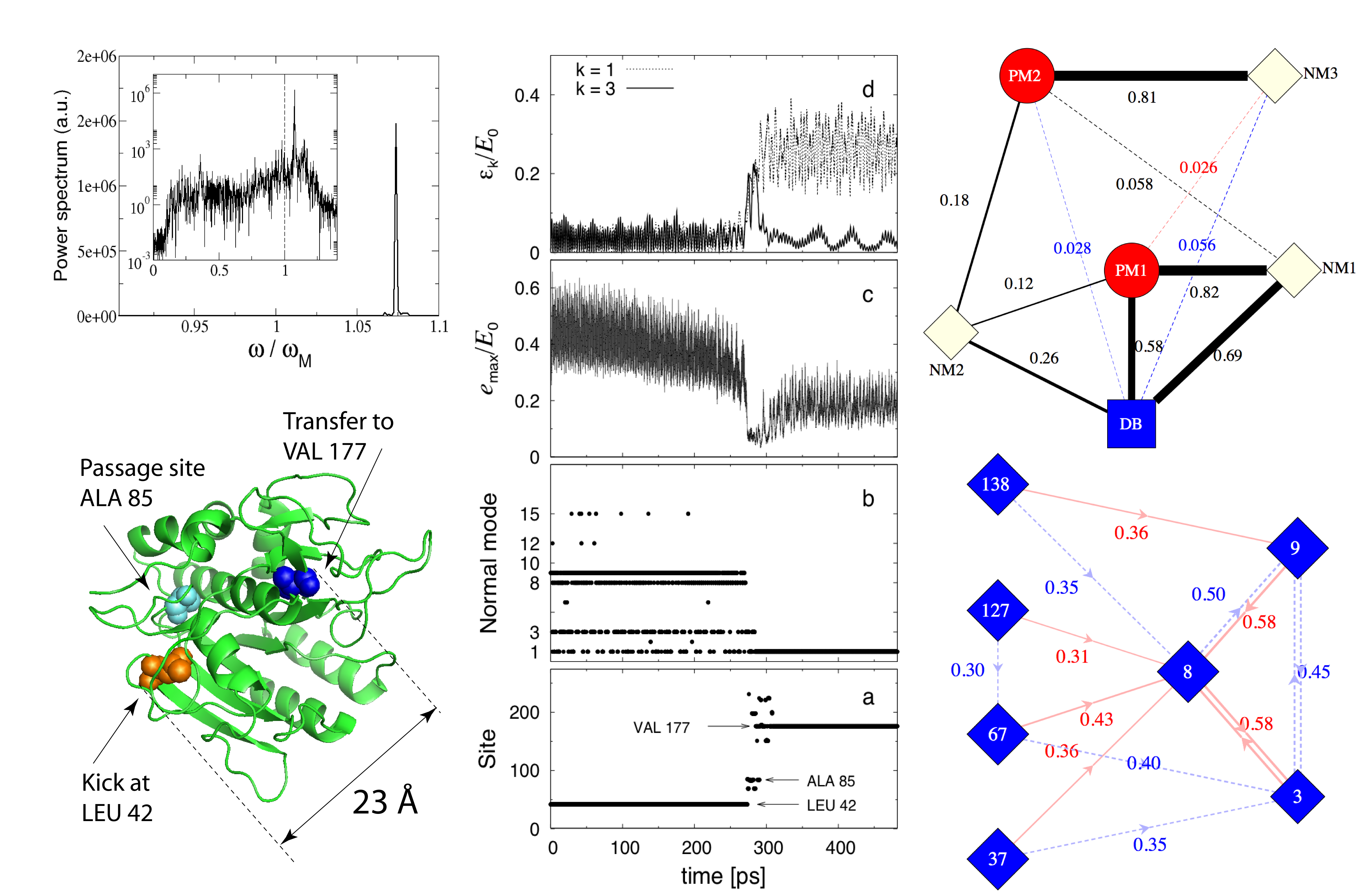
in detail the outcome of a single kick. Fig. 3 pictures a long-range transfer
event occurring when kicking at site LEU 42 in the enzyme Subtilisin. The middle-lower panel (a) shows
a plot of the most energetic site as a function of time, clearly illustrating the transfer
to site VAL 177, some 23 Å away, occurring at ps. The transfer process also involves
site ALA 85, as a passage site. Remarkably, a plot of the energy
of the most energetic site at time clearly shows that such passage coincides with a
redistribution of energy across the structure (see middle panel (c)). Subsequently, energy is
garnered from the neighborhood and stabilized in a localized mode centered at VAL 177, finally carrying about 20 %
of the total energy. This marks the true transfer event.
Such energy-harvesting, self-localized vibrations are generic in discrete non-linear systems
and are well-known as Discrete Breathers (DB) Flach and Willis (1998). These are robust, time-periodic
exponentially localized modes,
whose vibrational frequency lies outside the linear spectrum of the system.
In the context of the NNM, we have shown how accurate approximations of such periodic orbits can be calculated analytically,
reproducing the marked affinity of DB self-localization in topologically disordered media
for the stiffest spots Juanico et al. (2007); Piazza and Sanejouand (2008).
Here we have shown that DBs may also be excited as a consequence of localized impulses at considerable distances from
the excitation, playing the role of energy-accumulating transfer vectors.
In order to substantiate the above interpretation, we have performed Principal Component Analysis (PCA) on
an extended portion of the post-transfer dynamics. The power spectrum of the system trajectory projected
on the first principal mode (PM1) is shown in the upper left panel of Fig. 3, clearly revealing
the existence of a nonlinear, time-periodic excitation, reminiscent of chaotic DBs that self-localize
through modulational instability in nonlinear lattices Cretegny et al. (1998).
More insight as to why energy is transferred from LEU 42 a long distance away can be obtained
by turning to the space of Normal Modes (NM) .
Middle panel (b) of Fig. 3 reports the mode carrying
the highest energy
at time , where is the NM-transform
of the system coordinates ()
and are the NM frequencies. Before the transfer event,
energy is bounced among four high-frequency modes, NMs 1,3,8 and 9. This can be understood
by constructing the NM overlap network starting from the NMs with the largest projections on the
initial excitation unit vector are greatest (four modes in the first column of the
bottom graph in Fig. 3, NMs 138,127,67 and 37, making up about 60 % of ).
For a given NM , two links are drawn to the two-highest ranking NMs in the ordered list
of absolute overlap coefficients .
By doing this for the four NMs involved in the SMS vector,
a closed network emerges identifying the NM3
NM8 NM9
loop. Thus, in the presence of nonlinearity energy is immediately directed to a reduced group of NMs
via resonant overlap mechanisms. This finding agrees with results of atomistic simulations highlighting
the importance of spatial overlap for NM-NM energy transfer Moritsugu et al. (2000).
High-frequency NMs are strongly localized in space. In particular,
ALA 85 is the NM site (the site with largest displacement) in NM3 and the second NM site in NM8,
which explains the role of ALA 85 in the energy circulation process.
Before transfer, however, energy also bounces back and forth from NM1, the highest-frequency mode,
reflecting the nonlinear frequency shift on NM3 toward greater frequencies (see again panel b).
At energy starts departing the region around LEU 42 and a fluctuation pumping up
NM3 occurs (panel d), shifting its frequency upwards by virtue of nonlinearity. The energy at stake is
sufficient to trigger nonlinear localization and a DB finally installs at VAL 177, the NM site of NM1,
gathering vibrational energy from the background. Correspondingly, the energy on NM1 increases (see panel d).
To substantiate the above analysis, we have calculated analytically
the DB mode pattern centered at site VAL 177
with the technique described in Ref. Piazza and Sanejouand, 2008. Then we have built the
network connecting the first two principal modes, the first three NMs and the DB, where the links
are weighted by the normalized scalar products (upper graph in Fig. 3). As it shows, the
PMs essentially reflect the underlying competition between NM1 and NM3. In particular,
the first principal mode confirms the excitation of a DB emerging as a nonlinear continuation
of the edge normal mode, as predicted theoretically in Ref. Piazza and Sanejouand, 2008.
In agreement with this picture, kicks at ALA 42 of weaker energy resulted in a DB installing at MET 199,
the NM site of NM2. That is, less energy causes a smaller frequency shift and the DB branch
originating from the continuation of NM2 is excited instead. Reducing further, the transfer
is observed to halt at ALA 85, as explained by the NM overlap network.
In this paper we have shown how nonlinearity in a topologically non-regular system
boosts energy transfer to few specific locations. In enzyme structures, these coincide invariably
with the stiffest regions, also hosting the functionally relevant sites. Nonlinearity sharpens
the transfer selectivity, by reducing at the same time the transfer probability to generic locations.
The energy transferred by virtue of nonlinearity may be a conspicuous portion of the initial excitation,
in which cases localized vibrations akin to Discrete Breathers self-localize as energy-collecting centers,
often realizing amazingly efficient energy transfer channels across considerable distances.
References
- Best et al. (2007) R. B. Best, K. A. Merchant, J. M. Louis, I. V. Gopich, and W. A. Eaton, Biophysical Journal pp. 534A–534A (2007).
- Torchia and Ishima (2003) D. A. Torchia and R. Ishima, Pure and Applied Chemistry 75, 1371 (2003).
- Tirion (1996) M. M. Tirion, Physical Review Letters 77 (1996).
- Bahar et al. (1997) I. Bahar, A. R. Atilgan, and B. Erman, Folding & Design 2, 173 (1997).
- Hinsen (1998) K. Hinsen, Proteins 33, 417 (1998).
- Tama and Sanejouand (2001) F. Tama and Y. H. Sanejouand, Protein Engineering Design and Selection 14, 1 (2001).
- Micheletti et al. (2002) C. Micheletti, G. Lattanzi, and A. Maritan, J. Mol. Biol. 231, 909 (2002).
- Delarue and Sanejouand (2002) M. Delarue and Y.-H. Sanejouand, J. Mol. Biol. 320, 1011 (2002).
- Krebs et al. (2002) W. G. Krebs, V. Alexandrov, C. A. Wilson, N. Echols, H. Yu, and M. Gerstein, Proteins 48, 682 (2002).
- Kondrashov et al. (2007) D. Kondrashov, A. Van Wynsberghe, R. Bannen, Q. Cui, and G. Phillips, Structure 15, 169 (2007).
- Levy et al. (1982) R. Levy, D. Perahia, and M. Karplus, Proc. Natl. Acad. Sci. USA 79, 1346 (1982).
- Xie et al. (2000) A. H. Xie, L. van der Meer, W. Hoff, and R. H. Austin, Phys. Rev. Lett. 84, 5435 (2000).
- Edler et al. (2004) J. Edler, R. Pfister, V. Pouthier, C. Falvo, and P. Hamm, Phys. Rev. Lett. 93, 106405 (2004).
- Sagnella et al. (2000) D. Sagnella, J. Straub, and D. Thirumalai, J. Chem. Phys. 113, 7702 (2000).
- Leitner (2008) D. M. Leitner, Annual Review of Physical Chemistry 59, 233 (2008).
- Kopidakis et al. (2001) G. Kopidakis, S. Aubry, and G. P. Tsironis, Phys. Rev. Lett. 87, 165501 (2001).
- Archilla et al. (2002) J. F. R. Archilla, Y. B. Gaididei, P. L. Christiansen, and J. Cuevas, Journal of Physics A: Mathematical and General 35, 8885 (2002).
- Piazza and Sanejouand (2008) F. Piazza and Y. H. Sanejouand, Physical Biology 5 (2008).
- Juanico et al. (2007) B. Juanico, Y. H. Sanejouand, F. Piazza, and P. De Los Rios, Physical Review Letters 99 (2007).
- Brooks and Karplus (1985) B. R. Brooks and M. Karplus, Proc. Natl. Acad. Sci. USA 82, 4995 (1985).
- Marques and Sanejouand (1995) O. Marques and Y.-H. Sanejouand, Proteins 23, 557 (1995).
- Perahia and Mouawad (1995) D. Perahia and L. Mouawad, Comput. Chem. 19, 241 (1995).
- Flach and Willis (1998) S. Flach and C. R. Willis, Physics Reports 295, 181 (1998), ISSN 0370-1573.
- Cretegny et al. (1998) T. Cretegny, T. Dauxois, S. Ruffo, and A. Torcini, Physica D: Nonlinear Phenomena 121, 109 (1998).
- Moritsugu et al. (2000) K. Moritsugu, O. Miyashita, and A. Kidera, Physical Review Letters 85 (2000).