Experimental and theoretical study of oxygen adsorption structures on Ag(111)
Abstract
The oxidized Ag(111) surface has been studied by a combination of experimental and theoretical methods, scanning tunneling microscopy (STM), x-ray photoelectron spectroscopy (XPS), and density functional theory (DFT). A large variety of different surface structures is found, depending on the detailed preparation conditions. The observed structures fall into four classes: (a) individually chemisorbed atomic oxygen atoms, (b) three different oxygen overlayer structures, including the well-known phase, formed from the same Ag6 and Ag10 building blocks, (c) a structure not previously observed, and (d) at higher oxygen coverages structures characterized by stripes along the high-symmetry directions of the Ag(111) substrate. Our analysis provides a detailed explanation of the atomic-scale geometry of the Ag6/Ag10 building block structures, and the and stripe structures are discussed in detail. The observation of many different and co-existing structures implies that the O/Ag(111) system is characterized by a significantly larger degree of complexity than previously anticipated, and this will impact our understanding of oxidation catalysis processes on Ag catalysts.
pacs:
68.43.Bc, 68.37.EfI Introduction
The oxidation of the Ag(111) surface is a fascinating example of how a seemingly simple system can escape a detailed understanding for decades, in spite of repeated and thorough efforts. The history of surface science investigations of the oxidation of Ag(111), briefly reviewed recently,Michaelides et al. (2005) started back in the sixties and early seventies with a couple of single crystal studiesMüller (1966); Dégeilh (1969); Deydari and Mee (1973) and took a serious upswing with the investigations by Rovida et al. in the seventies,Rovida et al. (1972, 1974) who, in particular, reported on the now renowned phase. In the following much of the effort was directed towards this phase,Albers et al. (1977a); Campbell (1985a); Bare et al. (1995); Bukhtiyarov et al. (1999); Carlisle et al. (2000a, b); Li et al. (2003a, b, c); Michaelides et al. (2003); Bocquet et al. (2003a, b); Michaelides et al. (2005); Schnadt et al. (2006); Schmid et al. (2006); Reichelt et al. (2007a) but also numerous other studies of more general character appeared,Engelhardt and Menzel (1976); Albers et al. (1977b, a); Benndorf et al. (1983); Grant and Lambert (1984); Campbell (1985a); Schmeisser and Jacobi (1985); Campbell (1986); Spruit et al. (1989); Spruit and Kleyn (1989); Wang et al. (1991); Reijnen et al. (1991); Bao et al. (1993); Pettinger et al. (1994); de Mongeot et al. (1995); Raukema et al. (1996); Bao et al. (1996); Lacombe et al. (1996); Rocca et al. (1997); Schedel-Niedrig et al. (1997); Bukhtiyarov et al. (1999); Li et al. (2002, 2003a); Michaelides et al. (2003); Stegelmann and Stoltze (2004); Reichelt et al. (2007a); Reicho et al. (2007); Reichelt et al. (2007b) as well as works concerned with the reactivity of O/Ag(111) towards, in particular, the oxidation of ethene and methanol. Campbell (1985b, c); Tan et al. (1987); van Santen and Kuipers (1987); Hawker et al. (1989); Mukoid et al. (1990); Wu et al. (1994); Carley et al. (1998); Boronin et al. (1999); Avdeev and Zhidomirov (2001); Scheer et al. (2002); Stacchiola et al. (2001); Klust and Madix (2006); Gao et al. (2007); Gomes et al. (2007); Greeley and Mavrikakis (2007); Zhou and Madix (2008); Huang et al. (2008); Christopher and Linic (2008) Only recently the atomic-scale geometric model for the phase was completely revised on the basis of a combination of scanning tunnelling microscopy (STM), surface x-ray diffraction (SXRD), and x-ray photoemission spectroscopy (XPS) experiments and density functional theory calculations.Schnadt et al. (2006); Schmid et al. (2006) Subsequent, detailed low-energy electron diffraction and CO reaction studies supported this atomic model.Reichelt et al. (2007b); Klust and Madix (2007) At the same time an increasing number of studies have indicated that the phase diagram of the oxidized Ag(111) surface is more complex than previously anticipated. While earlier studies only stipulated the existence of the and an atomic adsorbate layer (including a phase with a local R symmetry), recent studies have reported phases containing both lessReicho et al. (2007), equally much,Carlisle et al. (2000b); Schnadt et al. (2006); Reichelt et al. (2007a) and moreSchnadt et al. (2006) oxygen than the phase (cf. Table 1). Many of the reactivity studies have assumed that the catalytical properties of Ag are associated with the structure due to the anticipation that this phase should be predominant under oxygen pressures similar to those under reaction conditions.Li et al. (2003c, b) These investigations have in general not taken into account the existence of further phases and, hence, a much more complex phase diagram for the Ag-O system will fundamentally change our picture of what happens in the catalytic process. At present, growing evidence exists that the static phases found in surface science studies may neither be responsible for the surface’s catalytic activity itself nor be preserved during catalytic operation.Stampfl et al. (2002); Hendriksen et al. (2004); Ackermann et al. (2005); Reuter and Scheffler (2006); Reichelt et al. (2007a) The surface is rather to be considered as a dynamic medium, the structure of which changes in response to the changing chemical environment. The formation and breakup of these static Ag-O phases are still of significant interest due to the phases’ role in the overall dynamics of the catalytic process. Thus surface science studies with their unprecedented ability of clarifying the atomic-scale structure still retain their value and validity.
Here we present a detailed and thorough experimental and theoretical study of oxygen adsorption on Ag(111), with data concerning both previously observed structures and hitherto new unobserved phases of oxidized Ag(111). The present study shows that the phase is embedded in a wider context of two additional phases, the rect and rect structures. It is also found that the structure, to our knowledge in contrast to the rect and rect phases, can host foreign atoms and molecules. Finally, the existence of two more phases with a and ”stripe” character is reported.
II Experimental and theoretical methods
The experiments were carried out in the STM laboratory and at the vacuum ultraviolet/soft x-ray synchrotron radiation facility ASTRID at Aarhus University. In the STM laboratory we used an ultra-high vacuum (UHV) chamber with a base pressure of Torr, equipped with standard instrumentation for sample cleaning, the Aarhus STM,Lægsgaard et al. (1988) a low-energy electron diffractometer, and a thermal gas cracker from Oxford Instruments for atomic oxygen exposure with a cracking efficiency which in our experiments varied from 30% at to 14% at Torr total pressure. Directly connected to the UHV chamber is a small and compact high pressure cell which makes it possible to dose molecular oxygen at pressures up to one atmosphere. After oxygen exposure the sample was characterized by STM and, in some cases, low-energy electron diffraction (LEED). The transfer between the high pressure cell and the UHV chamber could be accomplished within less than 15 min of oxygen exposure. Most of the STM experiments were performed at room temperature, although some of the STM images were recorded at liquid nitrogen temperature, which sometimes resulted in higher spatial resolution.
For the XPS measurements the surfaces were prepared and initially characterized in the STM chamber and then brought to the SX-700 beamlineUggerhøj (1995) of ASTRID in a vacuum suitcase with a base pressure better than Torr. The pressure during transfer from the vacuum chambers to the suitcase, which could be accomplished within a minute, rose to in between and Torr. The base pressure of the UHV chamber of the SX-700 beamline is Torr. After the XPS measurement the sample was transferred back to the STM chamber and the status of the sample reinvestigated by STM. The control experiments ensured that the examined structures were still present and clearly visible in the STM, although some minor deterioration of the surface might have occurred.
The Ag(111) surface was cleaned by repeated sputter/annealing cycles. It was then oxidized by exposing it to either atomic oxygen at partial atomic oxygen pressures of to Torr or molecular oxygen at pressures between 0.5 and 10 Torr. During oxygen exposure the Ag(111) sample was held at temperatures in between 420 and 600 K for an exposure time of 10 to 50 min. Following this recipe, the structure of the oxidized surface varied with oxygen pressure, sample temperature, and exposure time. As discussed below, the prepared surface typically exhibited co-existing domains of different surface phases. However, it was possible to prepare single phase surfaces for some of the structures presented below by carefully tuning the preparation parameters.
Bare et al.,Bare et al. (1995) have proposed an alternative preparation procedure, in which the surface is exposed to NO2 at pressures between to Torr, while the sample is kept at the same temperatures as described above. This method has also been used in a variety of subsequent studies.Carlisle et al. (2000a, b); Scheer et al. (2002); Huang and White (2002a, b); Bocquet et al. (2003c); Huang and White (2003); Webb et al. (2004); Alemozafar and Madix (2005); Reichelt et al. (2007b); Klust and Madix (2007); Zhou et al. (2008) Here we do not present any results obtained on samples prepared by this NO2 method, although we did carry out a limited number of such studies. Consistent with Carlisle et al.,Carlisle et al. (2000b) we were able to produce the atomic oxygen, rect-O, and -O phases in these studies, which indicates that the use of NO2 instead of atomic or molecular oxygen does not significantly change the picture drawn up here.
All the density functional theory (DFT) calculations reported here were carried out with the Perdew-Burke-Ernzerhof generalized gradient approximationPerdew et al. (1996) in periodic supercells within the plane-wave pseudopotential formalism as implemented in the CASTEP code.Segall et al. (2002) As discussed below, a variety of unit cells were used, all consisting of five layer Ag(111) slabs and with fine Monkhorst-Pack k-point meshes equivalent to at least 24241 per (11) surface unit cell. During structure optimizations the top layer of Ag atoms as well as the oxide overlayer atoms were allowed to relax, whilst the bottom four layers of Ag were held fixed. From the DFT results simulated STM images were obtained using the simple Tersoff-Hamann approximation.Tersoff and Hamann (1985) A “tip” height of 2.5 Å above the highest atom in each overlayer for occupied states within 2 eV of the Fermi level was used.
III Results and discussion
III.1 Overview of the observed surface structures
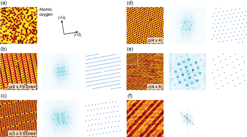
Figure 1 displays STM images of all the phases which we have observed experimentally. The symmetry assignments (rect, rect, and for the hitherto non-assigned phases; see below for a more detailed description) were derived on the basis of the STM results. The middle parts of the panels represent the fast Fourier transform (FFT) of the STM images, while the right-hand parts reproduce LEED simulations obtained from the symmetry assignments provided on the basis of the STM observations. These LEED results are in excellent agreement with the FFTs, which lends further credibility to the STM-based symmetry assignments for the oxygen-induced phases.
At the lowest oxygen coverages an apparently disordered arrangement of depressions is observed in the constant current STM images (Fig. 1(a)). The depressions are interpreted as atomic oxygen adsorbates, consistent with previous studiesCarlisle et al. (2000b) and the well-known fact that oxygen depletes the local density of states at the Fermi level, leading to a depression-like appearance of atomic oxygen on metal surfaces.Besenbacher (1996) Domains of this disordered structure are also frequently observed to co-exist with domains of the structures shown in Figs. 1(b) to (d).
| Structure | Oxygen | Atomic | Molecular | NO2 |
|---|---|---|---|---|
| coverage | oxygen | oxygen | ||
| Atomic oxygen | 0.05 ML111Reference Carlisle et al., 2000b. | 222Present work.,∗ | 333Reference Reichelt et al., 2007a.,∗ | 111Reference Carlisle et al., 2000b.,∗ |
| R30∘ | 0.33 ML444Based on the model in Ref. Bao et al., 1993. | 555A R30∘ overlayer was identified by MüllerMüller (1966) on epitaxially grown Ag(111) films for relatively small oxygen exposures (100 L). Later results on a Ag(111) single crystal, which show no formation of overlayers after oxygen exposure up to 3000 L under otherwise similar conditions, contradict these initial experiments.Engelhardt and Menzel (1976) An overlayer with a local R30∘ symmetry was once again reported by Bao et al. who used STM to study Ag samples exposed to pressures around one atmosphere for long times on the order of daysBao et al. (1993, 1996) (see also Ref. Schubert et al., 1995). Overall, the observed phase was argued to be of higher-order commensurability. | ||
| rect | 0.375 ML | 666Reference Schnadt et al., 2006.,∗ | 333Reference Reichelt et al., 2007a.,∗ | 111Reference Carlisle et al., 2000b.,∗ |
| rect | 0.4 ML | 666Reference Schnadt et al., 2006.,∗ | 222Present work.,∗ | |
| 0.375 ML | 666Reference Schnadt et al., 2006.,∗ | 777Reference Rovida et al., 1972.,∗ | 888Reference Bare et al., 1995.,∗ | |
| 0.37 ML999Based on the model in Ref. Reicho et al., 2007. | 101010Reference Reicho et al., 2007. | |||
| 0.25 ML111111Based on the tentative model in Section III.4. | 222Present work.,∗ | |||
| Stripe phase | ? | 222Present work.,∗ | 222Present work.,∗ |
| Structure | Oxidizing agent | Pressure | Sample temperature | Exposure time |
|---|---|---|---|---|
| rect | Atomic O | Torr | 500 K | 10 min |
| rect + bare Ag(111) | Atomic O | Torr | 450 - 500 K | 5 - 15 min |
| rect111Contained ”defects” of the rect phase (cf. Figure 1(c)). | Atomic O | Torr | 500 K | 10 - 20 min |
| O2 | 10 Torr | 500 K | 10 min | |
| Stripes | Atomic O | Torr | 500 K | 10 min |
The structure in Fig. 1(b) was previously observed by Carlisle et al.Carlisle et al. (2000b) and was assigned to the decomposition of the structure in panel (d). The structure, which we briefly mentioned in a previous report,Schnadt et al. (2006) has a rect unit cell in the convenient terminology for rectangular symmetries on hexagonal surfaces introduced by Biberian and van Hove.Biberian and van Hove (1984) In matrix notation the unit cell (the conventional and primitive unit cells are identical) is described by
The structure in Fig. 1(c) also possesses a rectangular symmetry with a conventional unit cell of rect symmetry. The brighter lines from left to right are units of the rect structure. We have observed that domains of the rect structure always contain at least small fractions of defects of rect symmetry, which in room temperature experiments sometimes have been observed to move along the [10] direction. The primitive unit cell of the rect phase is given byPre
Panel (d) of Fig. 1 shows the well-known structure, which has been the subject of surface science investigations since the early 1970s, whereas the structures in panels (e) and (f) are reported here for the first time. The first of these has a symmetry, while the appearance of the second structure is characterized by stripes of variable thickness along the {10} surface directions. In matrix notation the primitive unit cell of the structure is given by
Table 1 summarizes the conditions, which here and in other studies have been used to prepare the different surface structures. Both the atomic and molecular oxygen recipes are very versatile in terms of the variety of structures which can be produced. The NO2 recipe has so far primarily been used to prepare oxidized Ag(111) surfaces covered entirely by the structure.Bare et al. (1995); Carlisle et al. (2000a, b); Schmid et al. (2006); Reichelt et al. (2007b) The present results show that also the rect structure can be produced by exposing the Ag(111) crystal to NO2. Due to the similarity of the , rect, and rect structures (see below) it can be expected that it also is possible to form the latter using NO2, while it is more difficult to draw conclusions along these lines regarding the remaining structures.
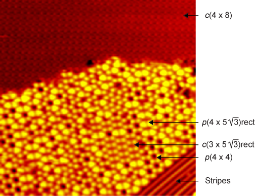
For most preparation conditions a variety of co-existing surface structures appear. Only for the preparation conditions summarized in Table 2 we were able to obtain single-phase or close-to single-phase surfaces. The otherwise observed co-existence of different surfaces structures is illustrated in Fig. 2, which displays an STM image recorded after exposing the surface to Torr atomic oxygen at 500 K for 40 min. In this STM image the rect, rect, , , and stripe structures are found to co-exist within a 250 Å 250 Å area of the surface. It is observed that the rect, rect, and structures have very similar STM appearances with respect to their height profiles (brightness), whereas the STM height profile for the the structure is much lower. As will be seen and discussed in further detail below this reflects that the atomic geometries of the rect, rect, and structures are indeed very similar.
III.2 Ag6/Ag10 building block structures
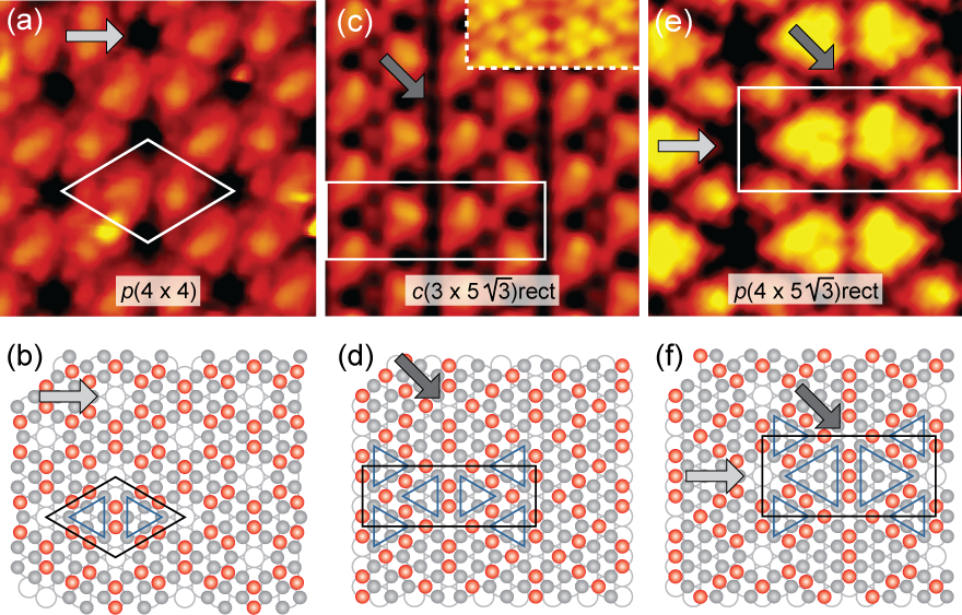
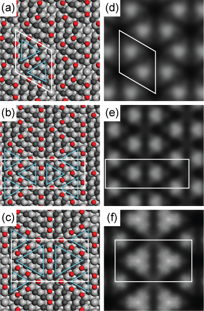
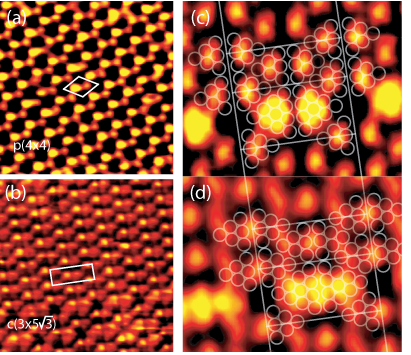
The STM images of the rect, rect, and phases in Figs. 1(b-d) are characterized by ordered arrays of unresolved bright features. Higher resolution images are depicted in Figs. 3(a), (c), and (e). From these it is seen that the bright features contain atomic sub-structure. In the case of the rect phase a particular tip state even allowed all Ag atoms in the overlayer to be resolved (inset of panel (c)). For the rect and phases the arrangement of the silver and oxygen atoms has already been discussed in detail in Ref. Schnadt et al., 2006. In brief, both structures are built from close-to identical elements of Ag6 triangles with the Ag atoms of a single Ag6 all located either in fcc or hcp sites. Each primitive unit cell contains one fcc triangle and one hcp triangle. The site difference implies a slight asymmetry between the two triangles of a dimer, which, at least for the phase, is by DFT shown to be further enhanced by small rotations in converse directions for the fcc and hcp trianglesSchmid et al. (2006); Reichelt et al. (2007b) [cf. Fig.4]. Indeed, for certain tip states fcc triangles and hcp triangles are imaged differently in STM, as is illustrated in Figure 5(a-b). On the basis of the STM measurements we thus propose the structural models depicted in Figures 3(b) and (d). It is difficult to provide a detailed assignment for the location of the oxygen atoms, which normally are not visible in STM due to the depletion of the local density of states at the Fermi level typical for oxygen (see, however, below for an example in which some oxygen density is visible). For the rect and phases the oxygen atom positions were derived by evaluating a large variety of different structures in DFT calculations.Schnadt et al. (2006); Schmid et al. (2006) The positions of the oxygen atoms in the thus derived structural models have been included in the models shown in Fig. 3(b) and (d), which were suggested on the basis of the experimental STM results. Figures 4(a) and (b) show the structural models emerging from the calculations. Particulary noteworthy are the converse rotations of the Ag6 triangles in the structure already mentioned above. The STM simulations, which correspond to the calculated structures, are shown in panels (d) and (e) and are found to be in good agreement with the experimental STM results in Figure 3(a) and (c). The DFT adsorption energies (eV/O) are provided in Table 3. They are seen to be the same to within the uncertainty of the DFT results.
The arrangement of the Ag atoms in the rect structure in Figure 3(e) can be understood from an extension of the Ag6 building block model for the rect and phases. The larger triangles constitute an additional Ag10 building block, which typically appears in dimer form, again with half of the silver atoms in fcc and the other half in hcp sites. In the pure rect phase the Ag6 and Ag10 dimers are then arranged as depicted in Fig. 3(f). It is noteworthy that the rect structure appears as a mixture of the and rect overlayers, with the voids between four Ag10 and two Ag6 triangles being rect-like (dark arrow in Fig. 3(f)) and those between four Ag6 and two Ag10 being -like (light arrow).
As in the cases of the and rect phases discussed above the oxygen atoms incorporated into the rect structure are normally not visible in STM. Again, we carried out DFT calculations for a large number of different arrangements of both oxygen and silver atoms to find the most stable arrangement of surface atoms. This search also included geometries which deviated from the experimentally derived Ag6/Ag10 building block structure. Among the investigated structures, the one displayed in Figure 3(f) was found to be the most stable one. The model is indeed based on the two different building blocks suggested above on the basis of the STM results. The oxygen atoms reside in the same sites as for the and rect phases, a finding which lends further credibility to the proposed model. The DFT oxygen adsorption energy (eV/O) (cf. Table 3) is seen to be the same, to within the DFT uncertainty, as that of the other two Ag6 building block structures.
Fig. 4 contains the final structural models for all three Ag6/Ag10 phases in panels (a)-(c). The Figure also shows that the Tersoff-Hamann (TH) STM simulations are in good agreement with the STM results. In particular, the TH STM simulations reproduce the experimentally observed high apparent height of the silver atoms in the centers of the Ag6/Ag10 triangles, which are characterized by a high silver coordination. The lower-Ag-coordinated edge atoms and even more so the corner atoms of the Ag triangles have a much lower local density of states intensity and therefore appear lower than the triangle center atoms. We also note that by slightly modifying the simulated tip apex height and bias conditions we are not only able to reproduce the experimentally observed triangular appearance of the building blocks shown in Figs. 4(d-f), but also the more spherical appearance of the bright features in Fig. 1(b-d).
The above derived models can explain the frequently observed alignment of defects of the rect type embedded in a rect domain. An example is shown in Fig. 1(c). A comparison of the structural models for the rect and rect phases shows that the local geometry of the Ag6 building blocks is very similar in both phases, and, in particular, the distance between the Ag6 triangles along the [11] surface direction is the same for both structures. Thus, an aligned line of Ag10 triangles, which is the second building block of the rect structure, fits very well into a domain of the rect phase. If now, in a thought experiment, a single Ag10 triangle is moved from an aligned line to an isolated position, the structural mismatch between the Ag10 and Ag6 triangles will either lead to a reduction of the number of oxygen atoms accommodated in the structure or, alternatively, to less favorable Ag-O bond geometries. Either possibility will be accompanied by an energetic cost, which, deeming from the experimental results, is not fully compensated by the gain in configurational entropy.
In Table 3 we compare the stoichiometries and top layer silver and oxygen atom densities of the three Ag6/Ag10 building block structures. Both the and rect phases have a Ag2O stoichiometry, but the rect has slightly higher Ag and O atom densities than the . Judging from the stoichiometry the rect phase seems to be more oxygen-deficient; however, the absolute oxygen density is the same for the and rect structures, while the Ag density is increased for the rect and reaches the same level as in the rect. It is thus not correct to view the rect phase as a decomposition phase of the , as suggested by Carlisle et al.Carlisle et al. (2000b) While this characterization applies to the rect in relationship to the rect phase, the rect structures rather corresponds to a Ag-rich reconstruction of the structure.
From the similar geometrical arrangements of the silver and oxygen atoms in the three Ag6/Ag10 building block structures as revealed in the STM images it seems likely that the electronic and chemical characteristics of these phases should be very similar. This is corroborated by the O 1s x-ray photoelectron spectra depicted in Fig. 6. Within the measuremental uncertainty of 0.1 eV the O 1s binding energies are the same for all three structures (528.5 eV) and they agree quite well with previous measurements on the phaseCampbell (1985a) (528.1 eV), although they are about 0.4 eV higher in energy (they are more similar to the recent measurement by Reichelt et al.,Reichelt et al. (2007a) who report a binding energy of 528.3 eV). However, the O 1s peak observed for the phase prepared from atomic oxygen is located at 528.7 eV, and this upwards shift in energy we cannot explain at present. It is further noted that all spectra contain a high energy component at energies between 530.1 and 530.5 eV. This peak is particularly pronounced for the phase prepared using molecular oxygen, i.e., at an elevated pressure. Previously, O 1s features at high binding energy have been assigned to both carbonatesRehren et al. (1991); Bukhtiyarov et al. (1999); Reichelt et al. (2007a), a reactive oxygen species or chemisorbed oxygenBare et al. (1995); Bukhtiyarov et al. (1999); Reichelt et al. (2007a), and – for the Ag(110), Ag(210), and Ag(111) surfaces as well as polycrystalline silver foil – to subsurface or bulk-dissolved oxygen.Rehren et al. (1991); Pawela-Crew and Madix (1995); Bao et al. (1996); Savio et al. (2006); Klust and Madix (2006) Considering that contaminations released from the reactor walls are common in high pressure experiments such as used in the molecular oxygen preparation of the phase (10 Torr in the present case) it seems most likely that a large part of the peak is related to carbonates. On the basis of the available data we cannot, however, provide a conclusive assignment of this high binding energy O 1s peak. Indeed, it is fully possible that this peak contains contributions from several different oxygen species.
Finally it is noted, that it is sometimes possible to directly image some of the surface oxygen density in the STM images (see, e.g., also Ref. Merte et al., 2009). As stated above, for normal STM tunneling conditions oxygen adsorbed on Ag is not easily identified in the STM images, but, as demonstrated in Figure 5(d) for a mixed rect/ rect surface, rare tip states seem to enhance those oxygen atoms which are coordinated by four top layer silver atoms. The oxygen atoms are identified from a comparison to the STM image in normal imaging mode in Figure 5(c) and the structural models in Figure 3(d-f). In this special tip state image the silver atoms of the smaller Ag6 triangles are not imaged at all, while the centers of the Ag10 triangles appear nearly as bright as the fourfold-coordinated oxygen atoms. The threefold-coordinated oxygens, however, remain invisible even for the special tip state.
| Stoichiometry | Ag density | O density | (eV/O) | |
|---|---|---|---|---|
| Ag2O | 0.104 Å-2 (0.75 ML) | 0.052 Å-2 (0.375 ML) | 0.46 | |
| rect | Ag2O | 0.111 Å-2 (0.80 ML) | 0.055 Å-2 (0.40 ML) | 0.39 |
| rect | Ag2O0.94 | 0.111 Å-2 (0.80 ML) | 0.052 Å-2 (0.375 ML) | 0.42 |
| Ag2O1.6 | 0.043 Å-2 (0.31 ML) | 0.035 Å-2 (0.25 ML) |


III.3 The phase as a host-guest structure
In Figure 7 STM images of the phase are shown, which were recorded at varying bias voltages. As shown in panel (a), at the lowest biases of approximately meV the STM image exhibits the typical honeycomb appearance with the structural voids displayed as depressions (cf. the atomic model in Fig. 3(b)). When the bias is increased to about mV a fraction of the voids appears filled (panel (b)). At the highest biases used, around mV, the fillings already visible at the intermediate bias are now the brightest features in the STM image, while other voids display fillings with a smaller apparent height in the image [panel (c)]. Only a very minor fraction of voids seem to be unfilled.
We frequently observed the presence of fillings in the voids of the structure; indeed, it is likely that they are an inherent feature of the phase. The fillings are most easily imaged at elevated biases of both tunneling polarities. At low biases they normally remain unobserved, and, thus, their absence in STM images recorded at low biases does not imply their absence from the surface. The fillings occur in preparations from both atomic and molecular oxygen, but we cannot clarify here whether they exist for preparations from NO2, as well. Interestingly, we have not seen any such filling for the rect phase, although it – in contrast to the rect structure – contains the same kind of voids as visible from the structural models in Fig. 3.
We suggest that the fillings are related to adsorbate species in the voids of the phase. Since we observed such fillings both directly after preparation of the surface by either atomic or molecular oxygen as well as after measurements for a time span of several hours, it seems unlikely that rest gas contamination can be made responsible for the fillings. Nonetheless, at present we cannot specify the chemical nature of the fillings and we can just note that the observation of the fillings suggests an interesting host-guest character of the phase, which would deserve a more systematic study.
III.4 The and stripe structure
Finally we would like to discuss two additional structures not observed previously. These are the and stripe phases of Figure 1(e-f). In both cases it is difficult to provide the exact details of the atomic scale structures. The reasons are that for the structure we have so far not been able to find a recipe for preparing a single phase surface, and for the stripe structure there exists a larger manifold of similar, co-existing structures. The difficulty in preparing surfaces covered by a single coherent structure have prevented the use of averaging techniques such as LEED, which would have resulted in useful additional information. In the following we therefore limit ourselves to primarily describing the characteristics of the structures to the extent they could be extracted from the STM data.
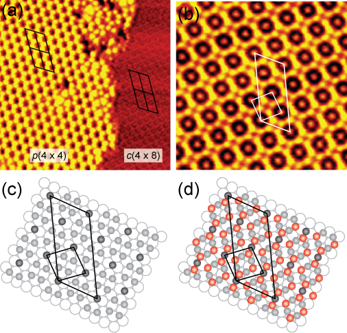
Starting with the structure, its symmetry assignment was derived from comparing its unit cell with those of other phases appearing in the same STM image. Such a comparison is shown in Fig. 8(a). The correctness of the assignment is supported by the good agreement between the FFT of the STM image and the LEED simulation based on the unit cell in Figure 1(e). Since oxygen typically is invisible in the STM images it is assumed that the bright features in the more highly resolved STM images of the structure correspond to silver atoms, cf. Fig. 8(b). This assignment leads us to suggest the tentative top layer Ag atom geometry depicted in Fig. 8(c). It is the only geometry for which the bright features in the experimental image can be brought into coincidence with lattice sites. In this model, 20% of the top layer silver atoms reside in bridging sites, while the remaining silver atoms are found in both hcp and fcc hollow sites.
The ring-formed depressions around the Ag atoms in bridging sites indicate that the Ag atoms are surrounded by oxygen atoms. The placement of four oxygen atoms in the dark ring leads to the detailed geometry shown in Figure 8(d). In this geometry the coordination of the silver atoms by other top layer silver atoms is much lower than in the building block structures described in section III.2. In these structures silver atoms with a low silver coordination such as the corner atoms of the Ag6/Ag10 triangles were imaged with a reduced apparent height as compared to the fully silver-coordinated silver atoms in the centers of the triangles. As seen clearly in Figure 8(a), also the phase is characterized by a much smaller apparent height than the brightest features of the phase. We take this as an indication that the structure indeed is more highly oxidized than any of the building block structures.
The suggested structure for the phase possesses a Ag5O4 (Ag2O1.6) stoichiometry (cf. Table 3). A very similar structure was observed previously for a Pd5O4 surface oxide.Lundgren et al. (2002) A major difference between the present and the Pd oxide case is, however, the full commensurability of the Ag5O4 unit cell, while the Pd5O4 is incommensurate in one surface direction. It is seen that the proposed stoichiometry for the is more reminiscent of AgO than Ag2O and that the density of Ag and O atoms is much lower in the as compared to the Ag6/Ag10 building block structures. This difference in stoichiometry and Ag and O densities in comparison to the , , and phases is surprising in view of the coexistence of these structures such as those in Figure 2. At present we cannot satisfactorily explain this finding.
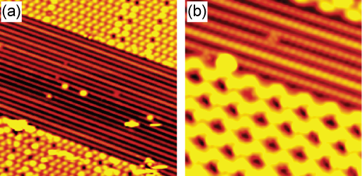
Now turning to the stripe phase, we would first like to again emphasize that there exists a larger number of co-existing structures which appear as stripes in the STM measurements. Probably these structures have similar, but not exactly equal atomic geometries. That the geometries indeed differ between different domains of the stripe phase is quite obvious from a comparison of the displayed domains in Figs. 1(f) and 9. The common denominator of the domains is the occurrence of stripes along {10}, but the arrangement and apparent height of the stripes are not always the same.
While it was difficult in general to obtain high resolution STM images on extended domains of stripe character, this was sometimes possible on smaller domains embedded in other structures. Figure 9 provides examples of such high-resolution STM images. We tentatively assume that the protruding features of these images are silver atoms. Along the {10} directions the distance between these features corresponds to the Ag(111) nearest-neighbor distance Å. In the perpendicular direction the distances of the rows is, however, not a multiple of , as might have been expected. By comparison to the surrounding surface covered by domains of the , rect, and rect phases it is estimated that the repetitive unit rather comprises four stripes and that this unit has a width of . In such a unit cell it is not possible to place all the silver atoms in the same lattice site. The differing brightness of adjacent stripes supports this conclusion.
The position of the oxygen atoms in the stripe phase cannot be specified in any detail from an analysis of the STM results. It seems likely that oxygen atoms reside in between the bright stripes. The XPS data in Figure 6 indicate that the oxygen content is considerably higher than in the and rect phases. Similar to the structure and the corner atoms of the triangles in the building block structures, the stripes are imaged at a smaller apparent height compared to that of the adjacent domains of primarily rect symmetry in panel (a) of Figure 9 and primarily in panel (b). This suggests that the silver coordination of the Ag atoms of the stripe phase is smaller than that of the Ag atoms in the centers of the triangles of the building block structures, which indicates a higher oxygen coordination and thus a higher oxygen content of the stripe phase. However, at present, we refrain from a more quantitative analysis, partly since it is unclear to what extent the XPS data represent fully covered surfaces and partly since the STM data cannot provide any more detailed information. It is noted, though, that metal/oxygen structure with quite a high oxygen content and exhibiting a stripe appearance have been observed on other metal surfaces such as Pt(110).Helveg et al. (2007)
IV Conclusions
We have shown from an interplay of STM and XPS experiments and DFT calculations that the oxidation of the Ag(111) surface may be structurally more complex than previously anticipated. Depending on the preparation conditions – oxidation agent, dose, and pressure, exposure time, and sample temperature during and after exposure – a large variety of different structures are found, essentially all of which may coexist. Among these phases we have identified a number of structures, which are formed from Ag6/Ag10 fundamental building blocks. This group of structures comprises both the renowned surface as well as two phases of rect and rect symmetry. The rect structure had been observed previously,Carlisle et al. (2000b) but we have devised a structural model which is in line with the geometries for the (Refs. Schnadt et al., 2006 and Schmid et al., 2006) and rect phases (Ref. Schnadt et al., 2006) proposed recently and discussed in more detail here. In addition, we have observed two additional structures of and stripe character.
The present results suggest that a complex coexistence of oxide and oxygen adsorbate overlayers with varying contents of oxygen may form under the conditions of industrial oxidation catalysis. This is in contrast to the prevalent perception that the phase alone represents an adequate model for the surface under such conditions. It is conceivable that the complexity of the phase diagram has a profound influence on the dynamics of the catalytic process. Hence, although an increased understanding of the structure of the oxidized silver surface is presently being obtained as demonstrated in this and other studies,Schnadt et al. (2006); Schmid et al. (2006); Klust and Madix (2007); Reicho et al. (2007); Reichelt et al. (2007a, b) we are still far from a detailed understanding of the oxidized silver surface under reaction conditions. It appears likely that the structure of the silver oxide surface may vary in response to changes in the gas composition and the chemical potential of the reaction intermediates.
Acknowledgements.
The competent assistance of the staff of the mechanical workshop of the Department of Physics and Astronomy in Aarhus is gratefully acknowledged. JS wishes to thank the European Commission for funding through a Marie Curie Intra-European Fellowship. AM is grateful to the Alexander von Humboldt foundation for partial support of this work and the European Science Foundation and EPSRC for a European Young Investigator Award (EURYI). Some of the calculations performed here were made possible by AM’s membership of the UK’s HPC Materials Chemistry Consortium, which is funded by EPSRC (EP/F067496).References
- Michaelides et al. (2005) A. Michaelides, K. Reuter, and M. Scheffler, J. Vac. Sci. Technol. A 23, 1487 (2005).
- Müller (1966) K. Müller, Z. Phys. 195, 105 (1966).
- Dégeilh (1969) R. Dégeilh, Vide 139, 29 (1969).
- Deydari and Mee (1973) A. W. Deydari and C. H. N. Mee, Phys. Status Solidi A 17, 247 (1973).
- Rovida et al. (1972) G. Rovida, F. Pratesi, M. Maglietta, and E. Ferroni, J. Vac. Sci. Technol. 9, 796 (1972).
- Rovida et al. (1974) G. Rovida, F. Pratesi, M. Maglietta, and E. Ferroni, Surf. Sci. 43, 230 (1974).
- Albers et al. (1977a) H. Albers, W. J. J. van der Wal, and G. A. Bootsma, Surf. Sci. 68, 47 (1977a).
- Campbell (1985a) C. T. Campbell, Surf. Sci. 157, 43 (1985a).
- Bare et al. (1995) S. R. Bare, K. Griffiths, W. N. Lennard, and H. T. Tang, Surf. Sci. 342, 185 (1995).
- Bukhtiyarov et al. (1999) V. I. Bukhtiyarov, V. V. Kaichev, and I. P. Prosvirin, J. Chem. Phys. 111, 2169 (1999).
- Carlisle et al. (2000a) C. I. Carlisle, D. A. King, M.-L. Bocquet, J. Cerdá, and P. Sautet, Phys. Rev. Lett. 84, 3899 (2000a).
- Carlisle et al. (2000b) C. I. Carlisle, T. Fujimoto, W. S. Sim, and D. A. King, Surf. Sci. 470, 15 (2000b).
- Li et al. (2003a) W.-X. Li, C. Stampfl, and M. Scheffler, Phys. Rev. B 67, 045408 (2003a).
- Li et al. (2003b) W.-X. Li, C. Stampfl, and M. Scheffler, Phys. Rev. B 68, 165412 (2003b).
- Li et al. (2003c) W.-X. Li, C. Stampfl, and M. Scheffler, Phys. Rev. Lett. 90, 256102 (2003c).
- Michaelides et al. (2003) A. Michaelides, M.-L. Bocquet, P. Sautet, A. Alavi, and D. A. King, Chem. Phys. Lett. 367, 344 (2003).
- Bocquet et al. (2003a) M.-L. Bocquet, A. Michaelides, D. Loffreda, P. Sautet, A. Alavi, and D. A. King, J. Am. Chem. Soc. 125, 5620 (2003a).
- Bocquet et al. (2003b) M.-L. Bocquet, A. Michaelides, P. Sautet, and D. A. King, Phys. Rev. B 68, 075413 (2003b).
- Schnadt et al. (2006) J. Schnadt, A. Michaelides, J. Knudsen, R. T. Vang, K. Reuter, E. Lægsgaard, M. Scheffler, and F. Besenbacher, Phys. Rev. Lett. 96, 146101 (2006).
- Schmid et al. (2006) M. Schmid, A. Reicho, A. Stierle, I. Costina, J. Klikovits, P. Kostelnik, O. Dubay, G. Kresse, J. Gustafson, E. Lundgren, et al., Phys. Rev. Lett. 96, 146102 (2006).
- Reichelt et al. (2007a) R. Reichelt, S. Günther, M. Rößler, J. Wintterlin, B. Kubias, B. Jakobi, and R. Schlögl, Phys. Chem. Chem. Phys. 9, 3590 (2007a).
- Engelhardt and Menzel (1976) H. A. Engelhardt and D. Menzel, Surf. Sci. 57, 591 (1976).
- Albers et al. (1977b) H. Albers, J. M. M. Droog, and G. A. Bootsma, Surf. Sci. 64, 1 (1977b).
- Benndorf et al. (1983) C. Benndorf, M. Franck, and F. Thieme, Surf. Sci. 128, 417 (1983).
- Grant and Lambert (1984) R. B. Grant and R. M. Lambert, Surf. Sci. 146, 256 (1984).
- Schmeisser and Jacobi (1985) D. Schmeisser and K. Jacobi, Surf. Sci. 156, 911 (1985).
- Campbell (1986) C. T. Campbell, Surf. Sci. 173, L641 (1986).
- Spruit et al. (1989) M. E. M. Spruit, E. W. Kuipers, F. H. Geuzebroek, and A. W. Kleyn, Surf. Sci. 215, 421 (1989).
- Spruit and Kleyn (1989) M. E. M. Spruit and A. W. Kleyn, Chem. Phys. Lett. 159, 342 (1989).
- Wang et al. (1991) X.-D. Wang, W. T. Tysoe, R. G. Greenler, and K. Truszkowska, Surf. Sci. 257, 335 (1991).
- Reijnen et al. (1991) P. H. F. Reijnen, A. Raukema, U. van Slooten, and A. W. Kleyn, Surf. Sci. 253, 24 (1991).
- Bao et al. (1993) X. Bao, J. V. Barth, G. Lehmpfuhl, R. Schuster, Y. Uchida, R. Schlögl, and G. Ertl, Surf. Sci. 284, 14 (1993).
- Pettinger et al. (1994) B. Pettinger, X. Bao, I. C. Wilcock, M. Muhler, and G. Ertl, Phys. Rev. Lett. 72, 1561 (1994).
- de Mongeot et al. (1995) F. B. de Mongeot, U. Valbusa, and M. Rocco, Surf. Sci. 339, 291 (1995).
- Raukema et al. (1996) A. Raukema, D. A. Butler, F. M. A. Box, and A. W. Kleyn, Surf. Sci. 347, 151 (1996).
- Bao et al. (1996) X. Bao, M. Muhler, T. Schedel-Niedrig, and R. Schlögl, Phys. Rev. B 54, 2249 (1996).
- Lacombe et al. (1996) S. Lacombe, F. Cemič, P. He, H. Dietrich, P. Geng, M. Rocca, and K. Jacobi, Surf. Sci. 368, 38 (1996).
- Rocca et al. (1997) M. Rocca, F. Cemič, F. Buatier de Mongeot, U. Valbusa, S. Lacombe, and K. Jacobi, Surf. Sci. 373, 125 (1997).
- Schedel-Niedrig et al. (1997) T. Schedel-Niedrig, X. Bao, M. Muhler, and R. Schlögl, Ber. Bunsenges. Phys. Chem. 101, 994 (1997).
- Li et al. (2002) W.-X. Li, C. Stampfl, and M. Scheffler, Phys. Rev. B 65, 075407 (2002).
- Stegelmann and Stoltze (2004) C. Stegelmann and P. Stoltze, Surf. Sci. 552, 260 (2004).
- Reicho et al. (2007) A. Reicho, A. Stierle, I. Costina, and H. Dosch, Surf. Sci. 601, L19 (2007).
- Reichelt et al. (2007b) R. Reichelt, S. Günther, J. Wintterlin, W. Moritz, L. Aballe, and T. O. Mentes, J. Chem. Phys. 127, 134706 (2007b).
- Campbell (1985b) C. T. Campbell, J. Phys. Chem. 89, 5789 (1985b).
- Campbell (1985c) C. T. Campbell, J. Catal. 94, 436 (1985c).
- Tan et al. (1987) S. A. Tan, R. B. Grant, and R. M. Lambert, J. Catal. 104, 156 (1987).
- van Santen and Kuipers (1987) R. A. van Santen and H. P. C. E. Kuipers, Adv. Catal. 35, 265 (1987).
- Hawker et al. (1989) S. Hawker, C. Mukoid, J. P. S. Badyal, and R. M. Lambert, Surf. Sci. 219, L615 (1989).
- Mukoid et al. (1990) C. Mukoid, S. Hawker, J. P. S. Badyal, and R. M. Lambert, Catal. Lett. 4, 57 (1990).
- Wu et al. (1994) K. Wu, X. Wei, Y. Cao, D. Wang, and X. Guo, Catal. Lett. 26, 109 (1994).
- Carley et al. (1998) A. F. Carley, P. R. Davies, and M. W. Roberts, Curr. Op. Sol. State Mat. Sci. 2, 525 (1998).
- Boronin et al. (1999) A. I. Boronin, V. I. Avdeev, S. V. Koshcheev, K. T. Murzakhmetov, S. F. Ruzankin, and G. M. Zhidomirov, Kinetics Catal. 40, 653 (1999).
- Avdeev and Zhidomirov (2001) V. I. Avdeev and G. M. Zhidomirov, Surf. Sci. 492, 137 (2001).
- Scheer et al. (2002) K. C. Scheer, A. Kis, J. Kiss, and J. M. White, Topics Catal. 20, 43 (2002).
- Stacchiola et al. (2001) D. Stacchiola, G. Wu, M. Kaltchev, and W. Tysoe, J. Mol. Catal. A: Chem. 167, 13 (2001).
- Klust and Madix (2006) A. Klust and R. J. Madix, Surf. Sci. 600, 5025 (2006).
- Gao et al. (2007) W. Gao, M. Zhao, and Q. Jiang, J. Phys. Chem. C 111, 4042 (2007).
- Gomes et al. (2007) J. R. B. Gomes, S. Gonzalez, D. Torres, and F. Illas, Russ. J. Phys. Chem. B 1, 292 (2007).
- Greeley and Mavrikakis (2007) J. Greeley and M. Mavrikakis, J. Phys. Chem. C. 111, 7992 (2007).
- Zhou and Madix (2008) L. Zhou and R. J. Madix, J. Phys. Chem. C 112, 4725 (2008).
- Huang et al. (2008) W. X. Huang, Z. Jiang, and J. M. White, Catal. Today 131, 360 (2008).
- Christopher and Linic (2008) P. Christopher and S. Linic, J. Am. Chem. Soc. 130, 11264 (2008).
- Klust and Madix (2007) A. Klust and R. J. Madix, J. Chem. Phys. 126, 084707 (2007).
- Stampfl et al. (2002) C. Stampfl, M. V. Ganduglia-Pirovano, K. Reuter, and M. Scheffler, Surf. Sci. 500, 368 (2002).
- Hendriksen et al. (2004) B. L. M. Hendriksen, S. C. Bobaru, and J. W. M. Frenken, Surf. Sci. 552, 229 (2004).
- Ackermann et al. (2005) M. D. Ackermann, T. M. Pedersen, B. L. M. Hendriksen, O. Robach, S. C. Bobaru, I. Popa, C. Quiros, H. Kim, B. Hammer, S. Ferrer, et al., Phys. Rev. Lett. 95, 255505 (2005).
- Reuter and Scheffler (2006) K. Reuter and M. Scheffler, Phys. Rev. B 73, 045433 (2006).
- Lægsgaard et al. (1988) E. Lægsgaard, F. Besenbacher, K. Mortensen, and I. Stensgaard, J. Microsc. Oxford 152, 663 (1988).
- Uggerhøj (1995) E. Uggerhøj, Nucl. Instrum. Meth. Phys. Res. B 99, 261 (1995).
- Zhou et al. (2008) L. Zhou, W. W. Gao, A. Klust, and R. J. Madix, J. Chem. Phys. 128, 054703 (2008).
- Huang and White (2002a) W. X. Huang and J. M. White, Catal. Lett. 84, 143 (2002a).
- Huang and White (2002b) W. X. Huang and J. M. White, Langmuir 18, 9622 (2002b).
- Bocquet et al. (2003c) M.-L. Bocquet, P. Sautet, J. Cerda, C. I. Carlisle, M. J. Webb, and D. A. King, J. Am. Chem. Soc. 125, 3119 (2003c).
- Huang and White (2003) W. X. Huang and J. M. White, Surf. Sci. 529, 455 (2003).
- Webb et al. (2004) M. J. Webb, S. M. Driver, and D. A. King, J. Phys. Chem. B 108, 1955 (2004).
- Alemozafar and Madix (2005) A. R. Alemozafar and R. J. Madix, Surf. Sci. 587, 193 (2005).
- Perdew et al. (1996) J. P. Perdew, K. Burke, and M. Ernzerhof, Phys. Rev. Lett. 77, 3865 (1996).
- Segall et al. (2002) M. D. Segall, P. L. D. Lindan, M. J. Probert, C. J. Pickard, P. J. Hasnip, S. J. Clark, and M. C. Payne, J. Phys.: Cond. Matt. 14, 2717 (2002).
- Tersoff and Hamann (1985) J. Tersoff and D. R. Hamann, Phys. Rev. B 31, 805 (1985).
- Besenbacher (1996) F. Besenbacher, Rep. Prog. Phys. 59, 1737 (1996).
- Schubert et al. (1995) H. Schubert, U. Tegtmeyer, D. Herein, X. Bao, M. Muhler, and R. Schlögl, Catal. Lett. 33, 305 (1995).
- Biberian and van Hove (1984) J. P. Biberian and M. A. van Hove, Surf. Sci. 138, 361 (1984).
- (83) In a previous communicationSchnadt et al. (2006) we based the matrix notation on a different set of substrate unit vectors with an inscribed angle of 60∘ between the two unit vectors. Here we instead assume 120∘. In the previously used notation the matrix is written .
- Rehren et al. (1991) C. Rehren, M. Muhler, X. Bao, R. Schlögl, and G. Ertl, Z. Phys. Chem. 174, 11 (1991).
- Pawela-Crew and Madix (1995) J. Pawela-Crew and R. J. Madix, J. Catal. 153, 158 (1995).
- Savio et al. (2006) L. Savio, A. Gerbi, L. Vattuone, A. Baraldi, G. Comelli, and M. Rocca, J. Phys. Chem. B 110, 942 (2006).
- Merte et al. (2009) L. R. Merte, J. Knudsen, L. C. Grabow, R. T. Vang, E. Lægsgaard, M. Mavrikakis, and F. Besenbacher, Surf. Sci. 603, L15 (2009).
- Lundgren et al. (2002) E. Lundgren, G. Kresse, C. Klein, M. Borg, J. N. Andersen, M. De Santis, Y. Gauthier, C. Konvicka, M. Schmid, and P. Varga, Phys. Rev. Lett. 88, 246103 (2002).
- Helveg et al. (2007) S. Helveg, W. X. Li, N. C. Bartelt, S. Horch, E. Lægsgaard, B. Hammer, and F. Besenbacher, Phys. Rev. Lett. 98, 115501 (2007).